Chapter 36
ORBITAL TRAUMA

The orbit is the cavity that houses, protects, and sustains the eyeball. It consists of seven bones (see Table 36–1), encompassing a pear-shaped area containing 30 mL3 of volume. Some of the bones of the orbit are among the thickest in the skull, while others among the thinnest.
HISTORY
In prehistoric times there was ample evidence of orbital trauma and its sequelae. The remains of an Incan sacrifice has been found frozen in the Andes Mountains with orbital fractures that were probably associated with human sacrifice. Archaeological evidence from Europe demonstrates that Neanderthals could sustain extensive orbital trauma, yet survive and live for many years.
With modern life, new mechanisms of injury have developed, including high-speed missiles (bullets) and high-velocity impacts (e.g., in MVCs). These new mechanisms of injury require new methods of prevention and repair.
| Strong | Weak | |
|---|---|---|
| Ethmoid | + | |
| Frontal | + | |
| Lacrimal | + | |
| Maxilla | + | |
| Palatine | + | |
| Sphenoid | + | |
| Zygomatic | + |
EPIDEMIOLOGY (USEIR DATA)
Rate of orbital involvement among all serious injuries: 15% ; breakdown:
◦ fracture: 78%;
◦ foreign body: 24%;
◦ hemorrhage: 1%.
Age (years):
• range: 0–103;
• average: 27;
• rate of 0- to 9-year-olds among the total: 8%;
• rate of 10- to 19-year-olds among the total: 22%;
• rate of ≥60-year-olds among the total: 7%.
Sex: 78% male.
Place of injury:
• street and highway: 29%;
• home: 28%;
• recreation and sport: 13%;
• industrial premises: 7%;
• public building: 5%.
Source of injury:
• various blunt objects: 36%;
• MVC: 23%;
• gunshot: 11%;
• BB/pellet gun: 11%;
• fall: 7%;
• various sharp objects: 4%.
Globe involvement among the total: 22%.
Safety laws often specify ocular and orbital protection. The use of safety glasses (see Chapter 27), the development of shatterproof windshieldsa for cars, and motorcycle/bicycle helmets have all been beneficial for the reduction of orbital injuries. Federal, state, and local governments have enacted laws designed to protect workers. Increased awareness through education, advertising, and publicity has elevated the public’s attention to accidents in general and orbital and ocular trauma in particular. Exercising common sense, however, remains the responsibility of the individual.
PATHOPHYSIOLOGY
The orbit’s primary role is to protect the eyeball. The thick bones of the lateral and superior orbital rims provide firm protection; conversely, bones constituting the medial wall and the floor of the orbit are the thinnest and weakest in the human skull.
PEARL… The combination of superior and lateral strength with medial and inferior wall weakness allows dissipation of energy when the orbit is struck.
The ability of the orbital floor to fracture selectively when the orbit is struck is an evolutionary masterpiece, a feature that is similar to a safety valve. When the energy from a blow to the orbit is dissipated through the fractured bone, it commonly saves the eyeball from rupturing.
PEARL… It must always be remembered that through the orbital bones course the nerves and vessels needed to sustain the orbit and the eye, and on the other side of the orbital bones are other, vital, organs, such as the brain.
EVALUATION
The evaluation of a patient with suspected orbital trauma begins with a general examination to determine whether the nervous system is intact and what the patient’s general medical condition is (see Chapter 10).
PITFALL
Injuries to the orbit are often associated with severe neurologic injuries, which are life threatening and take precedence over the orbital treatment.1,2
Presence or absence of enophthalmos or exophthalmos can be confirmed during physical examination. Evaluation of extraocular muscle movements may be performed and documented to check for restriction. Elements of an orbital examination include:
• Hertel exophthalmometry;
• extraocular muscle movement evaluation;
• visual acuity;
• sensory examination in the distribution of the supra- and infraorbital nerves;
• APD;
• palpation of the orbital rims; and
• auscultation for bruits.
A variety of imaging options (see Chapters 9 and 24) is available to assist with the evaluation of orbital trauma. They are always used in conjunction with an adequate physical examination. CT scan, MRI, and ultrasound all have advantages and disadvantages, and the initial physical examination will determine which tests to obtain and which are unnecessary (see Chapter 8).3
Orbital CT scans provide the best images of the relationship between the bone and soft tissue. This study is usually cheaper, faster, and more readily available than the MRI scan. The following are indications for ordering a CT scan:
• suspected orbital fractures;
• palpable bone step-offs;
• restricted extraocular muscle movements; and
• metallic orbital foreign bodies.
An MRI scan is best at differentiating soft tissue and may be the best diagnostic choice in cases of:
• associated neurologic damage; and
• wooden foreign bodies.4
PEARL… Specific contraindications for MRI scan include known or suspected ferrous intraorbital foreign bodies.5
An ultrasound examination is often not available in an ER setting, although it is helpful in identifying orbital abscess.
PEARL… The single most important test to perform when evaluating orbital trauma is visual acuity testing. With visual acuity loss, the orbital trauma becomes an orbital emergency (see Chapter 12).
The loss of vision implies pressure or impingement on the optic nerve or the eyeball. Emergency imaging and intervention should be anticipated. True orbital emergencies include:
• orbital abscess;
• optic nerve sheath hematoma;
• orbital foreign body in contact with the optic nerve; and
• open globe injury in conjunction with orbital injuries.
TYPES OF TRAUMA AND THEIR MANAGEMENT
Orbital Blowout Fracture
It is one of the most common orbital injuries encountered in an ER. Antecedent history often confirms a blow to the orbit with an object larger than the opening of the orbit itself, typically a fist or a softball. The classical clinical triad of a blowout fracture (Fig. 36–1) includes:
• diplopia caused by restrictive strabismus;
• infraorbital numbness caused by interruption of the inferior orbital nerve; and
• periocular ecchymosis.
Blowout fractures are caused by fractures of the bones in the inferior medial orbit.b There are two theories concerning the cause of blowout fractures.6
1. The direct injury theory describes a sudden compression of the globe with a fracture of the floor caused by increased orbital and ocular pressure.
2. The indirect injury theory postulates that a blow to the inferior orbital rim causes a ripple effect in the orbital bones, which leads to fracture at their weakest point.
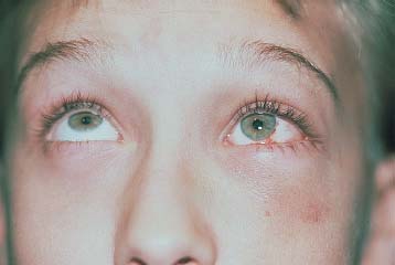
FIGURE 36–1 Left orbital blowout fracture; the patient presented with diplopia, infraorbital numbness, and periocular ecchymosis.
Both mechanisms of injury are probably involved in blowout fractures.
• The ecchymosis seen in conjunction with blowout fractures is caused by direct damage to the skin and orbicularis muscle.
• The restricted extraocular muscle movements typically seen with blowout fracture involve the bony entrapment of the inferior rectus muscle or suspensory ligaments. The entrapment leads to limited vertical eye muscle movements. Restrictive strabismus can be confirmed with a forced duction test.
 Forced duction testing requires topical anesthesia and small forceps. Once the conjunctiva is anesthetized, the insertion of the inferior rectus muscle is purchased with forceps.c
 A positive forced duction test indicates that there is palpable restriction to rotating the eyeball superiorly. A positive forced duction test is strong evidence that a blowout fracture is present with entrapment of the inferior rectus muscle.
PEARL… Entrapment is a clinical sign and cannot be diagnosed on CT examination.
Stay updated, free articles. Join our Telegram channel

Full access? Get Clinical Tree


