(1)
St. Johns, FL, USA
(2)
Helen Keller Foundation for Research and Education, International Society of Ocular Trauma, Birmingham, AL, USA
(3)
Consultant and Vitreoretinal Surgeon, Milos Eye Hospital, Belgrade, Serbia
(4)
Consultant and Vitreoretinal Surgeon, Zagórskiego Eye Hospital, Cracow, Poland
51.1 General Considerations
In this condition liquid is channeled through the optic pit1 (see Fig. 51.1) under the macula and into the retina itself2; it is most probably liquor. The subretinal fluid is different in its composition from that in RD; the visual acuity can chronically remain excellent despite the serous detachment. Eventually, however, there will be extensive RPE abnormalities and permanent loss of vision (Fig. 51.2).
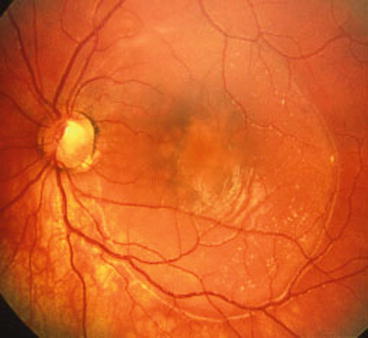
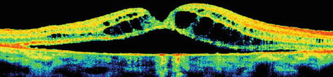
Fig. 51.1
Optic pit and related serous detachment of the macula. The pit, visible as small whitish-grayish area just temporal to the emergence of the blood vessels on the disc, caused a long-standing macular detachment. The border of the detached is clearly delineated; the chronicity of the condition is shown by the secondary pigmentary changes around the fovea and the presence of subretinal precipitates
Fig. 51.2
OCT image of the macular changes in an eye with optic pit. There is subretinal and intraretinal fluid. Obviously, the condition is not a true retinoschisis, simply large areas of (confluent) cystic retina
Stay updated, free articles. Join our Telegram channel

Full access? Get Clinical Tree


