Purpose
To evaluate the morphologic characteristics of optic nerve head drusen.
Design
Retrospective case series.
Methods
setting : Institutional (Seoul National University Bundang Hospital). patients : Sixty-one patients with optic nerve head drusen. observation procedure : Visible and buried optic nerve head drusen were identified using funduscopy, whereas homogenous and nonhomogenous optic nerve head drusen were identified using spectral-domain optical coherence tomography images. Buried optic nerve head drusen were classified according to the size. main outcome measures : Classification of optic nerve head drusen.
Results
Of 99 eyes in 61 patients, optic nerve head drusen were buried in 95 eyes and visible in 4 eyes. The patients with visible optic nerve head drusen were older on average than those with buried optic nerve head drusen (53.3 ± 8.6 years vs 13.5 ± 7.1 years; P < .001) and exhibited larger disc diameters (1643 ± 265 μm vs 1287 ± 185 μm; P = .016). All 4 eyes with visible optic nerve head drusen exhibited hyperreflective borders, which were not found in patients with buried optic nerve head drusen. Of 95 eyes with buried optic nerve head drusen, 64 eyes (67%) showed homogenous internal reflectivity, whereas 31 eyes (33%) showed nonhomogenous reflectivity with lobulations. Large optic nerve head drusen were associated with a small optic disc diameter, nonhomogenous internal reflectivity, a partial highly reflective border, intraretinal cysts, and increased temporal retinal nerve fiber layer thickness.
Conclusions
Optic nerve head drusen have a diverse spectrum of spectral-domain optical coherence tomography findings associated with patient age and disc size.
Differentiating optic nerve head drusen from true optic disc edema is important because optic nerve head drusen usually have a benign natural course. Ultrasonography, the past gold standard of diagnosing optic nerve head drusen, could detect only the calcification of optic nerve head drusen. The recent introduction of optical coherence tomography (OCT) has heralded a new era for the diagnosis of optic nerve head drusen. Johnson and associates characterized optic nerve head drusen as having a “lumpy-bumpy” internal contour with an abrupt decline in subretinal hyporeflective space when imaged by OCT. Choi and associates reported photoreceptor disruption in a patient with long-standing optic nerve head drusen who was evaluated using OCT. Differentiation of optic disc edema from optic nerve head drusen depended on the quantitative analysis of peripapillary retinal nerve fiber layer (RNFL) thickness. However, because of the low resolution of OCT scans, the direct visualization of optic nerve head drusen was not possible until the appearance of spectral-domain (SD) OCT.
We reported that optic nerve head drusen could exist as a focal mass without calcified borders posterior to the outer plexiform and outer nuclear layers when imaged with the SD OCT. However, there was controversy about the findings of optic nerve head drusen with SD OCT: both hyporeflective and hyperreflective appearances. We investigated various optic nerve head drusen characteristics with SD OCT after the completion of the previous study. We thought this controversy stemmed from the diverse spectrum of optic nerve head drusen. Furthermore, optic nerve head drusen evolve over the years. Therefore, optic nerve head drusen in patients of different ages will represent various stages of development. The SD OCT captures these differences. To the best of our knowledge, no published report has examined the SD OCT features of optic nerve head drusen in a large patient population representing multiple age groups. The purpose of this study was to investigate the morphologic characteristics of optic nerve head drusen as visualized using SD OCT. The results obtained were used to categorize the various kinds of optic nerve head drusen.
Methods
This study was approved by the Institutional Review Board of Seoul National University Bundang Hospital and adhered to the tenets of Declaration of Helsinki. This study was designed as a retrospective case series. Sixty-one patients with optic nerve head drusen seen at our institution from September 2009 through May 2012 were included in this study. All patients underwent a full ophthalmologic examination initially, including measurement of best-corrected visual acuity, funduscopic examination under mydriasis, fundus photography, and SD OCT (Spectralis OCT; Heidelberg Engineering, Heidelberg, Germany). In cases of definite or suspected optic disc edema, thorough neuro-ophthalmologic examinations including brain magnetic resonance imaging, lumbar puncture, neurologic examinations, visual field tests, and fluorescein angiography were performed for the differential diagnosis.
The diagnosis of optic nerve head drusen requires direct visualization by funduscopy or SD OCT. To be considered as a visible optic nerve head drusen, the drusen must be seen to protrude from the disc on funduscopic images. To be considered as a buried optic nerve head drusen, SD OCT should reveal the drusen to be localized as a mass between the outer retina and retinal pigment epithelium–choroid. For this evaluation, we used multiple horizontal and vertical SD OCT scans centered on the optic disc. One hundred OCT frames were averaged to obtain high-resolution horizontal scans dissecting the center of the optic nerve head, and 10 frames were averaged to obtain multiple horizontal or vertical scans. With SD OCT, optic nerve head drusen were classified by whether they had hyperreflective walls. Optic nerve head drusen without hyperreflective walls were classified as homogenous and nonhomogenous according to the internal reflectivity. The common peripapillary changes such as multiple intraretinal cysts and a partial hyperreflective border along the optic nerve head drusen also were assessed using SD OCT.
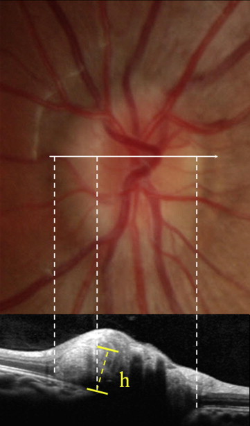
Buried optic nerve head drusen were classified further by vertical height ( Figure 1 ). Using the SD OCT caliper tool, we determined the height of the optic nerve head drusen as measured from the retinal pigment epithelium layer. On the basis of these measurements, the buried optic nerve head drusen were categorized as small (< 300 μm), medium (300 to 500 μm), or large (≥ 500 μm). The peripapillary retinal thickness (approximately 300 μm) was adopted as a cutoff value in determining small optic nerve head drusen. Disc diameter was measured in 2 different planes at the retinal surface by using confocal infrared images and at the choroidal plane by measuring the size of the opening in the Bruch membrane bordering the optic nerve. The mean horizontal and vertical disc diameters were calculated and used for intergroup comparison.
Demographics were compared using the Mann–Whitney U test and Kruskal–Wallis test. Correlations among optic disc size, optic nerve head drusen height, and RNFL thickness were obtained by calculating the Pearson correlation. RNFL thickness and disc diameter were compared against the nature of optic nerve head drusen (buried, visible) and the size of the optic nerve head drusen (small, medium, or large) using the Kruskal–Wallis test. In cases of bilateral optic nerve head drusen, similarities between the eyes in terms of drusen nature and size were determined using the κ index. Only 1 eye (the right eye) was selected for further comparison when significant concordance was observed. Intraindividual comparisons of disc diameter and drusen height were performed using the paired t test. The proportions of cases in each size class that had specific features (internal heterogeneity, highly reflective borders along the optic nerve head drusen, intraretinal cysts) were compared using the linear-by-linear association method. Statistical analysis was performed using SPSS software version 18.0 (SPSS Inc, Chicago, Illinois, USA).
Results
General Characteristics and Classification of Optic Nerve Head Drusen
Optic nerve head drusen were identified in 99 eyes from 61 patients and were not found in the other 23 eyes. Among this group, 44 patients (72%) were women and 38 patients (62%) had bilateral optic nerve head drusen. Optic nerve head drusen were visible in 4 eyes from 3 patients by funduscopic examination ( Figure 2 ), whereas buried optic nerve head drusen were detected in 95 eyes from 58 patients with SD OCT ( Figure 1 ; Table 1 ). Ninety-five eyes with buried optic nerve head drusen were classified further by vertical height of the drusen as measured using SD OCT. Optic nerve head drusen were small (< 300 μm) in 30 eyes, medium in 57 eyes (300 to 500 μm), and large in 8 eyes (≥ 500 μm; Figure 3 ). In patients with bilateral optic nerve head drusen (n = 38), drusen nature (buried or visible) was in perfect agreement (κ, 1.000; P < .001) between right and left eyes. Drusen size (small, medium, or large) was not similar between both eyes (κ = .232; P = .082). Optic nerve head drusen height differed between the 2 eyes (right eye, 374 ± 90 μm; left eye, 343 ± 84 μm; P = .017, paired t test), but was correlated closely ( r = 0.657; P < .001, Pearson correlation). Twenty-three patients had unilateral optic nerve head drusen (2 eyes with visible drusen, 21 eyes with buried drusen). Except for 1 fellow eye with optic disc atrophy, fellow eyes were larger than the eyes with drusen in patients with unilateral buried optic nerve head drusen (retinal surface plane: 1480 ± 198 μm vs 1336 ± 145 μm; P = .006; choroidal plane: 1549 ± 165 μm vs 1493 ± 168 μm; P = .279, paired t test). Optic disc edema was detected in only 1 patient who was diagnosed as having optic disc edema and concomitant optic nerve head drusen after a neuro-ophthalmologic examination and serial SD OCT follow-up. As the level of edema decreased, the buried optic nerve head drusen became apparent on the SD OCT images.
| Buried Drusen (n = 58) a | Visible Drusen (n = 3) a | P Value b | |
|---|---|---|---|
| Age | 13.5 ± 7.1 | 53.3 ± 7.1 | <.001 |
| Male-to-female ratio | 16:42 | 1:2 | |
| BCVA (logMAR) | −0.06 ± 0.22 | −0.06 ± 0.10 | .838 |
| Refraction (D) | −3.30 ± 2.60 | −3.92 ± 4.69 | .693 |
| Disc size R (μm) | 1287 ± 185 | 1643 ± 265 | .016 |
| Disc size C (μm) | 1525 ± 190 | 1950 ± 428 | .053 |
| RNFL thickness (μm) | |||
| Average | 119 ± 23 | 95 ± 31 | .155 |
| Superotemporal | 152 ± 42 | 112 ± 48 | .187 |
| Temporal | 110 ± 25 | 102 ± 34 | .765 |
| Inferotemporal | 182 ± 39 | 144 ± 50 | .250 |
| Inferonasal | 111 ± 33 | 93 ± 33 | .341 |
| Nasal | 54 ± 22 | 53 ± 16 | .841 |
| Superonasal | 108 ± 53 | 65 ± 21 | .069 |
a Only 1 eye (right eye) was selected for statistical analysis in patients with bilateral optic nerve head drusen.
The characteristic SD OCT features of optic nerve head drusen were the subretinal mass or spots, which contrasted with the low background reflectivity of the outer nuclear layer. The reflectivity of optic nerve head drusen was similar to that of retinal plexiform layers. Patients with visible optic nerve head drusen were older than patients with buried optic nerve head drusen (53.3 ± 8.6 years vs 13.5 ± 7.1 years; P < .001, Mann–Whitney U test). When eyes were classified into 2 groups according to age (10 eyes from patients 29 to 61 years of age and 89 eyes from patients 6 to 20 years of age), the older group had 4 visible optic nerve head drusen, 1 case of nonhomogenous buried optic nerve head drusen, and 5 cases of homogenous optic nerve head drusen. The buried optic nerve head drusen in the older group were small in 2 patients, medium in 3 patients, and large in 1 patient. Although all 4 cases of visible optic nerve head drusen were found in the older group, the cases with buried optic nerve head drusen in both age groups were similar in terms of internal heterogeneity ( P = .667, Fisher exact test) and optic nerve head drusen size ( P = .730, chi-square test).
Visible Optic Nerve Head Drusen
Four eyes from 3 patients (Patients 1, 2, and 3) had visible optic nerve head drusen. In all eyes with visible optic nerve head drusen, peripapillary chorioretinal atrophy encircled the entire disc ( Figure 2 , Top). In both eyes from Patient 1, the RNFL had atrophied, and optic nerve head drusen were scattered mostly inside but also outside the scleral inlets of the discs. The optic nerve head drusen were located in the superonasal or inferonasal side of the disc, which is where the buried optic nerve head drusen usually are located ( Figure 2 , Second row, Bottom). In Patient 2, RNFL thickness was within the normal range, and optic nerve head drusen existed as a single mass located around the superior quadrant and inside the scleral inlet of the disc. In Patient 3, optic nerve head drusen existed as multiple lumps located around the inferonasal quadrant of the disc.
In all 4 eyes with visible optic nerve head drusen, the optic nerve head drusen were characterized by hyperreflective walls contrasted with internal hyporeflective spaces or cysts ( Figure 2 ). Some deep cystic optic nerve head drusen were detectable only by SD OCT ( Figure 2 , Bottom). Multiple intraretinal cysts were observed as well ( Figure 2 , Third row). The average age of patients with visible optic nerve head drusen was greater than that of patients with buried optic nerve head drusen (53.3 ± 8.6 years vs 13.5 ± 7.1 years; P < .001, Mann–Whitney U test). Patients with visible optic nerve head drusen also exhibited larger optic disc diameters at the retinal surface plane (1643 ± 265 μm; vs 1287 ± 185 μm; P = .016, Mann–Whitney U test) and reduced RNFL thickness in the superotemporal and superonasal quadrants ( Table 1 ).
Buried Optic Nerve Head Drusen
Buried optic nerve head drusen were detected in 95 eyes from 58 patients. In all cases, optic nerve head drusen existed as a C-shaped lump that correlated with a halo adjacent to the nasal border of the optic disc ( Figure 1 ). In contrast to visible optic nerve head drusen, hyperreflective walls were not observed in all cases. On SD OCT images, the peripapillary retinal structures were deformed by the underlying optic nerve head drusen, and the outer plexiform layer and choroid diverged as they approached the optic nerve head. Because of the low background reflectivity of the outer nuclear layer, the margins of buried optic nerve head drusen could be delineated with ease ( Figure 1 ). In all cases of buried optic nerve head drusen, the RNFL exhibited pseudoedema, which corresponds to the halo in the fundus photographs ( Table 1 ).
Among 95 eyes with buried optic nerve head drusen, 28 eyes (30%) exhibited heterogeneous internal reflectivity and lobulations indicating the presence of nonhomogeneous material ( Figure 4 ). Thirty eyes (32%) had optic nerve head drusen with partial highly reflective borders ( Figure 5 , arrows). Thirteen eyes (14%) displayed multiple intraretinal cysts ( Figure 5 , arrowheads).
Stay updated, free articles. Join our Telegram channel

Full access? Get Clinical Tree


