Membranes on the macular surface can result from several pathogenic mechanisms with the common theme of tissue damage and subsequent repair (1–5). Epimacular membranes (EMMs) are hypocellular, largely collagen structures. EMMs are also called macular puckers, cellophane maculopathy, surface wrinkling retinopathy, and premacular fibrosis. Each of these names has certain deficiencies, hence the currently most widely accepted name, EMMs.
PATHOGENESIS
The so-called idiopathic type of EMM is caused by glial migration and proliferation from a defect in the internal limiting membrane (ILM) created by a posterior vitreous separation (6). Retinal breaks, retinopexy, photocoagulation, inflammation, and vascular disease (7) can lead to glial proliferation (8–12) on the retinal surface. Retinal pigment epithelial cells (13,14) can migrate through a retinal break and proliferate on the retinal surface just as they do in proliferative vitreoretinopathy (PVR). EMMs can be thought of as localized glial or retinal pigment epithelium (RPE)–induced PVR.
ETIOLOGY OF VISUAL LOSS
Hypocellular contraction of the EMM causes nonrhegmatogenous elevation of the macula thought by the authors to be responsible for a major fraction of the associated visual loss. Fluid under the macula is universally seen on optical coherence tomography (OCT). Reversible macular edema secondary to macular separation from the fluid pumping mechanism of the RPE contributes to visual loss as well. Although it is widely stated that traction on the ILM can produce macular edema, it is unclear what the mechanism would be and the concept remains unproven. Although the terms “macular pucker” and “surface wrinkling retinopathy” emphasize retinal distortion, some patients have marked improvement in postoperative vision in spite of persistent retinal distortion and metamorphopsia.
HISTORY
The typical EMM patient experiences a relatively rapid loss of vision accompanied by metamorphopsia over a period of several weeks, followed by relative stabilization of visual function. In spite of this typical history, it is common practice for doctors to advise a patient with a recent history of visual loss to, for example, the 20/50 level that he or she should wait until the vision is reduced to 20/80 or worse before considering surgery. In fact, the vision will usually stabilize at a visual level at or near that noted on initial presentation. Because visual results are better with better preoperative vision and shorter duration, it is better practice to make a decision on surgical intervention on the first visit.
CASE SELECTION
As with all surgical procedures, the decision to operate is a multifactorial process based on symptoms, extent of visual loss, visual needs, status of the other eye, age, duration, medical status, and the presence of other ocular diseases. There is no substitute for ethical, sound clinical judgment in making the decision to operate.
The principal author’s visual acuity threshold for surgery has moved from 20/200 to 20/40 in selected cases, as the methodology has improved. A patient with preoperative vision of 20/40 can and should be operated if the patient is significantly symptomatic, is in good health, is relatively young, and understands the issues. Specific visual acuity levels are less important than symptomatology and impact on activities of daily living for recommendation of surgery for EMMs. Duration is a relative rather than absolute criterion because cases of 10 years’ duration have had significant visual improvement following surgery. The visual improvement in long-duration cases is presumably because the minimal amount of subretinal fluid present in these cases leads to minimal irreversible photoreceptor degeneration, just as is the case in central serous retinopathy. Macular edema, except in the vascular disease subgroup, is probably secondary to macular elevation, typically reversible and not a contraindication to vitreoretinal surgery. Knowledge that the patient had poor vision before the membrane occurred is an absolute contraindication to surgery. The slow recovery of vision after retinal reattachment surgery coupled with the typical 1-month onset of EMM makes it difficult to make a surgical decision in this situation. Patients with severe hereditary photoreceptor degeneration or a previous central retinal artery occlusion frequently have wrinkling of the retinal surface without an epiretinal membrane because of marked decrease in retinal thickness. Surgery is contraindicated in these situations (15–27).
SURGICAL SEQUENCE AND TECHNIQUES
Vitreomacular Traction Syndrome
The posterior vitreous cortex (PVC) is rarely adherent to typical EMMs. Some patients have macular elevation secondary to hypocellular contraction of the PVC combined with marked adherence of the vitreous to the macula (28–30). This entity is known as vitreomacular traction syndrome. Spectral domain OCT invariably demonstrates vitreomacular traction in these cases. When operating on these cases, care must be taken to avoid tearing the fovea by imbrication of the vitreous into the port of the vitreous cutter. Fine curved scissors can be used to delaminate the PVC from the fovea prior to any removal of the vitreous (Fig. 17.1).
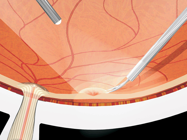
Figure 17.1  Curved scissors are used to resect the PVC in vitreomacular traction syndrome cases to prevent avulsion of the fovea caused by imbrication of the taut PVC into the cutter.
Nonrhegmatogenous Proliferative Vitreoretinopathy
Some patients have multiple star folds from PVR in addition to an epimacular component. Removing these additional epiretinal membranes is a stimulus for recurrent PVR and is unnecessary unless they are causing macular elevation or distortion.
Need for Vitrectomy at the Time of Membrane Peeling
The principal author initially suggested the concept of membrane peeling without vitreous removal but stopped advocating this approach after several hundred cases because the patients complain bitterly of floaters and in-office fluid-gas exchange cannot be performed if the patient develops a retinal detachment. If the vitreous has been removed, a postoperative retinal detachment can be managed by in-office, two-needle, fluid-gas exchange and laser retinopexy. Anterior vitreous cortex removal is probably correlated with an increased incidence of posterior subcapsular cataract probably related to fluid turbulence. Avoiding the anterior vitreous cortex may reduce postoperative posterior subcapsular cataract.
Epiretinal Membrane Removal
Machemer developed the concept of membrane peeling in 1972 soon after his introduction of vitrectomy. Originally, peeling was performed with a bent needle. O’Malley subsequently developed the concept of using a rounded, angulated instrument he called a pic to perform the peeling. The principal author and the late Ron Michels popularized the pic method. Bent needles and pics require the presence of a visible outer margin of the EMM, frequently called an “edge,” unless a slit is made in the membrane. Searching for an edge creates the risk of making a retinal break. The principal author developed the concept of inside-out forceps membrane peeling initially because of the difficulty of finding an edge in certain cases. In contrast to Machemer’s outside-in membrane peeling method, inside-out peeling is initiated by surface grasping the EMM with end-opening forceps (Fig. 17.2). Although the senior author formerly recommended making a slit in the apparent center of the EMM using the microvitreoretinal (MVR) blade or a sharp-tipped pic, this is not necessary with the Alcon 25-gauge ILM DSP forceps. The center of the membrane can be identified by noting the orientation of radial striae, the most elevated retinal region, the most opaque region of the membrane, and the relative movement of the membrane with respect to the retina induced by lateral movement of the forceps or MVR blade tip. End-opening forceps with diamond coating were developed by the senior author and Hans Grieshaber for surface grasping and must have precise alignment of the blade tips. Eckardt developed effective end-opening forceps that are preferred to diamond-coated forceps because the diamonds are recessed from the tips of the forceps blades. Conformal forceps were then developed by the principal author because Eckardt-type forceps have square corners that can tear the nerve fiber layer or cause bleeding. The tips of conformal forceps have a radius of curvature which matches that of the retina. The authors use 25-gauge Alcon disposable, DSP ILM forceps (Fig. 17.3) for all cases. The Alcon 25-gauge MVR pic is used by some surgeons if the membrane is very smooth and taut (so-called glassy), but the authors rarely find this step necessary when using DSP forceps. Membrane peeling should be accomplished by moving the forceps tangentially along the surface of the retina in a circular fashion (Fig. 17.4). If the membrane tears, it can be regrasped without removing any membrane from the forceps because microteeth will penetrate several layers of membrane and facilitate removal of the membrane through the pars plana as well. The surgeon should always observe the fovea during the peeling process rather than focusing on the forceps in order to prevent tearing the fovea. Areas of stronger adherence to the ILM can be detected by noting fine fibers being lifted from the retinal surface during the peeling process. If prominent striae are still noted after peeling the epiretinal membrane, ILM peeling with the 25-gauge DSP forceps must be performed. Kampik introduced the idea that ILM peeling reduces the incidence of recurrent membranes, and the authors have validated this observation. Scissors delamination with fine curved scissors rather than peeling is utilized if strong adherence to the fovea, vessels, or any region of the retina is noted during the peeling process (Fig. 17.5). If there are marked folds, the blunt, polished end of the vitrectomy instrument can be used to gently push the retinal folds into better position, a method referred to by the principal author as “burnishing.” Moderate-sized peeled or delaminated membrane pieces should be removed through the pars plana with the forceps. If the membrane is very large or dense, it should be removed with the vitrectomy probe.
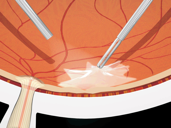
Figure 17.2  Inside-out forceps peeling is initiated at the epicenter, which is the densest portion of the EMM and the focal point of striae. Pics and looking for an edge can result in retinal damage and are not required with this method.
Stay updated, free articles. Join our Telegram channel

Full access? Get Clinical Tree


