and Mitrofanis Pavlidis2
(1)
Department of Ophthalmology, Uppsala University Hospital, Uppsala, Sweden
(2)
Augencentrum Köln, Cologne, Germany
Electronic supplementary material
The online version of this chapter (doi:10.1007/978-3-319-20236-5_10) contains supplementary material, which is available to authorized users.
Electronic supplementary material
for this chapter is accessible online at http://extras.springer.com/ by searching via the ISBN.
The intraoperative visualization of the retina opens new surgical and diagnostic possibilities. The anterior segment surgeon can assess the anterior segment (Fig. 10.1), the posterior segment surgeon can assess the retina and pathologic structures during surgery (Fig. 10.2) and the paediatric ophthalmologist can perform an OCT of children in general anaesthesia (Fig. 10.3). The companies Zeiss (Germany) and Haag-Streit (Switzerland) have developed a surgical microscope with integrated OCT. These microscopes are only available as floor microscopes.
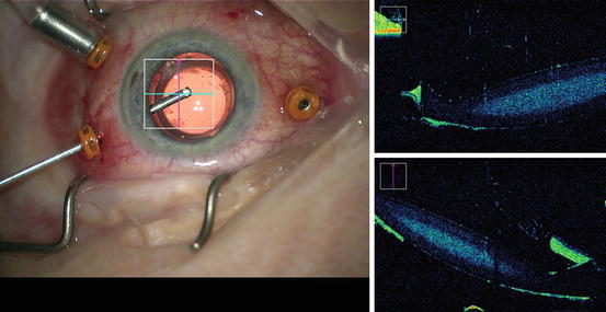
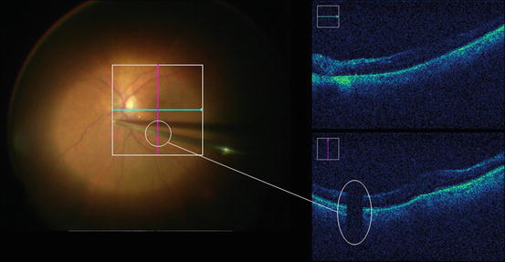
Fig. 10.1
Intraoperative OCT of the anterior segment with the Zeiss Callisto microscope. Note the IOL viewed by the OCT
Fig. 10.2


Intraoperative OCT with the Zeiss Callisto microscope during macular peeling. Note the vitreous cutter on the OCT image
Stay updated, free articles. Join our Telegram channel
Full access? Get Clinical Tree
