37 Mass in the Oral Cavity The oral cavity extends from the vermilion border of the lips anteriorly to the junction of the hard and soft palates and the circumvallate papillae of the tongue posteriorly. A wide variety of masses, both benign and malignant, can present in the oral cavity. Masses can arise from any of the various tissue types present in the oral cavity, including the mucosa, salivary glands, teeth, and bone. Most benign lesions are asymptomatic and present as slow-growing painless masses. Malignant lesions are more commonly associated with pain and ulceration, which can lead to difficulty eating and weight loss. More than 90% of malignant lesions of the oral cavity are squamous cell carcinomas, which are linked to tobacco and alcohol use. Therefore, assessment of these risk factors is important when evaluating a patient with an oral cavity mass. Infection with human papilloma virus (HPV) has been associated with oropharyngeal cancer and to a lesser degree with oral cavity cancer. This section reviews both benign and malignant oral cavity masses categorized by anatomical subsite. Each diagnosis is discussed according to the site of most common occurrence, with the exception of squamous cell carcinoma, which is detailed by subsite. Fig. 37.1 Squamous cell carcinoma of the left lateral tongue. Fig. 37.2 Irritation fibroma of the lateral tongue.
 Lip
Lip
 Mucus retention cysts: Due to an obstructed salivary duct, which leads to epithelial proliferation. As a result, they have an epithelial lining. They are submucosal, slow growing, fluctuant, and painless.
 Mucus escape reaction, or mucocele: Due to extravasation of mucus secondary to traumatic severance/interruption of a salivary gland duct. These are cystlike and painless and differ from mucus retention cysts in that they lack an epithelial lining. The lower lip is the most common site, followed by the buccal mucosa.
 Squamous cell carcinoma: Accounts for > 90% of cancers of the lips, of which > 90% occur on the lower lip. The most important risk factor for lip cancer is sun exposure, which likely explains the much higher rate of lower lip involvement. Although basal cell carcinomas are frequently described to involve the upper lip, these tumors likely arise from the cutaneous portion of the lip and are not true oral cavity lesions.
 Minor salivary gland tumors: See Palate section.
 Tongue
 Granular cell tumor: Small, benign, painless, submucosal mass thought to arise from Schwann cells. Tongue is the most common site. These tumors can be confused with squamous cell carcinoma on microscopic examination due to pseudoepitheliomatous hyperplasia.
 Papilloma: Benign squamous proliferation due to human papilloma virus (HPV). Different serotypes (HPV 6 and 11) account for benign papillomas than for HPV-related squamous cell carcinoma (HPV 16). Presents as a pedunculated mass with fingerlike projections. Usually painless and nonulcerated but may have a white surface change due to keratinization.
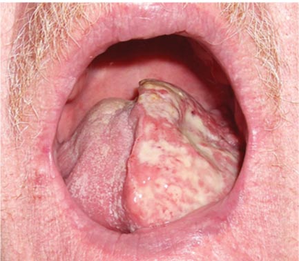
 Squamous cell carcinoma: Commonly occurs on the lateral border of the oral tongue (Fig. 37.1). Typically presents as a painless, nonhealing ulcer in the middle portion of the lateral tongue. Pain, referred otalgia, and dysarthria can develop with advanced lesions. Squamous cell carcinoma very rarely occurs in the midportion of the dorsum of the oral tongue. Diagnosis of squamous cell carcinoma in this location should raise suspicion of misdiagnosis and warrants a second histologic opinion.
 Leukoplakia: Clinical description for white plaque that cannot be rubbed off. Diagnosis of exclusion with up to 10% malignant transformation rate.
 Erythroplakia: Clinical description for a red mucosal plaque that cannot be explained by another clinical or pathological condition. Typically friable and has a much higher malignant transformation rate than leukoplakia.
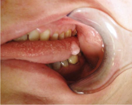
 Irritation fibroma: Firm lesion, on lateral border of oral tongue (Fig. 37.2)
 Verrucous carcinoma (see Buccal Mucosa section)
 Pyogenic granuloma (see Alveolar Ridge section)
 Floor of Mouth
 Wharton duct: Main salivary duct of the submandibular gland. The papilla opens in the anterior floor of the mouth, and the duct may be mistaken for a mass.
 Ranula: Mucocele occurring in the floor of the mouth in association with the sublingual gland. Named for its resemblance to the undersurface of a frog. Plunging ranula refers to a ranula that extends below the mylohyoid muscle into the neck.
 Sialolithiasis
![]()
Stay updated, free articles. Join our Telegram channel

Full access? Get Clinical Tree


