Mandibular Fractures
Mandibular fractures are uncommon in children, and the incidence increases with age. They usually occur as a result of severe trauma and are often associated with other injuries. It is necessary to search for a possible second fracture site when one is found. The most common fracture site is the condyle.
42-1 Treatment of Mandibular Fractures
Indications
Mandibular anatomy and function have to be reestablished via reduction of fractures based on normal occlusion. As the pediatric population has greater osteogenic capacity and faster healing rates compared to adults, anatomical reduction must be done sooner and immobilization times should be shorter (2–3 weeks for children). Immobilization can be accomplished with maxillomandibular fixation (MMF), open reduction and skeletal fixation with miniplates, or a combination of the two.
Close observation, a liquid to soft diet, and analgesics are used to manage mandibular fractures without displacement and malocclusion. Condylar fractures are treated with observation or closed reduction with MMF (7–10 days) followed by physical therapy, the latter being indicated for displaced condylar fractures. However, some would advocate early mobilization without MMF for even displaced condylar fractures—this is a debated topic. Subcondylar fractures without malocclusion may also be managed conservatively.
Fractures in other parts of the mandible usually need surgical intervention. Displaced mandibular fractures should be reduced and immobilized with MMF or open reduction and miniplate application. For open reduction and miniplate fixation, the miniplate can be placed at the inferior border of the mandible with the screws directed away from developing tooth buds. Resorbable or titanium plates can be used. Some advocate that the titanium plates should be removed in 2–3 months after reduction, although this is controversial (see Section 39-1). For skeletal fixation, 1.5- or 2.0-mm miniplates should be used with at least two secure screw holes on either side of the reduced fracture. Screw holes are monocortical to avoid damaging unerupted teeth.
Preoperative Evaluation
Some degree of malocclusion and trismus usually accompany mandibular fractures; after 3–4 days, muscle spasms generally improve. Radiographs or low-dose radiation computed tomography imaging should be obtained to document the suspected fractures and to search for the frequent second fracture. Panoramic dental films are useful to determine dental fractures. Most mandibular fractures do not require immediate reduction. They should be managed as soon as the patient’s general condition has stabilized (preferably within 48 hours).
Operative Technique
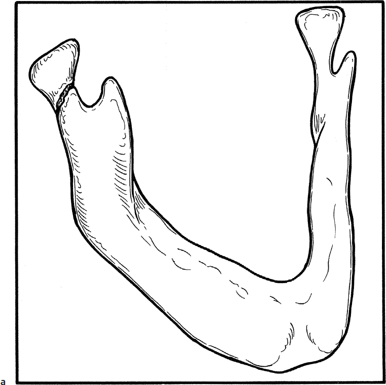
1. Uncomplicated condylar and subcondylar fractures in children do not require reduction if occlusion is reasonably good (Fig. 42.1a). Soft diet should be used for approximately 4–6 weeks until the child can eat without pain. An immobilization dressing (Barton bandage) may help to reduce discomfort by restricting jaw motion (Fig. 42.1b) but some would advocate only a brief use as muscle spasms improve after 3–4 days and this would impede efforts at keeping the jaw mobilized.
2. Mandible fractures in children are best reduced and immobilized utilizing general anesthesia.
3. If a fracture is present and the fragments are not displaced, simple immobilization for 1 to 3 weeks will allow the fracture to heal.
4. When teeth are present, Ivy loops fashioned from 24- or 26-gauge stainless steel wire are utilized to immobilize the mandible and may be used to reduce the fracture as well. Because of the narrow neck on primary dentition, the wire loops may become loose or may gradually pull the tooth from its socket.
5. A 24- or 26-gauge stainless steel wire is bent around a suction approximately 2 to 3 mm in diameter (Fig. 42.1c).
6. The wire is grasped with a clamp or needle holder and twisted clockwise with a needle holder (Fig. 42.1d).
7. The trailing wires are then passed between two teeth (molars or premolars) at the gum line. One end is passed back around one tooth and the second end is passed back around the other tooth (Fig. 42.1e).
8. The ends of the wires are tightened by twisting clockwise with a needle holder (Fig. 42.1f).
9. The twisted ends of the wire are bent toward the gingiva to prevent buccal lacerations (Fig. 42.1g).
10. The loops on the mandible and maxilla are then wired together with 26-gauge wire to create maxillomandibular fixation (MMF), which immobilizes the mandible (Fig. 42.1h).
11. When permanent dentition is available for reduction and fixation, arch bars may be used (Fig. 42.1i).
12. A 24- or 26-gauge wire is passed around each of the premolars and molars. The wires are twisted and tightened with a needle holder to hold the arch bars in place on the maxillary or mandibular teeth (Fig. 42.1i).
13. The arch bars are secured together with rubber bands or 26-gauge wire to immobilize the mandible (Fig. 42.1i).
14. MMF should be removed in approximately 2–3 weeks to avoid ankylosis.
15. Severely displaced fractures may require open reduction and fixation with 1.5-/2.0-mm miniplates (resorbable or titanium).
16. An incision is made below the lower edge of the mandible, taking care to avoid the marginal mandibular division of the facial nerve (Fig. 42.1j). One percent lidocaine with 1:100,000 epinephrine is injected at the incision site to aid in hemostasis.
17. The platysma muscle is divided with a knife (Fig. 42.1k).
18. The inferior edge of the mandible is exposed by cutting through masseter muscle or periosteum as needed (Fig. 42.1l).
19. The periosteum is reflected with an elevator and a hole is drilled in the mandible on each side of the fracture. The fragments are reduced with forceps (Fig. 42.1m). The plate must be in a solid portion of the bone and close enough to the lower border of the mandible to avoid injury to the tooth buds (Fig. 42.1m).
20. The fracture may be stabilized utilizing miniplates (resorbable vs. titanium). Two screws are placed on either side of the fracture along the inferior border.
21. The wound is closed in layers with absorbable subcutaneous and subcuticular sutures. The wound is drained with a small Penrose drain if needed.
22. Postoperative antibiotics are given for 10 days. The drain is removed in 24–36 hours.
Complications
1. Malunion/nonunion
2. Osteomyelitis
3. Infected plates
4. Hardware failure
5. Loosened teeth/damage to permanent dentition or unerupted teeth
6. Reduced range of motion of the temporomandibular joint