COMPLICATIONS ASSOCIATED
WITH KERATOPROSTHESIS
 The management of severe anterior segment disease and intractable corneal opacities has evolved rapidly in the recent years. Whereas in the late 1990s there was a push for limbal stem cell transplantation in conjunction with aggressive systemic immunosuppression, the complications and practical difficulties associated with immune therapy have decreased the interest in this technique. Keratoprosthetic devices have been in development for decades. Claes Dohlman, at the Massachusetts Eye and Ear Infirmary, has developed and perfected the Boston keratoprosthetic device. His relentless pursuit of technical improvements of the device has allowed the recent popularization of the Boston keratoprosthesis and its more common utilization in a variety of disorders of the cornea and anterior segment (1).
One may classify vitreoretinal disorders associated with keratoprosthesis implantation into
- Preexisting vitreoretinal conditions
Keratoprosthesis can be associated with various vitreoretinal disorders that may require vitreous microsurgery (2). The eyes that require keratoprosthetic implantations have commonly been operated on multiple times unsuccessfully and have sustained long-standing inflammation. These eyes may harbor preexisting retinal conditions that may be unmasked by the clear optics of the keratoprosthetic device, such as dense epiretinal membranes, retinal detachment, or macular holes, that may require surgery with the keratoprosthesis in situ.
- Disorders secondary to the presence of the keratoprosthetic device
Keratoprosthesis can commonly present with retroprosthetic membranes that severely limit visual outcomes. These membranes are usually more frequent and severe in children but can also present in adults. Retroprosthetic membranes are usually too thick and adherent to be amenable to YAG laser and frequently require vitrectomy techniques for removal of the retroprosthetic membrane.
In addition, since the Boston keratoprosthesis never integrates with the host cornea or sclera, there will always be a permanent open wound around the keratoprosthesis that may be the route for bacteria to develop endophthalmitis. The Alphacor keratoprosthesis and the osteo-odontogenic keratoprosthesis designs are attempts to increase integration of the device. Dohlman has noticed that placement of a contact lens over the keratoprosthesis and chronic topical antibiotics decreases endophthalmitis rates.
Glaucoma is very common after keratoprosthesis, and many surgeons recommend primary placement of Ahmed valve glaucoma implants (3). In eyes that have undergone iridectomy, lensectomy, and keratoprosthesis that also have an Ahmed valve, vitreous occlusion of the tip of the Ahmed valve can require emergent vitrectomy for relief of the acute glaucoma attack.
- Disorders associated with the surgical technique of keratoprosthesis implantation
Keratoprosthesis implantation is a difficult procedure that may require, beyond replacement of the cornea, total iridectomy, lensectomy, or intraocular lens explantation and open-sky vitrectomy. The iridectomy may cause intraoperative and postoperative vitreous hemorrhage, and the vitrectomy and lens removal may cause inadvertent retinal tears with subsequent retinal detachment. Early postoperative vitreous hemorrhages are often difficult to manage, since although the most likely cause for the hemorrhage is iridectomy in a hypotonic eye, one cannot usually rule out intraoperative retinal tear until surgery is undertaken. Another surgical complication of keratoprosthesis implantation is intraoperative suprachoroidal hemorrhage. Since these eyes undergo open-sky surgery, choroidal expulsive hemorrhages are a real risk. Unfortunately, it is almost impossible to repair these eyes even if the surgeon is able to suture the keratoprosthesis quickly enough to prevent expulsion of the intraocular contents, since the retina and anterior vitreous can become adherent to the corneal wound and the back plate of the keratoprosthetic device, making successful retinal detachment repair almost impossible.
TECHNICAL CONSIDERATIONS REGARDING OFFICE EXAMINATIONS IN KERATOPROSTHESIS PATIENTS
Office examinations of patients with keratoprosthesis can be challenging. While examination of the posterior pole with an indirect ophthalmoscope can usually be accomplished, examination of the retinal periphery may be more difficult. The authors prefer the use of high plus wide-angle lenses on the slit lamp for examination of the retinal periphery on patients with keratoprosthesis, since the oblique slit beam reduces reflections and glare from the surface of the keratoprosthesis. If B-scan ultrasonography is needed due to vitreous hemorrhage, the examiner may need to place the transducer directly adjacent to the keratoprosthesis and perform transscleral imaging, avoiding artifacts from the keratoprosthesis.
VITRECTOMY IN KERATOPROSTHESIS PATIENTS
The most significant technical considerations while doing vitrectomies in eyes with keratoprosthesis are (a) introduction of 25-gauge trocar-cannula systems, (b) peripheral retinal visualization and management, and (c) management of retroprosthetic membranes.
Introduction of the 25-Gauge Trocar Cannulas
Eyes that require keratoprosthesis may not have clearly identifiable limbal structures from which to measure posteriorly the location of the pars plana. Entry into the eye should obviously avoid the anterior retina but is constrained anteriorly by the presence of the back plate of the keratoprosthesis. The 25-gauge cannula needs to be at least 2 mm posterior to the edge of the back plate to allow clearance below the back plate, required to approach the posterior side of the optic in case of retroprosthetic membrane removal. The only identifiable structure on the anterior segment of keratoprostheses is the edge of the front plate. The back plate measures 8.5 mm in diameter, but 7-mm diameter back plates are also available for small pediatric eyes. It is important to remember that the edge of the back plate is not usually visible, so the surgeon should estimate its location from the edge of the anterior optic. Since the radius of the front plate is 2.5 mm, the edge of the back plate is 1.75 mm posterior to the edge of the optic. If 2 mm clearance posterior to the back plate is the target, then cannula entry should be performed 3.75 to 4 mm posterior to the edge of the optic (3 mm in case of smaller pediatric back plates). This entry location likely corresponds to pars plicata rather than pars plana. The authors do not recommend more posterior entry since the surgeon has no assurance of perfect centration of the keratoprosthetic device. If the device is decentered, posterior entry through the anterior retina can be unfortunately performed. If the surgeon can visualize the edge of the back plate, then direct measurement from this location overrides the previous discussion. The authors do not have experience with translid keratoprostheses but would recommend a similar analysis for sclerotomy placement if confronted with the situation. The authors find the 25-gauge system excellent of surgery in keratoprosthesis cases, since it avoids conjunctival dissection on these eyes that have preexisting ocular surface scarring. Transconjunctival sclerotomy closure with a single 8-0 Biosorb suture may be needed at the end of the surgery since the conjunctiva rarely covers the sclerotomies.
Peripheral Retinal Visualization and Management
The configuration of the optic is a cylinder of 3 mm diameter, with a thickness of slightly greater than 3 mm. Direct visualization of the posterior pole structures is usually excellent with the eye on primary position. Rotation of the globe, though, creates tilting of the optical cylinder and decreases the optical aperture for visualization. In essence, permanent keratoprosthetic vitrectomies require surgery in the primary position. Given this constraint, contact-based wide-angle lens (Volk) visualization is imperative for peripheral vitrectomy. Even in these conditions, visualization of the far periphery may be very difficult, and this is one of the reasons that rhegmatogenous detachments are difficult to repair. Endoscopic techniques may be required for complete retinal peripheral evaluation and laser.
If peripheral visualization impairs adequate management of a retinal detachment, a useful technique to consider is medium-term perfluorooctane with 360-degree peripheral retinal endophotocoagulation. Perfluoro-octane (PFO) can be injected over the optic nerve and used to fill the vitreous cavity and reattach the retina as described in Chapter 14. Posterior retinotomy should be avoided in these eyes. Once the retina is attached, the surgeon can proceed with laser of the retinal periphery circumferentially with at least three to four rows of confluent laser as far as visualization allows. The PFO can then be left in the vitreous cavity for 2 weeks and can be later removed. If permanent posterior pole reattachment is accomplished, despite peripheral subretinal fluid, this qualifies as a successful repair in an eye that would otherwise have proceeded to blindness. The authors discourage the use of silicone oil in keratoprosthesis retinal detachments. These eyes are already prone to glaucoma, intraocular pressure measurements are impossible to perform reliably, and the silicone oil can prevent fluid egress through the Ahmed valve.
Management of Retroprosthetic Membranes
Retroprosthetic membranes are often very thick and adherent to the posterior surface of the optic. Forceps, scissors, and the vitreous cutter are usually unable to start an opening on the membrane. The authors use a 25-gauge needle or 25-gauge microvitreoretinal (MVR) blade with the tip bent to form a sharp pick that is introduced through the 25-gauge cannula. The sharp bent needle tip is then used to engage the membrane away from the center of the optic (in case the needle scratches the posterior surface of the device) and an initial opening on the membrane is created. Once an edge is found, further removal with forceps, scissors (segmentation), or the vitreous cutter can be performed (Fig. 28.1). The authors attempt to remove the membrane beyond the edge of the optic along the surface of the back plate, with the goal of decreasing recurrence of the membrane over the optical surface of the device.
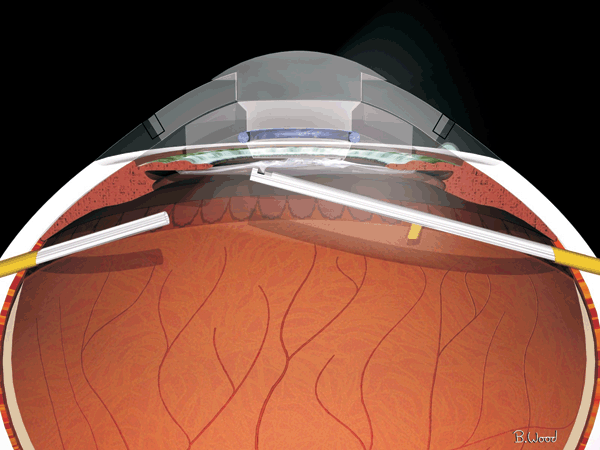
Figure 28.1  After an opening is made with an MVR blade, the retroprosthetic membrane can be removed with the vitreous cutter.
Stay updated, free articles. Join our Telegram channel

Full access? Get Clinical Tree


