Suprachoroidal hemorrhage is a devastating complication of intraocular surgery. This complication is very difficult to anticipate, prevent, and manage (1–5) (Fig. 30.1). Intraoperative management is complex, as is the decision to intervene in the postoperative period. These cases are also called choroidal hemorrhages because blood dissects into the spongy choroidal tissue. They are termed expulsive hemorrhages if the choroid and retina are forced out of the eye by high pressure in the suprachoroidal space.

Figure 30.1  Suprachoroidal hemorrhage pushes the retina centrally and anteriorly and compresses remaining vitreous.
INCIDENCE OF SUPRACHOROIDAL HEMORRHAGE
The incidence of suprachoroidal hemorrhage in extracapsular cataract surgery and phacoemulsification is approximately 0.15%, filtering procedures 0.15%, penetrating keratoplasty 0.56%, vitrectomy 0.41%, and the principal author’s vitrectomy series is 0.01% (5/26,000). Small-incision cataract surgery does not necessarily reduce the incidence of this complication as the intraocular pressure (IOP) must be reduced to atmospheric pressure during intraocular lens (IOL) insertion, although it certainly makes wound closure faster and safer. Clearly, the duration of low IOP is less with phacoemulsification than with intracapsular surgery. Small-incision surgery with self-sealing wounds construction facilitates rapid wound closure and normalization of the IOP. Filtering procedures such as trabeculectomy, setons, and valves remain a common cause of this complication. Late hemorrhages are common if antimetabolites (mitomycin) are used producing a sustained, very low IOP.
PATHOGENESIS AND PREVENTION
A key factor in the pathogenesis of suprachoroidal hemorrhages is a high trans–arterial wall pressure gradient due to acute lowering of the IOP to atmospheric pressure in the presence of hypertension. The bleeding is probably due to shearing of vessels traversing the suprachoroidal space caused by scleral stretch. Penetration of the eye is probably frequent and often unrecognized because of suprachoroidal hemorrhage. Hypertension and arterial disease are critical factors in the pathogenesis. Patients should be normotensive during cataract, penetrating keratoplasty, secondary IOL, and filtering procedures. If general anesthesia is being used, it is probably advisable to ask the anesthesiologist to use neuromuscular blockade for open eye procedures to prevent high blood pressure secondary to “bucking on the tube.”
COMPLICATIONS OF SUPRACHOROIDAL HEMORRHAGE
Bad outcomes in nonexpulsive cases are usually not directly due to the hemorrhage but are secondary to retinal detachment from hypocellular collagen contraction and adherence of vitreous to anterior structures (iris, wound, capsule). Vitreoretinal traction increases in the weeks and months after surgery after the choroidal blood absorbs and the buckle-like effect disappears ( Fig. 30.2). Many patients suffer optic nerve damage secondary to the acute increase in IOP or possibly high intrasheath pressures.
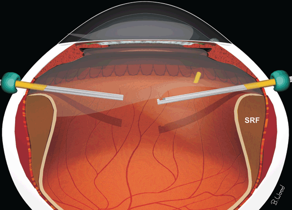
Figure 30.2  Retinal detachment may be caused by hypocellular contraction of the vitreous combined with adherence to anterior structures.
Stay updated, free articles. Join our Telegram channel

Full access? Get Clinical Tree


