Until 1991, it was thought that macular holes were untreatable and their pathogenesis was unknown. In that year, Kelly and Wendel developed the concept of using vitrectomy and fluid-gas exchange to treat these patients (1). Initially there was great skepticism about the treatment, but the facts prevailed. The initial goal was to “seal” the hole much as is done for rhegmatogenous retinal detachment and eliminate the cuff of subretinal fluid that surrounds the hole. When it was noted that many holes actually disappeared after surgery, and patients obtained near-normal vision, the skeptics again did not believe it. Fortunately, it is now accepted that complete disappearance of the hole is the usual outcome after surgery.
PATHOGENESIS
It was widely believed that a posterior vitreous detachment (PVD) pulled a full-thickness piece of tissue out of the macula, probably as a result of or during a saccade. Electron microscopy of the so-called operculum, which occurs in these cases, has shown that few photoreceptors are present (2,3). Gass published a hypothesis for the pathogenesis (4,5) of macular holes that has remained viable with minimal modifications since the introduction of high-resolution optical coherence tomography (OCT). The concept is that radial vitreous fibers remaining on the perimacular surface after apparent posterior vitreous separation contract and slowly tear the macula in a circumferential fashion. The Mueller cells may play a role as well according to Gass. Many observers have noted that vitreous is attached to the optic nerve after an apparent posterior vitreous separation characterized by the presence of a prepapillary (Weiss) ring. Because of these observations, the event might better be termed “delamination of the posterior vitreous cortex.” The senior author terms the cuff of fluid “the traction cuff.” Sjaarda has shown using scanning laser ophthalmoscope (SLO) microperimetry that the actual extent of scotoma extends far beyond the margin of the cuff (6). The vitreous that is attached to the inner margin of the macular hole is rarely contiguous with the vitreous attached to the midperipheral retina. Terminology represents a serious problem in discussing these cases with vitreoretinal surgeons.
Surgical success should be defined as clinical disappearance of the hole, reconstitution of the foveal anatomy on OCT, and marked improvement in vision.
INDICATIONS FOR MACULAR HOLE SURGERY
Freeman et al. have shown that smaller, more recent holes have the best prognosis. Size is much more important than duration with respect to closure rates; duration plays no role if size is controlled for. Duration probably plays a role in visual success in successful cases. Most surgeons do not suggest repairing holes secondary to chronic macular edema from diabetic retinopathy, venous occlusive disease, cystoid macular edema after cataract surgery, or secondary to uveitis. Traumatic macular holes are a complex decision-making process because of the high likelihood of associated photoreceptor, retinal pigment epithelium (RPE), and optic nerve damage and because many will spontaneously close within 1 to 2 months. If there is good evidence that the macular hole is the only significant damage, these cases can be considered for surgery after a reasonable period of observation.
OCULAR COHERENCE TOMOGRAPHY
OCT is invaluable in the evaluation of macular hole patients both preoperatively and after surgery. It can be difficult to distinguish partial-thickness holes from full-thickness holes clinically. Some holes are very small at the internal limiting membrane (ILM) level but much larger at the level of the outer retina; others have the opposite configuration. Vitreomacular traction and epimacular membranes can be seen with careful OCT examination. Some holes have no cuff, are elliptical, and are caused by typical epimacular membranes rather than vitreous. These holes may be thought of as secondary holes and round holes with a cuff above as primary or classic holes. The Gass classification system using Stage I, etc. is no longer used by the authors now that OCT is available. Macular holes are now classified as partial thickness or full thickness. Diameter is a major factor in driving outcomes and can be accurately measured with OCT. Whether there is a PVD or not is irrelevant clinically as an attempt must be made to create a PVD in all cases and ILM peeling removes any adherent posterior vitreous cortex. Some patients have apparent closure after surgery but modest visual recovery; some of these cases have edema or subretinal fluid as shown by Kaiser, while others have a persistent defect in the outer retina or disruption on the outer segment layer of the fovea on spectral domain OCT.
MACULAR HOLE SURGERY
Macular holes were untreatable until Neil Kelly developed the concept of core vitrectomy followed by fluid-gas exchange using an isoexpansive mixture of air and SF6 or C3F8 gas. His goal was to reattach the cuff of subretinal fluid around a full-thickness macular hole and therefore eliminate the relative scotoma that surrounds the absolute scotoma. Serendipitously he soon discovered that the macular hole often closed with remarkable improvement in vision. Many leading surgeons initially discounted his discovery, but fortunately it has become the standard of care. The prevailing thought at the time was that substantial neural tissue was avulsed by a PVD; an operculum was often seen, validating this notion. Examination of surgically removed operculums using the electron microscope revealed very little neural tissue; most of the operculum proved to be glial tissue, explaining why substantial visual improvement was possible. Subsequently, OCT demonstrated restoration of near-normal or normal foveal anatomy in successfully operated cases. Clearly, “closure” of macular hole is quite different than the term “closure” when used in the context of retinal detachment repair.
Much emphasis is placed on the substantial work of Don Gass, which involved clinical observation of the evolution of macular holes, theoretical considerations concerning the pathogenesis, and a classification system. Although all surgeons agree that the posterior vitreous cortex is somehow involved and the elasticity of the ILM plays a role, the pathogenesis of macular holes remains unknown. Presurgical classification, even using spectral domain OCT, is incapable of reliably determining if residual vitreous cortex is adherent to the retinal surface rendering the classification system virtually useless. Macular holes are three times as common in females as they are in males, but there is no explanation for this interesting observation.
OCT is essential to determine if the hole is partial thickness or full thickness; not infrequently clinical examination is inadequate to detect a very small, full-thickness hole at the base of a large-diameter partial-thickness hole. Occasionally, a very small, inner layer, full-thickness hole will overlie a large, full-thickness outer layer hole producing an appearance suggesting a macular cyst. Spectral domain OCT is essential in the evaluation of macular disease. Time domain OCT is no longer adequate. Charteris has shown that 30% to 50% of partial-thickness outer macular holes (Stage I on Gass’ Classification) spontaneously close if observed over the long term. A multicenter clinical trial done before the availability of OCT or ILM peeling did not demonstrate a benefit of operating on a partial-thickness hole to prevent progression to a full-thickness hole. Neither ultrasound nor OCT can predict whether a partial-thickness hole will progress to become a full-thickness hole, and status of the other eye is not helpful because holes are bilateral less than 10% of the time.
Size of the macular hole is the only preoperative factor that has been shown to drive surgical closure rates; duration is not a determining factor if size is controlled for. Although duration has an influence on visual outcomes, assessment of the subhole RPE using spectral domain OCT and confocal autofluorescence will probably prove to be more effective in predicting visual outcomes in longer duration holes. Some studies have shown reasonable visual outcomes after operating macular holes of relatively long duration.
Macular holes originating from macular cysts due to chronic macular edema typically have relatively poor visual outcomes because of macular ischemia related to underlying diabetic retinopathy or retinal vein occlusion. Similarly, macular holes arising from chronic inflammatory macular edema have a relatively poor visual prognosis.
Traumatic macular holes can spontaneously close in the first 4 to 6 weeks suggesting that a period of observation should precede the determination of operability. An afferent pupillary defect (APD) should be considered a relative contraindication to surgical repair because it indicates associated optic nerve damage. As mentioned above, assessment of the subhole RPE using spectral domain OCT and confocal autofluorescence will probably prove to be more effective in predicting visual outcomes. Presence of a choroidal rupture in the papillomacular bundle is a relative contraindication to repair of a traumatic macular hole; often these patients will have an APD.
The authors’ management of partial-thickness holes has changed over the years. The senior author began operating lamellar holes after Arthur Willis introduced the concept of macular hole prevention surgery. Macular hole prevention surgery was no longer performed after the results of the Macular Hole Study and the Charteris paper were published. The authors’ current practice is to determine the need for surgery on partial-thickness holes based on symptoms, particularly metamorphopsias from the epiretinal membranes (ERMs) that commonly surround lamellar macular holes. The goal is to improve visual function, not prevent a full-thickness hole because progression to a full-thickness hole cannot be predicted. The authors have determined in recent years that surface tension management using air or SF6 combined with ILM peeling is required to restore normal or near-normal foveal anatomy and improve or eliminate symptoms when operating on symptomatic partial-thickness holes.
HOLE CLOSURE MECHANISMS
The conventional explanation of the role of surgical steps in surgical hole closure is incomplete and, to an extent, probably incorrect. It is often stated that the role of PVD creation is to eliminate vitreous traction on the macula, but, in fact, the posterior vitreous is usually attached to the optic nerve head, nasal, and midperipheral retina but not attached to the macula or retina within the temporal arcades. Careful PVD creation reduces the likelihood of inferior retinal breaks caused by interaction of the bubble with residual vitreous similar to what may occur with pneumatic retinopexy. The role of core vitrectomy is to enable the exchange of vitreous for a large gas bubble and to ensure that a thin layer of vitreous does not prevent contact of the gas bubble with the hole.
Virtually all surgeons agree that ILM peeling improves closure rates, but why this is true has a complex answer. ILM peeling ensures removal of tangential traction due to residual vitreous on the retinal surface, which, although rare, can occur even with an apparent PVD as evidenced by a Weiss ring. In addition, ILM peeling guarantees successful removal of ERMs that are occasionally present. ILM peeling increases retinal elasticity by over 50%, which enables lateral surface tension forces to close the hole as soon as the bubble comes into contact with the hole. It is also likely that ILM peeling also initiates mechanical signaling to the astrocytes to heal the hole margins days after it is closed by lateral surface tension.
Most surgeons use the term “tamponade” when describing the mechanism of action of air, gas, and silicone oil bubbles. To some, tamponade means to “press,” but tamponade is from the French and means to “seal.” It is more accurate and descriptive to use the term “surface tension management” or “interfacial tension management” to describe the function of the bubble. It is obvious that a bubble eliminates transhole flow, which is the reason it is used in retinal detachment repair. An additional and crucial function of the bubble as Reppucci has pointed out is the lateral surface tension effect. Surface tension forces act along an interface of two immiscible substances effectively pulling the surface inward. Surface tension makes a droplet nearly spherical as it falls away from a faucet and a soap bubble spherical. Reppucci describes the bubble as bridging the hole, but the key concept is that the bubble pulls the hole margins together immediately after surgery as soon as the patient is positioned with bubble in contact with the hole. Yet another function of the bubble, as the senior author has pointed out, is the prevention of transretinal flow (uveal-scleral outflow). Tornambe has introduced the hydration hypothesis making a note of the marked edema surrounding the hole as seen on OCT; edema-mediated retinal thickening coupled with the elasticity of the ILM causes eversion of the hole margins. Elimination of the edema facilitates restoration of near-normal foveal anatomy, resolution of the edge eversion, and approximation of the hole margins. The bubble dries out the retinal surface, which probably signals the astrocytes to heal the hole days after approximation of hole margins via the lateral surface tension effect.
POSTERIOR VITREOUS DETACHMENT CREATION METHODS
Many surgeons use a lateral (tangential) motion of the extrusion cannula or vitreous cutter to create a PVD. This method creates shear force at the vitreous base, potentially leading to iatrogenic retinal breaks. A better method is to position the vitreous cutter at the nasal, superior and inferior disk margins with the port oriented away from the center of the disk and pull anteriorly (toward the cornea) using the vacuum-only mode (Fig. 18.1). The anterior-pull, disk-margin method safely, reliably, and quickly produces a PVD using 25-gauge cutters. It is a misconception that higher flow rates help produce PVDs or that 25-gauge cutters cannot produce a PVD; it is all about technique.
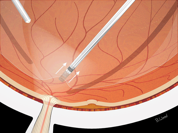
Figure 18.1  After core vitrectomy, a PVD is created using suction-only mode on the vitreous cutter, pulling anteriorly (not tangentially) over the optic nerve. Peeling should extend to the midperiphery, and the peripheral retina should be examined for peeling-induced retinal breaks.
Stay updated, free articles. Join our Telegram channel

Full access? Get Clinical Tree


