, Maneli Mozaffarieh1 and Hans Bebie2
(1)
Department of Ophthalmology, University of Basel, Basel, Switzerland
(2)
Institute for Theoretical Physics, University of Bern, Bern, Switzerland
Abstract
Lipids are a heterogeneous group of hydrophobic or lipophilic molecules, respectively. Some of them are amphiphilic in nature, which allows them to form structures such as vesicles, liposomes, or membranes in an aqueous environment. Biochemically, lipids are made up of two subunits (or “building blocks”): ketoacyl and isoprene groups. Lipids may be classified into eight categories: fatty acids, glycerolipids, glycerophospholipids, sphingolipids, saccharolipids, polyketides, sterol lipids, and prenol lipids (Fig. 17.1). The main biological functions of lipids include energy storage, maintenance of the structure of cell membranes, and signaling. In the eye, lipids have numerous different functions. One example is the vitamin A-derived retinal, which is involved in the visual cycle (Chap. 16). Another example is the prostaglandins, which are often involved in both physiological and pathophysiological processes. In this chapter, we will focus on the role of lipids in the tear film and in the retina.
Lipids are a heterogeneous group of hydrophobic or lipophilic molecules, respectively. Some of them are amphiphilic in nature, which allows them to form structures such as vesicles, liposomes, or membranes in an aqueous environment. Biochemically, lipids are made up of two subunits (or “building blocks”): ketoacyl and isoprene groups. Lipids may be classified into eight categories: fatty acids, glycerolipids, glycerophospholipids, sphingolipids, saccharolipids, polyketides, sterol lipids, and prenol lipids (Fig. 17.1). The main biological functions of lipids include energy storage, maintenance of the structure of cell membranes, and signaling. In the eye, lipids have numerous different functions. One example is the vitamin A-derived retinal, which is involved in the visual cycle (Chap. 16). Another example is the prostaglandins, which are often involved in both physiological and pathophysiological processes. In this chapter, we will focus on the role of lipids in the tear film and in the retina.
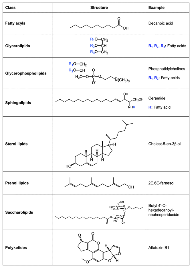
Fig. 17.1
The eight categories of lipids
17.1 Tear Film
In humans, the tear film of the eye, known as the precorneal film, has three distinct layers (Fig. 17.2).
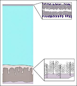
Fig. 17.2
Tear film. Schematic drawing of the tear film showing the outermost lipid layer, with the polar and non-polar heads, middle aqueous layer, and inner mucus layer containing transmembrane glycoproteins and mucins
The lipid layer is the outermost surface and provides a smooth tear surface; the aqueous layer, consisting mainly of water, promotes spreading of the tear film, whereas the innermost mucus layer increases the wettability of the corneal surface. The tear film creates a smooth surface for light to pass through the eye, nourishes the front of the eye, and provides protection from infection.
The lipid layer of the tear film contains nonpolar lipids as well as lipids with a polar head. The polar heads of the phospholipids form the interface between the aqueous–lipid layers (Fig. 17.2). The nonpolar lipid components allow other lipids, such as cholesterol, to dissolve in the tear film. Tear lipids are not susceptible to lipid peroxidation because they contain extremely low levels of polyunsaturated fatty acids (Sect. 13.3). The lipids in the tear film are secreted primarily by the meibomian glands. There are approximately 30–40 meibomian glands in the upper lid and 20–30 smaller glands in the lower eyelid. As shown in the illustration (Fig. 17.3), each gland opens onto the skin of the eyelid margin, between the tarsal gray line and the mucocutaneous junction. A second group of glands also secrete lipids: the sebaceous glands of Zeis. These glands are located at the lid margin in relation to the lash roots and the secreted lipids are also partly incorporated into the tear film. The lipids in the tear film have several functions. They provide a smooth surface, reduce the evaporation of water, prevent tear overflow onto the lids, and provide a watertight seal during lid closure when sleeping.
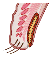
Fig. 17.3
Schematic drawing of the meibomian glands of the lid. The orifices of the ducts for the glands are located along the posterior margin of the eyelid
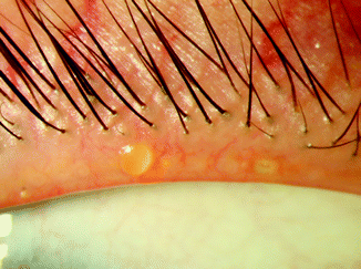
Various different diseases such as meibomitis (Fig. 17.4) and blepharitis (Fig. 17.5) can lead to lipid alteration.
 < div class='tao-gold-member'>


Only gold members can continue reading. Log In or Register to continue
Stay updated, free articles. Join our Telegram channel
Full access? Get Clinical Tree
