54 Lesion (Nonpigmented or Pigmented) A thorough skin examination is an essential component of the complete head and neck evaluation. Whether the chief complaint or an incidental finding, the otolaryngologist often encounters cutaneous lesions and is faced with the diagnostic challenge. Care must be taken to differentiate benign skin lesions from premalignant or malignant lesions that require further attention. Cutaneous lesions may also alert the physician to a broader systemic illnesses or genetic condition. In terms of physical findings, facial lesions can be broadly classified into pigmented and nonpigmented lesions. Examination involves observing the location, color, texture, consistency, and extent of the lesion. Palpation may reveal certain attributes regarding its subcutaneous extent, change in color with manipulation, and composition. All of these factors must be taken into consideration when making a diagnosis. Melanocytes give pigmented skin lesions their characteristic color. The melanocyte is a pigment-producing cell normally found in the basal layer of the epidermis. Pigmented lesions appear secondary to an increase in the number of melanocytes (melanocytic neoplasms) or through a change in melanin production. The majority of pigmented lesions are benign; however, care must be taken to rule out the possibility of malignant disease. Fig. 54.1 Malignant melanoma. Fig. 54.2 Intradermal nevus.
 Pigmented Lesions
 Melanocytic neoplasms
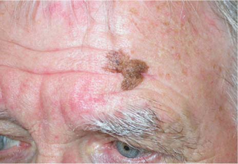
 Malignant melanoma (Fig. 54.1): Approximately 25% of melanomas are found in the head and neck. Suspicious lesions are defined by their physical appearance and growth characteristics. This constitutes the “ABCD” of melanoma. Asymmetry—If folded in half, the two halves wouldn’t match. Border—Melanoma often has an irregular and scalloped border. Color—Presence of nonuniform pigment, with a variegated red, white, and blue color. Diameter—Enlarging pigmented lesions, and those greater then 6 mm must be scrutinized. Early diagnosis is paramount to the successful treatment of malignant melanoma.
 Nevocellular nevus: Commonly referred to as moles, these benign lesions are histologically characterized by the presence of melanocytic nests. Nomenclature is based upon the histologic location of the nests within the skin.
 Junctional nevus: Located in the epidermal–dermal junction, these lesions are usually 1 to 6 mm flat to slightly raised, with symmetric borders and a uniform brown-black color. They are frequently found in children over the age of 2.
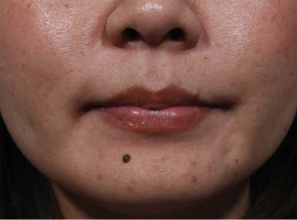
 Intradermal nevus (Fig. 54.2): Located within the dermis, these are the most common nevi seen on the adult face. Facial lesions are usually dome-shaped, smooth, and symmetric, and terminal hairs may grow through the nevi. They can be up to a few centimeters in size. Color may vary from a brown-black solid or speckled pigment to a translucent appearance. They are occasionally mistaken for basal cell carcinoma.
 Compound nevus: Located in both the epidermal–dermal junction and within the dermis, these nevi appear similar to intradermal nevi.
 Blue nevus: Located deep within or past the dermis, the pigment appears blue on the surface. They are less then 1 cm in size and are often indurated and palpable. They typically do not change appearance.
 Spitz nevus: A compound nevus most frequently found in children. It is commonly dome-shaped with a pink-red color. Histologically these may be difficult to distinguish from melanoma.
 Dysplastic nevus: Larger then the typical nevi at 5 to 15 mm. May be associated with atypical nevus syndrome wherein a patient has greater than 100 nevi and is at a greater risk of developing melanoma. These patients must be referred to a dermatologist for annual examinations.
 Nevus of Ota: A congenital macular brown-blue pigmentation of the skin and mucous membranes that follows the distribution of the first and second trigeminal nerve branches. Also known as nevus fusoceruleus ophthalmomaxillaris. This is most commonly seen in Asian patients.
 Disorders of pigmentation
 Solar lentigo
![]()
Stay updated, free articles. Join our Telegram channel


Full access? Get Clinical Tree


