Fig. 9.1
Anatomy of the larynx. Lateral view (a), sagittal view (b), posterior view (c), and superior view (d)
The cricoid cartilage is the only complete ring-like cartilage in the larynx. It is located at the level of the transverse process of the C6 vertebrate and has a wider posterior surface (lamina) compared to its anterior arch. This cartilage has a pair of cricoarytenoid joints on the upper surface of the posterior lamina and is supported by posterior cricoarytenoid ligaments [1]. The arytenoid cartilages are paired, three-sided pyramid-shaped cartilages that articulate with the cricoid cartilages. The arytenoid cartilage has an anterior vocal process for attachment of the vocal ligament and a lateral muscular process. The arytenoid cartilage pivots and slides at the cricoarytenoid joint, which allows for adduction and abduction of the vocal process.
The epiglottis is a teardrop-shaped, thin plate of elastic fibrocartilage expanding superiorly and attaches to the laryngeal prominence and the hyoid by the thyroepiglottic ligament and hyoepiglottic ligament, respectively. Laterally, the epiglottis is attached to the arytenoid cartilages by the aryepiglottic folds. This structure provides the epiglottis a degree of flexibility to bend posteriorly during swallowing.
The ligaments and membranes of the larynx include the conus elasticus (cricovocal membrane), the vocal ligament, the cricothyroid ligament/membrane, the thyrohyoid membrane, the hyoepiglottic ligament, the thyroepiglottic ligament, the quadrangular membrane, and the vestibular ligament (Fig. 9.1a, b). These structures serve as the support for the cartilages and function as anatomical barriers in the larynx.
The cricovocal membrane is the anterior portion of the cricothyroid membrane. It attaches to the vocal process of the arytenoid cartilage posteriorly and to the angle of the thyroid cartilage anteriorly. At the superior boundary, the membrane is thickened between these two points and is known as the vocal ligament, which is covered by the vocal fold (true vocal cord). The quadrangular membrane and the vestibular ligament form the aryepiglottic fold and the vestibular fold, respectively. The paired vestibular folds are referred to as the false vocal cords [3], and the ventricles are located between the vestibular folds and the vocal folds.
The muscles of the larynx are categorized as extrinsic or intrinsic. The extrinsic laryngeal muscles have an attachment outside of the larynx. They are responsible for movements of the larynx, and are divided into elevators and depressors. The thyrohyoid, palatopharyngeus, stylopharyngeus, and inferior pharyngeal constrictor muscles elevate the larynx, while the omohyoid, sternohyoid, and sternothyroid muscles depress the larynx. The intrinsic laryngeal muscles attach between laryngeal cartilages, and include the thyroepiglottic muscle, the aryepiglottic muscle, the lateral/posterior cricoarytenoid muscle, the transverse/oblique arytenoid muscle, the vocalis muscle, the thyroarytenoid muscle, and the cricothyroid muscle (Fig. 9.1c, d). Of these muscles, only the posterior cricoarytenoid muscles open the glottis, while all remaining muscles that attach to the arytenoid cartilage close the glottis. The cricothyroid muscles tense the vocal cords and the vocalis muscle attunes the vocal cords finely by relaxing specific portions of the cords [4].
The larynx is innervated by the superior and recurrent laryngeal branches of the vagus nerve. The superior laryngeal nerve divides into an internal and external branch. The internal branch of the superior laryngeal nerve enters the larynx running through the thyrohyoid membrane with the superior laryngeal artery. This branch carries sensory fibers to the larynx above the vocal cords, autonomic fibers to the laryngeal glands, and taste fibers to the epiglottis and adjacent root of the tongue [1]. The external branch of the superior laryngeal nerve runs with the superior thyroid artery. They separate at the superior pole of the thyroid, and the external branch of the nerve passes anteriorly and inferiorly to innervate the cricothyroid muscle, a portion of the inferior pharyngeal constrictor muscle, and the cricopharyngeus muscle. The recurrent laryngeal nerve enters the larynx with a branch of the inferior thyroid artery called the inferior laryngeal artery (Fig. 9.1c). All or the intrinsic laryngeal muscles, except for the cricothyroid muscle, are innervated by motor and sensory fibers of the recurrent laryngeal nerve. The sensory fibers provide general sensation to the subglottic area [1].
9.2 Laryngeal Development Overview
Laryngeal development has been studied for over a century, and different concepts have developed concerning the developmental processes. However, new technologies, including computer-generated 3D reconstructions of the human embryo or organized human embryonic collections, allow researchers to reexamine embryologic events, the origin of laryngeal components, and their anatomic relationships over time [2]. In this section, we review laryngeal development during the embryonic period.
The laryngotracheal groove, the respiratory primordium, appears as an outgrowth in the floor of the caudal end of the pharyngeal foregut during the 4th week of human gestation (Fig. 9.2a). Initially, this primordium of the tracheobronchial tree is separated from the hepatic primordium by the septum transversum, and is observed as a longitudinal fissure caudal to the fourth pair of pharyngeal pouches (Fig. 9.3a). By the end of the 4th week, the laryngotracheal groove has deepened to form a pouch-like laryngotracheal diverticulum located ventral to the caudal portion of the foregut. This respiratory diverticulum is separated from the pharyngeal floor by the primitive laryngopharynx. The primitive pharyngeal floor and the primitive laryngopharynx eventually develop into the infraglottic and the supraglottic region of the adult larynx, respectively [5]. With elongation of the diverticulum, it becomes surrounded by splanchnic mesenchyme (primordial embryonic connective tissue), and, during the 5th week, its distal end enlarges to form a bronchopulmonary bud that eventually develops into the lower respiratory tract. Later in the 5th week, the respiratory diverticulum migrates superiorly, and its cranial portion develops as the infraglottis [5]. The pulmonary epithelium and glands of the larynx, trachea, and bronchi develop from the endoderm lining the laryngotracheal groove. The connective tissue, cartilaginous components, and smooth muscle of the lower respiratory system are derived from the splanchnic mesoderm surrounding the laryngotracheal tube [6].
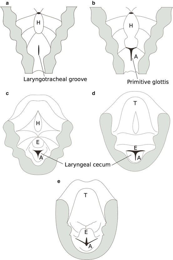
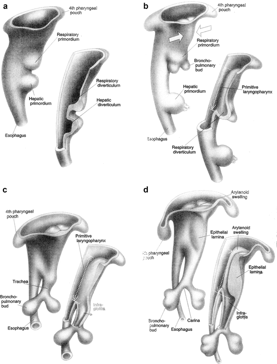
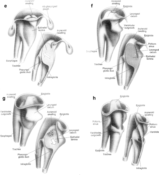
Fig. 9.3
Laryngeal development at Carnegie stage 11 (a), stage 12 (b), stage 13/14 (c), stage 15 (d), stage 16 (e), stage 17/18 (f), stage 19/23 (g), and fetal period (h) (Reproduced from Ferlito [5])
Initially, the laryngotracheal diverticulum communicates with the foregut. With the descending outgrowth of the diverticulum, it becomes separated from the primordial pharynx by the development of tracheoesophageal ridges. The ridges gradually approach each other and form the tracheoesophageal septum by the end of the 5th week. This septum divides the cranial portion of the foregut into ventral and dorsal regions. The ventral region is the laryngotracheal tube that eventually becomes the larynx, trachea, bronchi, and lungs, and the dorsal region is the primordium of the oropharynx and esophagus [6]. The respiratory primordium maintains communication with the pharynx via an opening of the laryngotracheal tube into the pharynx called the laryngeal orifice [7].
The laryngeal orifice becomes the primitive glottis during the 5th week (Fig. 9.2b). During this week, the mesenchyme at the cranial end of the laryngotracheal tube proliferates rapidly and forms the paired arytenoid swellings at the same anatomic level as the fourth pharyngeal pouch. Initially, this primitive glottis is shown to be a slit-like aperture, it becomes narrow as the arytenoids swellings grow towards the tongue. Further, there is a prominence at the ventral ends of the third and fourth pharyngeal arches, the hypobranchial eminence, that develops caudal to the copula with the proliferation of mesenchyme. The caudal part of this eminence develops as the epiglottis by the 10th week, and the rostral part forms the pharyngeal portion of the tongue. At the beginning of the 6th week, the primitive glottis surrounded by the hypobranchial eminence and arytenoid swellings forms a T-shaped laryngeal aditus that reduces the laryngeal lumen to a narrow slit with the development of the epithelial lamina, a condensation of mesenchymal tissue outlining the hyoid, thyroid, and cricoid bodies (Fig. 9.3d) [5, 8]. Later in the 6th week, the primitive glottis is obliterated completely except for the pharyngoglottic duct, and the laryngeal cecum begins to develop between the arytenoid swellings and the epiglottis, forming the vestibular outgrowth (Figs. 9.3d and 9.4e) [9]. The laryngeal cecum descends and reaches to the level of the glottic region during the 7th week (Fig. 9.4f, g) [5].
During the 8th week, the epithelial lamina begins to recanalize in a dorso-cephalic to ventro-caudal direction, resulting in a communication between the ventral laryngeal cecum and the dorsal pharyngoglottic duct. Complete recanalization is necessary for the communication between the supraglottis and the infraglottis, and incomplete recanalization may lead to supraglottic and glottic webs and atresia [5]. Recanalization of the laryngeal lumen is completed by the 10th week and creates a pair of lateral recesses called the laryngeal ventricles (Figs. 9.3e and 9.4h). The ventricles are bounded by the vocal folds and ventricular folds, which appear as outgrowths from the pharyngeal floor related to the third pharyngeal pouches at the end of the 7th week. During 8th to 9th week, they modify their morphology as laryngeal framework enlarges [10]. At the 8th week, the epithelium of the larynx is pseudostratified columnar and showing a clear basement membrane [11].
9.3 Development of the Laryngeal Framework
The laryngeal framework includes the thyroid, cricoid, and epiglottic cartilages and the paired arytenoid, corniculate, and cuneiform cartilages. These cartilages begin to chondrify at the beginning of the 8th week [12]. The thyroid, cricoid, and arytenoid cartilages are composed of hyaline cartilage and may exhibit calcification during the young adult ages. However, the other cartilages are composed of fibroelastic cartilage with little tendency of ossification over time.
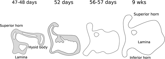
Each pharyngeal arch has cartilaginous component, and the fourth and sixth arch cartilages fuse to form the thyroid, cricoid, arytenoid, corniculate, and cuneiform cartilages, but not the epiglottis. The thyroid cartilage develops from the ventral ends of the cartilages of the fourth pharyngeal arch. Initially, the rudiment of this cartilage appears as two plates in front of the fourth pouch and cleft, and lies between the constrictors at the end of the 1st month. Bilateral small foci of cells appear in the laminae of the thyroid cartilage at the 7th week indicating the subsequent chondrocyte differentiation (Fig. 9.4). A week later, they joined cranially and caudally [9, 13], and complete confluence of these foci and formation of the definitive cartilages occurs postnatally [14]. Bilateral centers for the cricoid arch and the arytenoids appear at almost same time as those for the thyroid cartilage.
The thyroid cartilage is composed of two laminae, and they join the hyoid bone at the upper edges. The superior horns of the thyroid cartilage are a direct prolongation of the greater horns of the hyoid. By the end of the 2nd trimester (21st week), the hyoid and the thyroid horns become separated by the thyrohyoid membrane, and the thyroid cartilage shows a neonatal form [13


Stay updated, free articles. Join our Telegram channel
Full access? Get Clinical Tree
