Keratoconjunctivitis Sicca: Physiology and Biochemistry of the Tear Film


Keratoconjunctivitis Sicca: Physiology and Biochemistry of the Tear Film


Robin R. Hodges

Darlene A. Dartt


FUNCTIONS OF THE TEAR FILM

The tear film is a complex fluid secreted by multiple adnexal glands surrounding the orbit as well as by the ocular surface epithelia. The tears form the first refractive surface encountered by light on its path to the retina. For clear vision, it is critical that the tears themselves are transparent. Tears, in addition, function to maintain the transparency of the second refractive surface that rays of light encounter, the cornea. Therefore, the key role of tears is to protect the ocular surface, the cornea, and conjunctiva, and to maintain their health and normal functions. The tear film protects the ocular surface from the external environment by responding dynamically to a wide range of external conditions and potentially damaging situations. These external stresses include desiccation, bright light, cold, mechanical stimulation, physical injury, noxious chemicals, and bacterial, viral, and parasitic infection. Tears also provide for a smooth and reflective surface and lubrication to avoid mechanical damage to the surface of the eye from the surprisingly high pressures generated by the blink. Tears transport oxygen and a limited number of other nutrients to the avascular cornea, regulate their electrolyte composition and pH, and function to remove waste products. Finally, tears contain a multitude of proteins and other molecules that not only protect the ocular surface but also can regulate a myriad of cellular functions of both the conjunctiva and cornea. Some of the main physical characteristics of tears are listed in Table 27-1. To respond to changes in the external environment and in the internal requirements of the cornea and conjunctiva, the volume, composition, and structure of the tears are exquisitely regulated. This control regulates the tear film primarily by coordinately regulating secretion from the adnexa, cornea, and conjunctiva, rather than by controlling tear drainage.


STRUCTURE OF THE TEAR FILM

The classical view of the tear film is as a stratified fluid consisting of three layers: an outer lipid layer, a middle aqueous layer, and an inner mucous layer. In addition there is a mucin-containing glycocalyx that extends from the apical membrane of the superficial cells of both the corneal and conjunctival epithelium, which acts as an interface between the ocular surface cells and the tears (Fig. 27-1). The different layers of the tear film have been measured; the lipid layer is 0.1 μm thick, the aqueous layer is 7 to 10 μm thick, and the mucous layer is 0.2 to 1.0 μm thick (1). Currently, there is controversy about the thickness and structure of the tear film. Measurements by Prydal and co-workers (2) indicated a far thicker mucous layer of about 30 μm and an aqueous layer that was 10 to 11 μm (2). Recent measurements, however, found a prelens tear film layer averages 2.7 μm (3,4). Still another hypothesis is that the mucous and aqueous layers are not distinct, but rather are a gradient of decreasing mucous and increasing aqueous content from the ocular surface to the lipid layer. Finally, McCulley and Shine (5) have suggested that the lipid layer is a monolayer consisting of two phases, a polar phase adjacent to the aqueous layer and a nonpolar phase at the tear film-air interface. The controversy over the structure and thickness of the tear film has yet to be resolved.


ADNEXA OF THE OCULAR SURFACE EPITHELIA THAT PRODUCE TEARS

Distinct tissues secrete the different layers of the tear film. The meibomian glands secrete the outer lipid portion of tears (Fig. 27-2). The glands of Zeis and Moll are minor contributors to this layer. The main lacrimal gland is the major gland that secretes the aqueous layer of tears. A minor amount of this layer is produced by the accessory lacrimal glands (the glands of Krause and Wolfring). There are two additional sources to the aqueous layer: the corneal and conjunctival epithelia. The cornea secretes a limited amount of electrolytes and water into tears, but the conjunctiva is a major source that has only recently been recognized. The conjunctival epithelium can additionally function to absorb specific components from the tears
(Fig. 27-2) (6,7). The goblet cells of the conjunctiva and the stratified squamous cells of the cornea and conjunctiva secrete the mucous layer of the tear film (Fig. 27-2). These stratified squamous cells also produce the glycocalyx. The lacrimal gland also secretes a soluble mucin.


TABLE 27-1. PHYSICAL CHARACTERISTICS OF TEARS


Characteristic


Value


pH


6.5-7.6


Osmolarity


302 ± 6.3 mOsm/L


Volume


6.5 ± 0.3 μL


Evaporation rate


10.1 × 10⁻⁷ g/cm−2/sec−1


Flow rate


1.2 μL/min−1


Refractive index


1.336


Surface tension


40.1 ± 1.5 dyne/cm


From Lamberts DW. Physiology of the tear film. In: Smolin G, Thoft RA, eds. The cornea, 3rd ed. Boston: Little, Brown, 1994, 439-455.


Over the past few years, secretion from the individual ocular adnexa and the ocular surface epithelia has been extensively studied. This is in contrast to earlier studies of secretion from these tissues, especially from the main lacrimal gland, in which tear fluid was used. Tears are a mixture of secretions from multiple tissues, and analysis of tears does not present an accurate picture of secretion from any of the individual glands or other tissues. All the tissues that secrete tears respond to stimuli and thus need to be studied individually. Animal models to study lacrimal gland and corneal secretion have been available for many years and the majority of information on the secretion of tears has been derived from animals. Recently, cell cultures and immortalized cells of the individual cell types from the conjunctiva have been developed (8, 9, 10, 11, 12). Many of these cell types are of human origin. Culture of meibomian gland cells is ongoing (13). Study of the individual cells of the epithelia that secrete tears is elucidating the cellular mechanisms that control the secretion of the multiple components of the tear film.


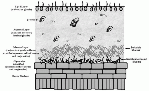
FIGURE 27-1. Schematic drawing of the three layers of the tear film. The tear film covers the cells of the ocular surface (cornea and conjunctiva). The upper lipid layer is secreted by the meibomian glands; the middle aqueous layer is secreted by the main and accessory lacrimal glands and the conjunctival epithelium; the inner mucous layer is secreted by conjunctival goblet cells and stratified squamous cells of the conjunctiva and cornea and the glycocalyx of the cornea and conjunctiva. (From Hodges and Dartt. Regulatory pathways in lacrimal gland epitheliom. Int Rev Cytol 2003;231:129-196.)


LIPID LAYER


Meibomian Glands

The lipid layer is secreted primarily by the meibomian glands located in the upper and lower lids as described in Chapter 1. These glands secrete a complex fluid containing
a plethora of lipids, which melts at about 35°C and thus is always fluid in the living eye. The lipid layer behaves as a film essentially independent of the aqueous layer underneath. It is anchored at the orifices of the meibomian glands above and below and does not take part in the flow of tears from the lateral canthus to the lacrimal puncta (1). When the lids close during a blink, the lipid layer is compressed over the aqueous layer (14). When the lids open, the lipid layer begins to spread again over the aqueous layer. The spreading front can move faster than the opening lid, so the aqueous layer is never exposed (14). During lid closure the lipid layer thickens, and during eye opening it thins. This has been elegantly shown by Doane (15) using a specially designed interferometer that detects colors produced by interference patterns as the light passes through an oily layer whose thickness is changing.


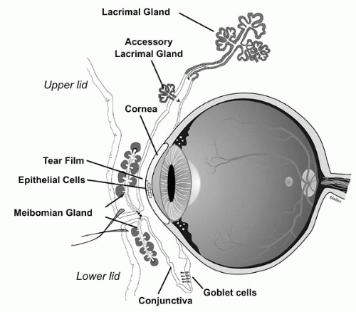
FIGURE 27-2. Schematic representation of the tear-producing glands. Cross section of the eye and the glands that produce the three layers of the tear film. (From Dartt DA. Regulation of mucin and fluid secretion by conjunctival epithelial cells. Prog Retin Eye Res 2002;21:555-576.)


Functional Anatomy of Meibomian Glands

Meibomian glands lie in a row in the superior and inferior eyelids. Each gland consists of a single, straight duct that opens directly onto the inner margin of the eyelids adjacent to the mucocutaneous junction (Fig. 27-3). The duct is lined with four cell layers of ductal epithelial cells and branches off into smaller ducts that each terminate in an acinus (16). Each acinus is composed of several layers of epithelial cells with the outer single layer of cells consisting of germinal basal cells that do not contain lipid droplets (Fig. 27-3). As the cells mature they migrate toward the center of the acinus at a rate of 0.62 μm per day (17,18). During maturation the endoplasmic reticulum of each acinar cell develops and begins to synthesize lipids that are stored in secretory granules surrounded by a membrane. The closer the cell to the center of the acinus, the greater the number of lipid secretory granules. It takes an average of 4.1 days for the cells to mature and migrate (18). Secretion occurs when the mature cells in the center of the acinus disintegrate, known as holocrine secretion. The lipid content of the secretory granules as well as the secretory granule membranes and the remaining content of the cell is released into the duct. Thus meibomian gland fluid contains lipid secretory products as well as normal cellular contents.
This accounts for the complex nature of the meibomian gland fluid. This fluid is stored in the ducts until released by the action of the blink.


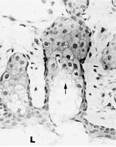
FIGURE 27-3. Light micrograph of alveoli of a rabbit meibomian gland. Cells in the outer layer (arrow) of the alveolus do not contain lipid droplets. Clear lipid droplets are visible in the middle layers of cells. Cells in the inner layer disintegrate into the alveolar duct, releasing the lipid droplets to form the secretory product that diffuses into the lumen (L) of the central duct. (H&E ×350.) (From Jester JV, Nicolaides N, Smith RE. Meibomian gland dysfunction. I: Keratin protein expression in normal human and rabbit meibomian glands. Invest Ophthalmol Vis Sci 1989;30:927.)

Meibomian glands are richly innervated as described in Chapter 1. There is parasympathetic, sympathetic, and sensory innervation of these glands with parasympathetic innervation predominating (Fig. 27-4) (17,19, 20, 21). The parasympathetic nerves contain acetylcholine and vasoactive intestinal polypeptide (VIP) as neurotransmitters (20). The sympathetic nerves contain norepiephrine and the sensory nerves contain substance P and calcitonin gene-related peptide (CGRP). Although neuropeptide Y (NPY) is usually found in sympathetic nerves, in meibomian glands, it was found in nerves with a parasympathetic nerve-like distribution (21). Despite the fact that the meibomian gland is well innervated, the functional role of the nerves in this gland remains unknown.

The meibomian glands contain receptors for the sex hormones, which are important for the regulation of meibomian gland secretion. Androgen, progesterone, and estrogen receptors are present in the acinar cell nuclei of these glands (Fig. 27-5) (22, 23, 24). Androgens increase the size, activity, and lipid production in meibomian glands, whereas estrogens and progestins decrease them (25). Thus multiple functions of the meibomian glands are regulated by sex steroids and there are substantial gender-based differences between the meibomian glands of males and females.


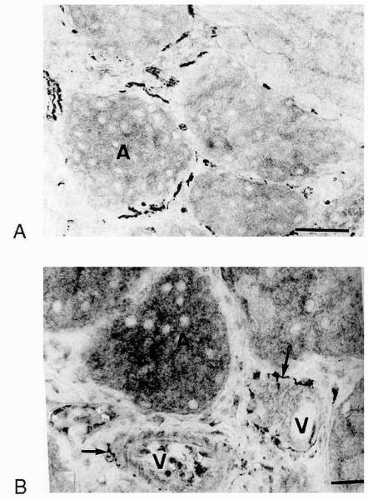
FIGURE 27-4. Innervation of the rat meibomian glands. Immunohistochemical microscopy showing localization of (A) sympathetic nerves using an antibody against tyrosine hydroxylase, which is one of the enzymes that coverts dopamine to norepinephrine, and (B) parasympathetic nerves using an antibody against choline acetyltransferase. (From Seifert P, Spitznas M. Immunocytochemical and ultrastructural evaluation of the distribution of nervous tissue and neuropeptides in the meibomian gland. Graefes Arch Clin Exp Ophthalmol 1996;234:648-656.)


Lipids Secreted by the Meibomian Glands

Meibomian glands secrete a complex fluid containing hydrocarbons, wax esters, triglycerides, diesters, free sterols,
sterol esters, free fatty acids, and polar lipids (for a review see ref. 26). The complexity of meibomian gland fluid reflects the products from the disintegrating cells as well as from the synthesized lipids. Meibomian gland lipids differ from cellular lipids in the acyl chain types and the sterol types. The major classes of lipids are the wax monoesters and sterol esters that make up about 60% to 70% of the meibomian gland fluid (26). A third type of lipid is the diesters that form ester linkages with fatty acids, fatty alcohols, or sterols. These diester compounds make up about 8% of the fluid.


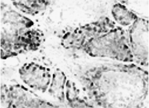
FIGURE 27-5. Presence of androgen receptor protein in acinar cells of the rat meibomian gland. Immunoperoxidase microscopy of meibomian gland showing the location of androgen receptor protein in acinar cells. (From Sullivan DA, Sullivan BD, Ullman MD, et al. Androgen influence on the Meibomian gland. Invest Ophthalmol Vis Sci 2000;41:3732-3742.)(see color image)


Regulation of Meibomian Gland Secretion

The steps in the secretion of meibomian gland fluid include synthesis of lipids, release of lipids from the cells, and release of secretory fluid from the ducts. Synthesis of meibomian lipid is coordinated by differentiation of the acinar cells with the more differentiated cells having synthesized more lipid secretory product. Androgens control the differentiation of the acinar cells and their lipid production. A review by Sullivan et al. (22) summarizes this evidence. Androgens increase the transcription of genes that produce proteins to stimulate the synthesis and secretion of lipids. Androgen action is enhanced by the presence of an enzyme that converts testosterone into a more potent androgen. Furthermore, androgens modulate genes that induce fatty acid and cholesterol synthesis, the degree of fatty acid saturation and branching, incorporation of fatty acids into phospholipids and neutral lipids, the total amount of lipids, the secretion of wax esters and other lipids, and the metabolism of lipoproteins (22). Androgens also stimulate expression of transcription factors (sterol regulatory binding proteins) that coordinate regulation of the enzymes that regulate lipid synthesis. Meibomian gland lipids are secreted by disintegration of the acinar cells. The compounds that regulate this process are unknown. The final step in the secretion of meibomian gland fluid is the release of preformed meibomian gland fluid from the ducts where it has been stored. Evidence suggests that the blinking process itself regulates this step.

An additional candidate for the regulation of meibomian gland secretion is the nerves that innervate the gland and surround the acini at their basement membranes. Their role in regulating meibomian gland function is unknown. Nerves could potentially regulate the release of lipids stored in the secretory granules by stimulating fusion of the secretory granule membranes with the apical membrane or by inducing the disruption of the entire cell.


Functions of the Lipid Layer

As evaluated by Tiffany (26), the major functions of the lipid layer are to prevent the spillover of tears and contain the tears within the palpebral opening, prevent damage of the lid margin skin by tears, and form a seal over the exposed portion of the eye during sleep. The lipid layer may possibly reduce evaporation in the open eye, but opposing results have been obtained in studies of tear evaporation. Another possible function of the tear film was suggested by McCulley and Shine (5), who hypothesized that the lipid layer may protect the eye from microorganisms, pollen, and other organic matter by trapping these particles in the monolayer. There is no evidence for this function. Tiffany suggests that the lipid layer does not prevent sebaceous lipids from entering tears and does not increase the stability of the tear film.


AQUEOUS LAYER


Lacrimal Gland


Anatomy

The main lacrimal gland is the major contributor to the aqueous layer of the tear film and as such synthesizes, stores, and secretes proteins, water, and electrolytes. It is an almond-shaped gland located on the anterior and lateral parts of the roof of the orbit of the eye. This gland is a multilobed, tubuloacinar structure with ducts that terminate at the surface of the eye in front of the lateral portion of the superior fornix. The major cell type is the acinar cell, which composes about 80% of the gland. These cells appear as a ring in cross section in a structure termed an acinus (Fig. 27-6). The tubules containing the acinar cells converge into interlobular ducts, which empty into larger ducts, and eventually to the ocular surface.

Similar to other exocrine glands, lacrimal gland acinar cells are surrounded by tight junctions at the lumen dividing the plasma membrane into apical (lumen) and basolateral (blood) membranes (Fig. 27-6). This has the effect of generating a polarized secretory cell to allow for unidirectional secretion of proteins and secretion of electrolytes and water into the lumen. Nerves surround the acinar cells on the basolateral side. As a result, the basolateral membranes of acinar cells have receptors for the neurotransmitters and neuropeptides released from the nerves surrounding them.

In addition to acinar cells, the lacrimal gland also contains ductal cells, myoepithelial cells, and lymphocytes. Ductal cells line the ducts one to two cell layers thick. These cells secrete water and electrolytes and a small amount of protein. Myoepithelial cells surround the acinar and ductal cells on the basal side with multiple processes containing smooth muscle actin. Based on the similarities to myoepithelial cells in other tissues and studies in the lacrimal gland, it is believed that myoepithelial cells in the lacrimal gland are involved in contraction of the acinar and
ductal cells to eject the secretory product (27). Many types of lymphocytes producing immunoglobulin (Ig) A, IgG, IgE, IgM, and IgD, plasma cells, mast cells, and macrophages are also present in the lacrimal gland.


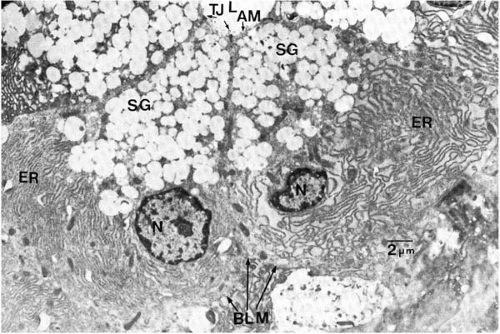
FIGURE 27-6. Electron micrograph of adjacent acinar cells from the rat lacrimal gland. The nucleus (N) lies in the basal portion of the cell and is surrounded by endoplasmic reticulum (ER). Secretory granules (SG) lie in the apical portion of the cell. Tight junctions (TJs) are present adjacent to the lumen (L) separate the apical membrane (AM) from the basolateral membrane (BLM). (From Dartt DA. Signal transduction and control of lacrimal gland protein secretion: a review. Curr Eye Res 1989;8:619-636.)


Proteins Secreted by the Lacrimal Gland

Most of the proteins secreted by the lacrimal gland are antibacterial, antiviral, or otherwise involved in protection of the eye (Table 27-2). These proteins include lactoferrin, lysozyme, peroxidase, and group II secretory phospholipase A2 as well as IgG, IgM, monomeric and polymeric IgA, secretory IgA (SIgA), and IgE. Other proteins such as cysteine-rich secretory proteins 1, 2, and 3, defensins, and convertase decay-accelerating factor are also secreted. In addition, the lacrimal gland produces many growth factors such as epidermal growth factor (EGF), transforming growth factor-α and –β, basic fibroblast growth factor, hepatocyte growth factor, and platelet-derived growth factor (28,29). The lacrimal gland has also been identified as the source of the retinol and retinal-binding protein found in tears (30,31). Recently, a novel protein has been detected in human tears that is secreted by the lacrimal gland. This protein, a glycoprotein, has been called lacritin and its function is unknown (32). Lacritin is expressed at high levels in the lacrimal gland and at lower levels in the salivary glands, and is not expressed in any other tissue (32).


Gender Differences

Many hormones from the hypothalamic-pituitary-gonadal axis have significant effects on the lacrimal gland (Table 27-3). These effects include influencing the growth and differentiation of the gland as well as secretion from the gland. Disruption of this axis via either hypophysectomy or anterior pituitary ablation causes gland atrophy, a decrease in tissue protein and messenger RNA (mRNA) levels and a subsequent decrease in fluid and protein secretion. One of the major hormones to exert effects on the lacrimal gland is androgens. Androgens are known to stimulate the secretion of SIgA and cystatin-related protein and are responsible for many sex-related differences seen in the lacrimal gland (33). It has been shown that male rats secrete higher levels of secretory component (SC), IgA, and cystatin-related protein and peroxidase than female rats and in general secrete more tears (33). In all species studied thus far, lacrimal glands from males were larger than those from females. Major gender-related differences have also been observed in the morphology, histochemistry, and biochemistry of lacrimal glands (Table 27-3) (33).


TABLE 27-2. PROTEINS KNOWN TO BE SECRETED BY THE LACRIMAL GLAND


Apolipoprotein D


β-Amyloid protein precursor


Basic fibroblast growth factor


Convertase decay-accelerating factor


Cystatin


Cystatin-related protein


Defensins


Endothelin-1


Epidermal growth factor


Granulocyte-monocyte-colony stimulating factor


Group II phospholipase A2


Hepatocyte growth factor


Immunoglobulin G


Immunoglobulin M


Interleukin-1α


Interleukin-1β


Lacritin


Lactoferrin


Lysozyme


Monomeric immunoglobulin A


Peroxidase


Plasminogen activator


Platelet-derived growth factors


Polymeric immunoglobulin A


Prolactin


Retinoic acid


Secretory component


Secretory immunoglobulin A


Tear lipocalins


Transforming growth factor-α


Transforming growth factor-β1


Transforming growth factor-β2


Tumor necrosis factor-α


Modified from Dartt DA and Sullivan DA. Wetting of the ocular surface. In: Albert D, Jakobiec F, eds. Principals and practices of opthalmology. Philadelphia: WB Saunders, 1994, 1043-1049.


Innervation of the Lacrimal Gland

It is well established that lacrimal gland protein secretion is under hormonal and neural control. Hormones play a major role in stimulation of constitutively secreted proteins, i.e., proteins that are secreted immediately upon synthesis. Nerves play a crucial role in the secretion of regulated protein secretion, i.e., proteins that are stored in secretory vesicles after synthesis and released upon the appropriate stimulus. Not surprisingly, stimuli of regulated and constitutive secretory proteins differ, as do the signaling pathways activated by these stimuli.

The lacrimal gland is innervated with parasympathetic, sympathetic, and sensory nerves (34). Parasympathetic nerves contain the neurotransmitter acetylcholine (ACh) and the neuropeptide VIP (Fig. 27-7A). Some VIP-containing nerves also contain pituitary adenylate cyclase activating peptide (35). There is additional evidence to suggest that parasympathetic nerves contain proenkephalin A-derived peptides (36).

Sympathetic nerves and sensory nerves are much less densely distributed than parasympathetic nerves (34) (Fig. 27-7B). Sympathetic nerves contain the neurotransmitter norepinephrine and may also contain the neuropeptide NPY (34). Sensory nerves originate from the trigeminal ganglion and contain substance P, galanin, and calcitonin gene-related protein (34). Sensory nerves do not participate in reflexes from the ocular surface, nor do their neurotransmitters stimulate protein secretion.

It is interesting to note that not all acinar cells are directly innervated. Gap junctions present within an acinus allow each cell to be electrically and chemically connected to adjacent cells. This allows for noninnervated cells to also be activated by neural stimulation (37).


Regulation of Lacrimal Gland Protein Secretion


Regulated Protein Secretion

Because the amount and composition of lacrimal gland fluid are critical to maintaining a healthy ocular surface, nerves tightly control the secretory process. Parasympathetic and sympathetic nerves are major stimuli of lacrimal gland protein secretion. The parasympathetic neurotransmitters ACh and VIP and the sympathetic neurotransmitter norepinephrine are potent stimuli of lacrimal gland secretion. The sympathetic neuropeptide NPY has a minor stimulatory effect on protein secretion.


Cholinergic Pathway

ACh released from parasympathetic nerves activates muscarinic receptors located on the basolateral membranes of acinar and the plasma membranes of myoepithelial cells (Fig. 27-8) (28). The M3 or glandular subtype of muscarinic receptors is the only muscarinic receptor identified in the lacrimal gland (28). The activated M3 receptor interacts with a Gαq/11 G protein (28). Activated G proteins in turn activate phospholipase C (PLC) to breakdown phosphatidylinositol bisphosphate (PIP2) into 1,4,5-inositol trisphosphate (1,4,5-IP3) and diacylglycerol (DAG) (28). The 1,4,5-IP3 released binds to and activates the IP3 receptors located on intracellular Ca2+ (Ca2+i) stores such as the endoplasmic reticulum. This interaction releases Ca2+ into the cytosol (38). The Ca2+i response is biphasic and includes a rapid peak followed by a slower sustained phase. It has been demonstrated that the rapid response is due to the release of Ca2+ from intracellular stores by 1,4,5-IP3 whereas the slow, sustained phase is due to the entry of extracellular Ca2+ into the cell. Emptying of the Ca2+i stores subsequently activates Ca2+ influx across the plasma membrane (38) (Fig. 27-8).

In addition to 1,4,5-IP3, activation of PLC also produces DAG. DAG activates a family of enzymes known as protein kinase C (PKC). The role of PKC in lacrimal gland secretion
is complicated due to the fact that 11 different isoforms of PKC have been identified. The lacrimal gland contains PKC-α, δ, ε, and λ. In general, PKC isoforms have cell and tissue specific localizations, and the lacrimal gland is no exception (28). The PKC isoforms detected in the lacrimal gland have specific although somewhat overlapping distribution (28).


TABLE 27-3. GENDER-RELATED DIFFERENCES IN THE LACRIMAL GLAND AND TEAR FILM


Male


Female


Morphology


Large acini


Smaller acini


Centrally located nucleus varying in size and shape


Basally located nucleus uniform in size and shape


Cell borders indistinct


Cell borders distinct


Biochemistry


Greater number of β-adrenergic receptors


Greater peroxidase activity


Greater level of androgen receptor protein


Greater amounts of melatonin


Greater activity of carbonic anhydrase


Greater amounts of N-acetyltransferase


Immunology


Increased synthesis of secretory component


Increased incidence of autoimmune disease


Greater production of immunoglobulin A (IgA)


Secretion and tears


Greater amount of secretion


Smaller amount of secretion


Greater tear levels of secretory component (SC), IgA, and cystatin-related protein


Higher amounts of pancreatic lipase-related protein1


Higher tear levels of total protein


Greater amounts of epidermal growth factor (EGF) and transforming growth factor-α (TGF-α)


Modified from Dartt DA and Sullivan DA. Wetting of the ocular surface. In: Albert D, Jakobiec F, eds. Principals and practices of opthalmology. Philadelphia: WB Saunders, 1994, 1043-1049.


To determine the role of PKC isoforms in secretion, PKC isoform selective inhibitory peptides can be used. When such peptides were used, cholinergic agonist-stimulated secretion was inhibited the most by the PKC-α inhibitory peptide, followed by the PKC-ε inhibitory peptide with the least inhibition obtained using the PKC-δ inhibitory peptide. Confirming the role of these PKC isoforms in lacrimal gland secretion, treatment with phorbol esters downregulated PKC-α the most followed by PKC-δ and PKC-ε with cholinergic agonist-stimulated secretion inhibited over 90% (28). Thus, these PKC isoforms play important, but differential, roles in lacrimal gland secretion stimulated by cholinergic agonists.


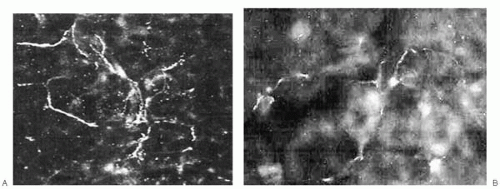
FIGURE 27-7. Innervation of the rat lacrimal gland. Immunofluorescent microscopy showing localization of (A) parasympathetic nerves using an antibody against vasoactive intestinal peptide (VIP). B. Sympathetic nerves using an antibody against dopamine β-hydroxylase which is the enzyme that coverts dopamine to norepinephrine.

In addition to activating PLC, cholinergic agonists also activate phospholipase D (PLD) in the lacrimal gland. PLD hydrolyzes phospholipids preferring phosphatidylcholine as a substrate, which produces phosphatidic acid and free polar head group. The phosphatidic acid can be a signaling molecule itself or can be degraded to generate DAG. Cholinergic agonists, activation of PKC, and increasing the [Ca2+]i all stimulated PLD activity in lacrimal gland acini (28). Because cholinergic agonists produced phosphatidic acid, but, surprisingly, activation of PKC and increasing the [Ca2+]i did not, it appears that the lacrimal gland contained two different types of PLD activity.


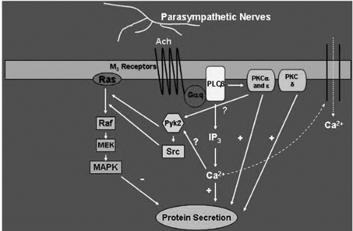
FIGURE 27-8. Schematic drawing of the signal transduction pathways activated by cholinergic agonists in the lacrimal gland. Ach, acetylcholine; M3, muscarinic receptor subtype 3; Gαq, alpha subunit of Gq G protein; PLC, phospholipase C; Pyk2 and Src, nonreceptor tyrosine kinases; raf, mitogen-activated protein kinase kinase kinase; MEK, mitogen-activated protein kinase kinase; MAPK, p44/p42 mitogen-activated protein kinase; InsP3, inositol trisphosphate; PKC, protein kinase C. (Modified from Dartt DA. Regulation of lacrimal gland secretion by neurotransmitters and the EGF family of growth factors. Exp Eye Res 2001;73:741-752.)

In summary, the lacrimal gland contains M3 muscarinic receptors. Upon ligand binding, the receptor, through Gαq/11 G proteins, activates PLC and PLD. The two second messengers, DAG and IP3, are generated from PLC hydrolysis of PIP2 and stimulate secretion, whereas the role of PLD is protein secretion is not known. DAG activates the PKC isoforms PKC-α, -δ, and -ε, whereas 1,4,5-IP3 releases Ca2+ from intracellular stores. Both increasing [Ca2+]i and activation of PKC-α, -δ, and -ε play integral roles in stimulation of cholinergic-induced protein secretion.


α1-Adrenergic Pathway

In the lacrimal gland norepinephrine, released from sympathetic nerves, activates α1-adrenergic receptors to stimulate lacrimal gland protein secretion (Fig. 27-9) (39,40). In fact stimulation by the α1-adrenergic agonist phenylephrine was as potent and effective as cholinergic agonists in stimulating protein secretion. The types of α1-adrenergic receptors (α1A, B, or D) present in the lacrimal gland are not known. α1-Adrenergic agonists use the Gαq/11 subunit G protein to stimulate about one third of its secretion (28). The G protein activating the remaining secretion remains unidentified.

The next step in the α1-adrenergic agonist-signaling pathway, activation of a phospholipase, is unclear (Fig. 27-9). α1-Adrenergic agonists in the lacrimal gland do not appear to activate either PLC or PLD in the lacrimal gland. The most likely candidate for α1-adrenergic agonist activation is PLA2, which generates arachidonic acid that can be converted to DAG. It is known that α1-adrenergic agonists do cause a small increase in [Ca2+]i, which is about 20% of the cholinergic agonist response in isolated acini (28). In addition, activation of PKC isoforms plays a role in α1-adrenergic agonist stimulated protein secretion. Although no translocation of any PKC isoforms was detected with stimulation of α1-adrenergic agonists, when PKC isoforms were downregulated with a long-term phorbol ester treatment phenylephrine-induced secretion was increased (28). This suggests that activation of PKC inhibits α1-adrenergic agonist-induced protein secretion. Using the PKC isoform selective inhibitor peptides, inhibition of PKC-α and -δ stimulated α1-adrenergic agonist-induced protein secretion, whereas inhibition of PKC-ε blocked α1-adrenergic agonist-stimulated protein secretion (28). It appears then that activation of PKC-α and -δ inhibits α1-adrenergic agonist-induced protein secretion whereas activation of PKC-ε stimulates it. Thus cholinergic and α1-adrenergic agonists are activating the same PKC isozymes PKC-α, -δ, and -ε, but are doing so differently. This suggests compartmentalization or targeting, because the same PKC isoforms either stimulate or inhibit secretion depending on the agonist. The targeting proteins in the lacrimal gland are unknown.


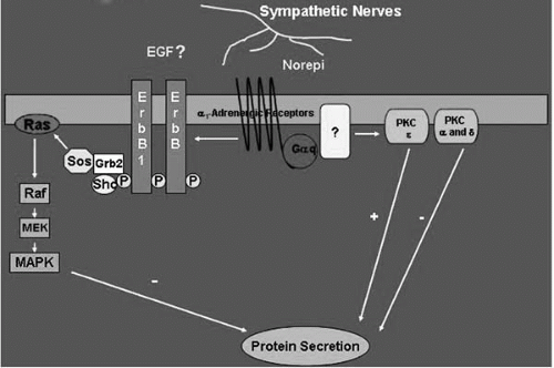
FIGURE 27-9. Schematic drawing of the signal transduction pathways activated by α1-adrenergic agonists in the lacrimal gland. Norepi, norepinephrine; PKC, protein kinase C; ErbB1, the epidermal growth factor (EGF) receptor; ErbB, the EGF family of growth factor receptor; P, phosphorylated tyrosine residues on the ErbBs; SOS, Grb2 and Shc, adaptor proteins; raf, mitogen-activated protein kinase kinase kinase; MEK, mitogen-activated protein kinase kinase; MAPK, p44/p42 mitogen-activated protein kinase. (Modified from Dartt DA. Regulation of lacrimal gland secretion by neurotransmitters and the EGF family of growth factors. Exp Eye Res 2001;73:741-752.)

Only gold members can continue reading. Log In or Register to continue

Stay updated, free articles. Join our Telegram channel

Sep 18, 2016 | Posted by in OPHTHALMOLOGY | Comments Off on Keratoconjunctivitis Sicca: Physiology and Biochemistry of the Tear Film

Full access? Get Clinical Tree

Get Clinical Tree app for offline access