Keratoconjunctivitis Sicca: Clinical Aspects


Keratoconjunctivitis Sicca: Clinical Aspects


Abha Gulati

Reza Dana


Keratoconjunctivitis sicca (KCS) or dry-eye syndrome (DES) is composed of a variety of ocular surface disorders of diverse pathogenesis that share, as a common manifestation, signs and symptoms of ocular surface damage (1). According to the “global definition” of DES by the National Eye Institute/Industry Workshop on Clinical Trials in Dry Eyes, dry eye is a disorder of the tear film due to tear deficiency or excessive tear evaporation that causes damage to the interpalpebral ocular surface and is associated with symptoms of ocular discomfort (2). The healthy tear film and ocular surface form a complex and stable system that can lose its equilibrium through numerous disturbing factors. A sufficient quantity of tears, a normal composition of tear film, a normal lid closure, and regular blinking are the factors necessary for maintaining a stable preocular tear film. Disturbance in one or more of these factors leads to an unstable tear film, ocular surface damage, and dry eye disease. Most symptoms in patients with KCS result from an abnormal, nonlubricative ocular surface that increases shear forces under the eyelids, and diminishes the ability of the ocular surface to respond normally to environmental challenges. Increasingly, it has become apparent that KCS is associated with ocular surface inflammation and that the inflammatory component contributes to the symptoms as well as the disease process itself.

KCS is a chronic condition that currently has no cure. Early diagnosis and timely therapy usually helps in averting the complications, which may be severe and if inadequately treated, may lead to severe ocular surface damage and blindness (2). The use of artificial tears, which was the mainstay of therapy for dry eye disease in the past, is mainly palliative, resulting in a reduction of ocular surface eyelid shear forces and some transient symptomatic relief. Antiinflammatory therapies that target one or more of the components of the inflammatory response in dry eye are now increasingly being used in treating signs and symptoms of dry eye. Surgical intervention is required in treating end-stage dry eye disease and for visual rehabilitation in patients with severe corneal surface damage.


EPIDEMIOLOGY

An estimated 2.2 million women aged 55 and older have dry eye disease in the United States (3). In a community study by Jacobsson et al. (4) in Sweden, a prevalence rate of 15% was found among 705 members of the general population aged 55 to 72 years, based on the presence of dry eye symptoms and positive findings on Schirmer test, tear film breakup time, or rose bengal staining.

In the Salisbury Study (5), dry eye was assessed by questionnaire and clinical tests. The questionnaire queried subjects about six symptoms: dryness, grittiness/sandiness, burning, redness, crusting on the eyelashes, and eyes being stuck shut in the morning. Each subject was asked to describe the frequency with which he or she experienced each symptom as being “all of the time,” “often,” “sometimes,” or “rarely.” Based on the symptoms alone, a subject was considered to have DES if he or she experienced one of the six symptoms at least “often.” In this study, 41% reported no symptoms, 45% reported one or more symptoms at least rarely or sometimes and 14% reported one or more symptoms at least often. According to the Melbourne Visual Impairment Project dry eye sub-study (6), the most commonly reported severe symptom of DES was photophobia.

Clinical dogma has long suggested that DES becomes more common with age, and some evidence suggests an age-related decrease in tear production (7). Schaumberg et al. (3) have summarized data on the relationship of dry eye prevalence with age in the four large epidemiologic studies (5,6,8,9) conducted to date. The results of these studies taken together were most consistent with a trend toward higher prevalence of DES in the older age groups. However, currently no information is available on the incidence, either generally or with regard to age, of dry eye in a population.

It has been observed that DES is more common in women, particularly after menopause. Androgens probably account for many gender-specific differences observed in lacrimal glands of numerous species (10). In the
Melbourne Study (6), women were nearly two times more likely to report severe symptoms of dry eye. In World Health Studies (WHS), 66% to 70% higher prevalence of severe dry eye symptoms or clinically diagnosed DES has been shown among postmenopausal women who use estrogen alone compared to women who never used hormone replacement therapy (11).

Various other factors that have shown statistically significant independent associations are arthritis history, cigarette smoking, caffeine use, thyroid disease, history of gout, and diabetes mellitus (9). Thus, to summarize, DES is a relatively common condition among middle-aged and older adults with higher prevalence among women than men.


ETIOLOGY

The etiology of KCS is multifactorial. Based on the concept of the “lacrimal functional unit” (12), which consists of the ocular surface, main and accessory lacrimal glands, interconnecting nerves, and neuroendocrine factors that regulate the function of these nerves, alterations at any of these levels that may disturb this functional unit can result in KCS.

The preocular tear film that covers the ocular surface consists of three layers. The superficial lipid layer, which is the outermost layer of the tear film, is believed to retard or decrease evaporation of tears. The middle aqueous layer is secreted by the main and accessory lacrimal glands and contains water-soluble factors and electrolytes, and helps to wet the conjunctival and corneal epithelial cells, causes mechanical flushing of debris and organisms, and its constituents help inhibit the growth of microorganisms on the ocular surface. It also serves as a permeability barrier and is essential for maintaining a smooth and regular optical surface. The deep mucinous layer is composed mainly of glycoproteins (mucin) secreted by goblet cells and epithelial cells of conjunctiva and cornea. It helps in lowering the surface tension of tears, thus enhancing its spreading and stability. Thus, integrity of the tear film can be affected by disorders involving epithelial cells, goblet cells, meibomian glands, lacrimal gland, and neuronal innervation.

Because the preocular tear film and the ocular surface exist in a state of equilibrium, broadly speaking, all those conditions that may directly or indirectly influence tear secretion or evaporation can result in an unstable tear film, ocular surface damage, and dry eye disease. For classification purposes, KCS is classified into (a) lacrimal- or tear-deficient dry eye, and (b) evaporative dry eye (2). However, in clinical practice, KCS is often seen as a combination of both tear-deficient and evaporative types. Various causes of KCS are enumerated in Table 28-1.


TABLE 28-1. ETIOLOGICAL CLASSIFICATION OF DRY-EYE SYNDROME


  1. Tear-deficient dry eye


    1. Sjögren’s syndrome dry eye


      1. Primary Sjögren’s syndrome


      2. Secondary Sjögren’s syndrome


        1. Rheumatoid arthritis


        2. Systemic lupus erythematosus


        3. Polyarteritis


        4. Wegener’s granulomatosis


        5. Systemic sclerosis


        6. Primary biliary cirrhosis


        7. Mixed connective tissue disease


    2. Non-Sjögren’s syndrome dry eye


      1. Congenital


        1. Familial dysautonomia (Riley-Day syndrome)


        2. Congenital alacrima


        3. Trigeminal nerve aplasia


        4. Ectodermal dysplasia


      2. Acquired


        1. Injury


          • Trauma


          • Lacrimal gland ablation


        2. Infections


          • Trachoma


          • Mumps


          • HIV


          • Mononucleosis


          • Other infections


        3. Immune mediated


          • Graft-versus-host disease


        4. Lacrimal obstructive


          • Cicatricial pemphigoid*


          • Burns*


          • Stevens-Johnson syndrome*


        5. Lymphoproliferative


          • Lymphoma


          • Leukemia


        6. Reflex hyposecretion


          • Neuropathic-neuroparalytic disorders


          • Contact lens wear


          • Diabetes


          • Aging


          • Corneal surgery


          • Herpetic keratitis


        7. Medications


          • Antihistamines


          • Antimuscarinics


          • Beta-adrenergic blockers


        8. Infiltrative


          • Sarcoidosis


          • Amyloidosis


          • Hemochromatosis


  2. Evaporative dry eye


    1. Lipid deficiency


      1. Primary


        1. Absent meibomian glands


        2. Distichiasis


      2. Secondary


        1. Blepharitis


        2. Meibomian gland dysfunction (MGD)


        3. Medications: Accutane


    2. Lid abnormalities


      1. Blink abnormalities


        1. Parkinson’s disease


        2. Facial nerve palsy


        3. Leprosy


        4. Contact lens wear


      2. Aperture abnormalities


        1. Proptosis


        2. Lagophthalmos


      3. Lid surface abnormalities


        1. Coloboma


        2. Ectropion


        3. Entropion


    3. Ocular surface abnormalities


      1. Vitamin A deficiency


      2. Pterygium


      3. Symblepharon


      4. Scars and nodules


* Associated mucin deficiency.


Tear-Deficient Dry Eye

The aqueous layer forms the greatest bulk of the precorneal tear film. Aqueous tears are produced in the main lacrimal glands, with a lesser contribution from the accessory glands of Wolfring and Krause. Abnormalities of stimulation of tear secretion, destruction of lacrimal gland and accessory lacrimal glands, or scarring or occlusion of lacrimal gland secretory ducts may be responsible for the aqueous-deficient dry eye. Sjögren’s syndrome, which is an immune-mediated disorder and the commonest type of aqueous tear deficient type of dry eye disease (see Chapter 29).

The most severe forms of KCS are due to the destruction, or rarely absence, of the lacrimal gland, and include Sjögren’s syndrome, the acquired immune deficiency syndrome (AIDS) (13), graft-versus-host disease (GVHD) (14), and congenital and surgical removal of lacrimal gland. Less severe forms of KCS occur due to abnormalities of the regulation of tear secretion, such as that brought on by aging, alterations in hormone levels (menopause), and systemic medications. Ocular surface diseases such as ocular cicatricial pemphigoid, lichen planus, Stevens-Johnson syndrome, chemical burns, and GVHD can cause KCS through the scarring, narrowing, and obliteration of the lacrimal and accessory lacrimal gland secretory ducts.

Many systemic medications can cause dryness of eyes by altering tear flow and influencing production of natural tear substances, or by penetrating and combining with natural components of the tear film (15). Oral antihistamines, antihypertensives, antiemetics, antidepressants, and diuretics may cause DES (16). Antihistamines have antimuscarinic activity that may affect the aqueous layer by decreasing tear production, and the mucous layer by decreasing mucin output of the conjunctival goblet cells (15). Dry eye associated with long-standing contact lens wear is proposed to be due to decreased corneal sensation and blinking (17). Reduced sensory function facilitates drying by decreasing tear secretion and reducing the blink rate. A similar mechanism is operative in dry eye following herpes infections and LASIK (18). Neurotrophic keratitis, caused by sensory loss in the distribution of the first division of the fifth cranial nerve, is associated with severe ocular surface disorder, which is also partly due to the loss of the trophic function of the trigeminal nerve.


Evaporative Dry Eye

A well-structured lipid layer of the tear film decreases evaporation of tears from the ocular surface (19) and also prevents the overflow from the aqueous-mucin layer, such as might occur during a blink. Deficiency of the lipid layer permits more rapid evaporation of moisture from the eye surface, and, in the absence of an adequate increase of tear production by the lacrimal glands, gives rise to evaporative form of dry eye (20).

Meibomian gland dysfunction (MGD) is believed to be a predominant cause of evaporative dry eye (21). MGD may result from local pathology (e.g., chronic blepharitis), from dermatologic disease (e.g., ocular rosacea), or from iatrogenic etiology such as drugs including isotretinoin (Accutane). Ocular rosacea produces blepharitis and MGD, which involves chronic inflammation of eyelids and ocular surface, with tear film, conjunctival, and occasional corneal impairment.

One of the most important functions of the lid is to resurface the eye with tears through blinking. Any break in the integrity of the lid or its close apposition to the ocular surface leads to increased tear evaporation and areas of dryness. Lid surface abnormalities including ectropion, entropion, and lid coloboma can cause dry eye due to excessive evaporation. The preocular tear film is dynamic such that it changes continuously and is thinned and disrupted after each blink, and blinking helps resurface and reestablish its integrity. Regular, frequent blinking prevents the formation of dry spots on the surface of the cornea and conjunctiva. Blink disorders such as seen in Parkinson’s disease, in which there is infrequent blinking, can cause drying of the ocular surface.

An intact tear film, because it is very thin, is dependent on a smooth, uninterrupted epithelial surface. Any irregularity in the surface will cause an associated irregularity in the tear film. Such an irregular or elevated area will predispose the tear film to break up instantly at that spot, which may be one of the mechanisms involved in dry eye associated with pterygium and also vitamin A deficiency. Increased evaporation of tears can also occur secondary to increased palpebral fissure width. Palpebral fissure width may be large due to heredity, lid surgery, or thyroid eye disease. It has been recognized that tear film evaporation is proportional to the ocular surface area exposed (22). Indeed, increased rates of evaporation from the tear film can increase tear film osmolarity and create dry eye disease independent of abnormalities of aqueous tear secretion.


Combined-Mechanism Dry Eye

It has been seen that aqueous-deficient and evaporative types of dry eye usually occur in conjunction with one another, as lacrimal gland deficiency may often be accompanied by meibomian gland and goblet cell deficiency. For example, cicatricial conjunctival disease may cause dry eye both by occlusion of the lacrimal gland ductules, and by causing lid incongruity, which interferes with tear resurfacing with each blink. Additionally, studies have shown that in patients with Sjögren’s syndrome, MGD may contribute to the ocular surface disease in addition to the aqueous tear deficiency (23).

Lack of aqueous tear secretion may predispose to blepharitis as a consequence of increased levels of bacterial flora resulting from decreased levels of tear immunoglobulin A (IgA), lactoferrin, and lysozyme. Blepharitis itself can also lead to abnormalities of meibomian gland secretion as well as disruption of the lipid layer, resultant tear film instability, and increased evaporation (24). Another explanation of the combined mechanism is that prolonged stimulation of enhanced secretion by the lacrimal gland, caused by the chronic increased evaporation of water from tear film, could result in neurogenically induced inflammation to the lacrimal gland itself, slowly transforming an evaporative dry eye into a tear deficient dry eye (20,25).


Mucin Deficiency and Dry Eye

The mucin layer lies immediately above the keratoconjunctival epithelium and plays an important role in helping spread the tear film over the anterior ocular surface and in providing the biophysical properties needed to interface with the hydrophobic epithelial layer of the eye surface. The ocular surface epithelium expresses at least three major mucin genes. In the conjunctiva, goblet cells are responsible for secreting gel-forming mucin MUC5AC, whereas the stratified epithelium produces the cell membrane-spanning mucins MUC1 and MUC4 (26, 27, 28).

Pathologies that damage the conjunctiva such as ocular cicatricial pemphigoid, and Stevens-Johnson syndrome, result in mucin deficiency due to loss of goblet cells, thereby causing increase in surface tension, increasing evaporation, ocular surface damage, and dry eye disease.

Alterations in the overall quantity and quality of ocular surface mucins may occur as a result of, as well as lead to, KCS (29,30). It has been suggested that elevated proinflammatory cytokine levels within the tear film, perhaps combined with reduced concentrations of essential lacrimal gland derived factors (i.e., epidermal growth factor, retinol), create an environment in which terminal differentiation of the ocular surface epithelium is impaired (30). As a consequence, the epithelium becomes hyperplastic, displaying increased mitotic activity, and loses the ability to express mature protective surface molecules including the membrane-spanning mucin, MUC-1, which is secreted by epithelial cells to form the glycocalyx, deficiency of which will lead to an unstable tear film and dry eye disease. We have recently found a significant reduction of the goblet cell-specific mucin MUC5AC in the tears of patients with Sjögren’s syndrome, corresponding to a decrease in the RNA transcripts of MUC5AC in the conjunctival epithelium (31). Thus, depletion of goblet cell MUC5AC mucin may be an additional mechanism contributing to tear film instability in Sjögren’s syndrome (31).


PATHOGENESIS


Role of Immunity

Clinically significant KCS is associated with a variable degree of ocular surface inflammation characterized by the red eye. The first step in the generation of inflammation is an inciting stimulus, which may lead to expression of proinflammatory cytokines and a variety of other mediators (e.g., chemokines and adhesion factors) that in the aggregate signal the host that the normal physiology and microenvironment have been altered. In response to these signals, the second step in the cascade of events occurs when local (resident) tissue cells activate signal transduction pathways, e.g., NFκB (Nuclear factor Kappa B) that augment or down-modulate these cells’ expression of cytokine gene and/or cytokine receptor genes, which in turn dictate the response of these resident cells to paracrine signals in the microenvironment by other cells in close proximity (32). The inflammatory immune response to ocular surface injury may be innate as well as adaptive. Adaptive (acquired) immunity is the more evolved arm of the immune response that is characterized by a delayed, stimulus-specific response. The generation of an adaptive immune response requires activation of local antigen-presenting cells to stimulate T cells (33,34).

Lymphocytic infiltration of the lacrimal gland by T cells has been described in both Sjögren’s syndrome (35,36), and non-Sjögren’s KCS (37,38). This infiltration is believed to be responsible for the dysfunction and/or destruction of normal secretory function. Immunopathologic studies of the lacrimal gland in patients with Sjögren’s syndrome show that the lymphocytic infiltrate primarily consists of CD4+ T cells. Various studies have also shown that conjunctival inflammation is also present in more than 80% of patients with KCS (39,40). Certain proinflammatory cytokines, such as interleukin (IL)-1, IL-6, and tumor necrosis factor-α (TNF-α), have been detected at increased concentration in the tear fluid and conjunctival epithelium of patients with dry eye (41). Ocular rosacea, which is a prominent cause of KCS, has been found to be associated with differential increase in the levels of the proinflammatory cytokine IL-1α in the tear fluid (42), and an overexpression of immune inflammatory markers such as human
leukocyte antigen (HLA)-DR and intercellular adhesion molecule-1 (ICAM-1) on conjunctival epithelium (43). All of these factors are thought to act in concert to promote the immunoinflammatory response in the ocular surface seen so commonly in severe DES.

Tear hyperosmolarity has been proposed as a possible mechanism of ocular surface inflammation in both tear-deficient (44) and evaporative forms of dry eye (45). It has been suggested that tear hyperosmolarity, together with the microabrasive effects of blinking in the dry eye state, leads to an upregulation of pro-inflammatory cytokines such as TNF-α, IL-1, and IL-6, which may be the critical intermediaries involved in maintaining and perpetuating the ocular surface inflammation seen in KCS (46). Finally, inflammatory cytokines have been implicated in the regulation of the expression of epithelial mucins (47).

Inflammation of the lacrimal gland and/or ocular surface leads to the anomalous production of secretory growth factors or cytokines that modulate gene expression within the conjunctival and lacrimal epithelium (48), leading to an altered cellular phenotype that can promote immune responsiveness. It has been shown that epithelial cells of the ocular surface or lacrimal glands overexpress major histocompatibility complex (MHC) class II antigens (40,49) in dry eye syndrome. A possible explanation of this phenomenon is that epithelial cells may acquire antigen-presenting capability, and that these immunologically activated epithelial cells may be the target of lymphocytes (49) or that they may directly participate in recruitment of inflammatory cells, thus perpetuating inflammation and immune responsiveness. However, to date, conclusive proof regarding the role of ocular surface epithelial cells in orchestrating T cell-mediated immunity has been lacking.


Role of Neuronal Regulation

The ocular surface is richly innervated, with the cornea having a density of free nerve endings approximately 60 times that of tooth pulp. It is well known that corneal stimulation results in a rapid reflex including immediate blinking, profuse reflex tearing, and withdrawal of the head. Sensory impulses from the cornea and conjunctiva travel via the ophthalmic branch of the trigeminal nerve. Efferent fibers from the pons extend (facial nerve) to the lacrimal gland, where they influence the secretomotor function of the gland (modulation of water and protein transport). The lacrimal gland is also densely innervated with parasympathetic nerves, and to a lesser extent with sympathetic and sensory nerves (50). Accessory lacrimal glands (51) and goblets cells are also innervated (52). Acetylcholine (parasympathetic mediator) acts through muscarinic receptors to stimulate secretion of water, proteins, and electrolytes from the lacrimal glands. Cholinergic stimulation also induces increased concentrations of lactoferrin and epidermal growth factor (EGF) in glandular tissues and tears. The routes by which the signals from these various systems are transmitted are interconnected and closely related. An abnormality in any one of the steps in this complex cascade is likely to result in overall dysregulation of lacrimal function.

Parasympathetic neural transmission can be inhibited by cytokines (53). Studies by Zoukhri and associates (54) on the mouse model of Sjögren’s syndrome led to the hypothesis that the impaired secretory function of lacrimal glands is most likely a functional blockage caused by inflammatory cytokines, either in neurotransmitter release from nerve endings, or in the cellular response to neuromediators, leading in both cases to a denervation-like supersensitivity. Recently, it has been demonstrated that there is upregulation of IL-1β in the lacrimal gland of the mouse model of Sjögren’s syndrome, suggesting that proinflammatory cytokines such as IL-1β may be responsible for the impaired secretory function in the exocrine glands, in response to neuronal stimulation, as seen in Sjögren’s syndrome (55).


Role of Hormones

There is evidence to support that the eye, like many other tissues, is a target organ for sex hormones, and that these hormones control certain trophic functions and homeostasis of the lacrimal system. The receptors of various steroid hormones have been identified in rat and human ocular tissues, particularly in the lacrimal gland, meibomian glands, conjunctiva, and cornea (56). In addition, messenger RNA (mRNA) for 5α-reductase, the key enzyme involved in the metabolism of androgens to their biologically active derivative 5α-dihydrotestosterone, has been found in these ocular structures (57).

Sex steroids have been implicated in the pathogenesis of Sjögren’s syndrome through their action on the immune system (58). The mechanism of androgen action in lacrimal tissue appears to be mediated through hormone interaction with receptors in nuclei of epithelial cells (not lymphocytes) (59), which then leads to alterations in the expression of cytokines, proto-oncogenes, and apoptotic factors (60). These androgen-induced changes include enhancement of levels of transforming growth factor-β (TGF-β), a potent immunomodulatory and antiinflammatory cytokine, and suppression of mRNA content of proinflammatory cytokines (e.g., IL-1β and TNF-α) in the lacrimal glands (10). Moreover, apoptosis has been shown to occur in the lacrimal glands following withdrawal of androgen in the ovariectomized rabbits (61), which has led to the suggestion that these apoptotic fragments may be a source of potential autoantigens and could be subsequently presented by either interstitial antigen-presenting cells or acinar cells to initiate the autoimmune response.

The meibomian gland is also an androgen target organ, and androgens regulate this tissue’s lipid profile (62,63).


FIGURE 28-1. Paradigm for dry-eye pathogenesis.


Pathogenic Model for Dry Eye

The exact sequence of cellular and molecular events that leads to KCS remains unknown. It is possible that lacrimal dysfunction leads to decrease in tear volume and secretion of trophic factors with resultant ocular surface distress. The ocular surface distress and related hyperosmolarity and microabrasive effect of blinking on a surface that is not adequately protected by the proper mix of tear film constituents (mucins, lipids, etc.) leads to inflammation that can in turn add to the lacrimal insufficiency by affecting accessory lacrimal gland function. With reduced tear secretion, there is decreased tear clearance (64) that results in increased retention of inflammatory cytokines, released into the tear film from the ocular surface epithelium or lacrimal gland (65), thus perpetuating the inflammatory response. Additionally, with inflammation, there is abnormal lipid secretion onto the surface due to changes in the meibomian gland orifices, resulting in decreased tear film stability. Moreover, ocular surface changes as a result of the cytokine microenvironment may lead to relative hypesthesia and changes in blink rate that may in turn amplify the ocular surface disease by increasing exposure, evaporation, and altering the feedback to the lacrimal gland. Such a paradigm is illustrated in Fig. 28-1.


PATHOLOGY

Tears are necessary for the continued health of the ocular surface, maintaining the nonkeratinized surface essential for corneal transparency and lubrication required for movement of the lids on the globe. Morphologically, in KCS the conjunctiva is affected before the cornea is. Initially the conjunctival epithelium appears normal (66), but there is loss of conjunctival goblet cells (67), with subsequent edema in the conjunctival stroma (68). As the disease becomes more advanced, intracellular edema appears, manifested by decreased cytoplasmic density (69). Conjunctival epithelial cells with decreased cytoplasmic density demonstrate blunting and loss of cell surface microplicae. As fluid moves between superficial conjunctival epithelial cells, there is an increase in conjunctival epithelial cell desquamation (66,68). With time, and as the disease progresses, squamous metaplasia of the conjunctiva develops (70), with further decrease in conjunctival goblet cell density (67), increase in the surface area and flattening of conjunctival epithelial cells (66), and eventually disappearance of intercellular and stromal edema (66).

The cornea is more resistant than the conjunctiva to disease in KCS. The corneal epithelium forms a protective barrier between the environment and underlying ocular structures. Using wide field color specular microscopy, a shift toward smaller superficial corneal epithelial cells has been shown in patients with KCS (71), a reflection of accelerated corneal desquamation.


CLINICAL FEATURES

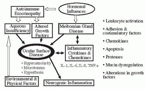
In spite of their rather diverse origins, the clinical presentation of patients with dry eye diseases is similar. Tear film instability is a component of all forms of dry eye disease, and tear hyperosmolarity is a key mechanism for the ocular surface damage. Aqueous-deficient dry eye is associated with reduced lacrimal function, but lacrimal function may be reduced as part of the aging process without producing the signs or symptoms of dry eye. The various factors to be considered in the diagnosis of KCS are (a) presence or absence of inflammation, (b) assessment of the function of lacrimal tissues, (c) determination of causes that could lead to decreased reflex tearing, (d) assessment of meibomian glands, and (e) environmental factors.


History

The patient’s history is extremely important in diagnosing dry-eye syndromes. Symptoms are a hallmark of the disease, and the most frequently encountered symptoms in patients with dry eyes are dryness, foreign-body or sandy sensation, burning, and photophobia. Patients may also complain of itching, excessive mucus secretions, heaviness of the eyelids, tight eyelids, inability to produce tears, pain, and redness. Moreover, as dry eye can coexist with other disorders (e.g., allergy), it is not unusual for patients to have symptoms due to multiple pathogenic mechanisms. The principal function of the tear film is to maintain a smooth, clear, refractive optical surface in a hostile external environment. Any adverse effect on the corneal regularity and clarity will interfere with vision and may cause symptoms. Patients often use the term “dryness” to describe their condition but will have difficulty defining exactly what this means. The term “discomfort” may be a more accurate summation of all the patient’s symptoms. Various questionnaires have been developed (72, 73, 74) to assess symptoms in dry eye patients. These questionnaires
are useful tools for clinical treatment trials in dry eye patients.

Dry eye patients are exquisitely sensitive to drafts and winds. Often they volunteer information regarding their intolerance to air conditioning or driving in the car with the windows down. Patients with KCS feel worse in a dry, cold environment and under conditions of increased evaporation. Reading is often difficult for dry eye sufferers. This probably occurs because the blink frequency decreases during tasks requiring concentration. Patients often complain that nighttime or awakening is the worst part of their day. Sleep (like general anesthesia) decreases tear production. If the eye is already compromised with regard to tear flow, further reduction during sleep may be enough to produce nocturnal symptoms. This is especially true if concurrent blepharitis or lagophthalmos is present. Smoke is almost universally intolerable to tear-deficient patients. Because smoke is actually a suspension of solids in air, the particulate bombardment of the ocular surface produces discomfort. Determining whether symptoms are worse or better indoors or out, at work or at home, will aid in identifying environments that need to be modified to improve the patient’s symptoms.

Patients should be asked whether they are able to produce irritant and emotional tears. “Do you get tears when you peel onions? Can you cry when you feel sad or hurt? Affirmative responses suggest some lacrimal gland function remains, whereas negative answers suggest that the lacrimal gland is incapable of secreting tear fluid in response to any stimuli. In patients with KCS, the ability to generate irritant tears is lost before the ability to generate emotional tears.

“What medications do you take?” should also be asked. Systemic antihistaminics, antidepressants, anticholinergics, and diuretics are most responsible for decreased tear production. The use of systemic steroids and other immunosuppressives, such as hydroxychloroquine (Plaquenil), methotrexate, and cyclophosphamide (Cytoxan), which may be used in treating patients with Sjögren’s syndrome and other collagen vascular diseases should be noted.

It is important to determine whether the patient has any associated systemic symptoms or diseases. Important questions should be directed toward detecting a history of dry mouth (xerostomia) or dental and gum disease. Patients with Sjögren’s syndrome and xerostomia are at greater risk of dental and gum disease owing to lack of saliva. Questions that help in determining whether the patient has significant xerostomia include the following: “Can you feel saliva in your mouth?” “Can you swallow bread or meat without additional fluids?” Women should be asked whether they have experienced a noticeable decrease of vaginal secretions. Patients should be asked if they have ever been told they have Sjögren’s syndrome, lupus, rheumatoid arthritis, systemic sclerosis, vasculitis, thyroid disease, lymphoma, or AIDS. If the patient has had a Bone Marrow Transplant, a history of rejection or graft-versus-host disease should be noted.

“Do you have any skin problems?” is a question that should always be asked. Although primary dermatologic illness is rarely the cause of dry eye, looking for such illnesses often provides useful clues. Examples are numerous: scleroderma, scurvy, thrombotic thrombocytopenic purpura, the facial rash in lupus (all these are seen in association with Sjögren’s syndrome), skin lesions in pemphigoid, old scars from Stevens-Johnson syndrome, and acne rosacea (associated with lipid abnormalities). An equally important reason for exploring skin problems during history taking is to discover entities that may mimic tear deficiency. Many skin disorders are associated with superficial punctate keratopathy and may present a picture that could be confused with sicca. Some of these are seborrheic dermatitis, psoriasis, ichthyosis, and keratosis follicularis (Darier’s disease). Family history should be elicited as to whether there is any blood relative with KCS, Sjögren’s syndrome, collagen vascular disease, and other eye diseases.

Information should be obtained regarding use of lubricants and medications, including drops as well as ointments. Inquire about these specifically, as most patients will not consider topical treatment when asked generally about medications. “Are the topical lubricants preserved or unpreserved?” “How long have they been used and how often?” Most patients with KCS improve with topical lubricant therapy. The severity of a patient’s KCS often can be determined by how frequently topical lubricants are used. Patients with severe KCS use topical lubricants more frequently than those with mild KCS. It is also important to remember that patients with moderate to severe KCS can be made worse by topical lubricants containing preservatives. Furthermore, any topical medication is potentially toxic due to the patient’s inability to dilute it because of a lack of aqueous tear secretion. Patients should be asked whether they have had previous placement of temporary collagen plugs or silicone plugs, or whether previous permanent occlusion with laser or cautery has been performed. If so, then one should ask whether there was an improvement in symptoms and whether epiphora occurred.


Physical Examination


Nonocular Examination

A limited physical examination must be done before the eyes are examined. The facial skin must be examined for evidence of acne rosacea and for the malar rash of systemic lupus erythematosus (SLE). The parotid, submandibular, and submaxillary glands should be palpated for the presence of enlargement or masses. Salivary gland enlargement may be seen in patients with Sjögren’s syndrome. The thyroid gland is palpated for enlargement and nodules. Thyroid disorders are commonly seen in patients with Sjögren’s syndrome and with superior limbic keratitis. Exophthalmos and decreased blinking, which occur in Graves’
disease, can cause symptoms and findings of dry eye due to increased evaporation. The mouth is examined for presence or absence of saliva and for oral candidiasis. The tongue is examined for evidence of dryness. The hands are assessed for joint inflammation and changes indicative of rheumatoid arthritis and scleroderma, in which case lacrimal insufficiency may be suspected. Petechial rashes and eczema must be looked for on extremities.


Ocular Examination

In early KCS, the eye may appear to be perfectly normal. Eyelids should be examined for presence and severity of dermatochalasis. Function of the eyelids, completeness of the blink, and blink rate must be assessed. The size of the lacrimal gland is evaluated by asking the patient to look down while the upper eyelid is retracted. Patients with Sjögren’s syndrome, leukemia or sarcoidosis may show enlargement of the lacrimal glands.

The most characteristic finding on slit-lamp biomicroscopy examination is abnormality of the inferior tear meniscus. A height of 0.2 to 0.3 mm is most common, found in about 85% of normal subjects (75). Meniscus “floaters” are extremely common in dry eye patients. They can be seen as tiny bits of debris being carried along in the upper and lower tear menisci. These probably have two origins. Some are dead epithelial cells that have fallen off the surface of the cornea, and some are small fibrils of lipid-contaminated mucin. Although almost always present in dry eyes, they are not pathognomonic, because patients with conjunctival infections or blepharitis may also show these.

Mucous strands may be seen in some cases of dry eye syndrome. They are actually strings of lipid-contaminated mucus that have rolled up and been pushed into the cul-de-sac by the shearing action of the lids. Although common in aqueous-deficient states, they can become significant in the mucin-deficient diseases. If the aqueous layer of tears is thick enough to prevent diffusing lipid from contaminating the mucous layer before a blink, or if mucin is available in excess to absorb the lipid molecules before a blink, then mucous strands will not become a problem. On the other hand, if mucin and excess lipid become intermingled, mucous strands form.

Corneal filaments are commonly associated with dry eye conditions. Filaments are short (usually <2 mm long) “tails” that hang from the surface of the cornea (Fig. 28-2). Although the exact pathogenesis of filament formation is not known, it has been proposed that when the cornea dries to a point that is incompatible with a healthy epithelial layer, some surface cells become desiccated and are shed. This creates a small pit on the corneal surface that is hydrophobic compared with the mucus-coated normal surface. Lipid-contaminated mucus will become attached to these pits by hydrophobic bonding. Within a short time, surface epithelium will grow down these mucous cores, and a true filament will thus be born. Because filaments are anchored to epithelial cells, pulling on them can be very painful. Unfortunately, this is exactly what happens during blinking, with the resultant symptoms not unlike those produced by a foreign body.


FIGURE 28-2. Filamentary keratopathy. Fluorescein staining of the cornea showing filaments that represent strands of epithelial cells attached to the surface of the cornea over a core of mucus.

The eyelid margins should be examined for thickening, telangiectasia, and irregularity, which suggest chronic blepharitis. Broken and missing eyelashes are found in cases of chronic blepharitis. Trichiasis is seen in more severe form of eyelid inflammation. The health of meibomian glands must be assessed. Pouting, plugged, or missing meibomian gland orifices, toothpaste-like thick turbid secretions (Fig. 28-3), or the presence of oil or foam suggests MGD. Segmental inflammation of the posterior eyelid margin is seen in meibomianitis. The bulbar conjunctiva may lose its normal luster and may be thickened, edematous, and hyperemic, or show slight folding inferiorly. Papillary conjunctivitis, which is a nonspecific reaction of the conjunctiva to irritation, may also be seen in dry eye conditions.


FIGURE 28-3. Meibomian gland dysfunction. Thick turbid secretions expressed at the mouth of meibomian glands.(see color image)


FIGURE 28-4. Schirmer test. The amount of wetting of the paper strip is a measure of tear flow.


Clinical Diagnostic Tests


Measurement of Tear Secretion


Schirmer and Jones’s Test

The Schirmer I test without anesthetic is a test of tear secretion in response to conjunctival stimulation and basal, nonreflex secretion (76). It is by far the simplest test for assessing aqueous tear production. Less than 6 mm of wetting after 5 minutes indicates a diagnosis of tear deficiency, although the reliability of this test may be affected by environmental conditions such as temperature or humidity.

Historically, a wide variety of paper types (filter paper, litmus paper, cigarette paper, blotting paper) have been used for the Schirmer test. In 1961, Halberg and Berens (77) described their standardized Schirmer paper strip manufactured from No. 589 Black Ribbon filter paper (similar to Whatman No. 41 paper) and prepared in pre-cut strips, 5 by 35 mm (SMP Division, Cooper Laboratories, San German, Puerto Rico). The test is performed without touching the paper strip directly with the finger to avoid contamination of skin oils. The strip is placed at the junction of the middle and lateral one third of the lower eyelid (Fig. 28-4). The patient is told to look forward and to blink normally. Strips are removed after 5 minutes and the wetting is recorded in millimeters. The Schirmer test may be done with the eyes open or closed.

Jones’s basal secretion test after topical anesthesia is used to eliminate conjunctival reflex stimulation of tearing (78). The Schirmer I test is recommended to determine the amount of tear secretion without anesthesia; to determine the minimum (basal) amount of tear secretion using the Jones’s basal secretion test. Therefore, for example, in patients with moderate to severe KCS, where it is important to determine whether any functional lacrimal gland is present, Schirmer’s test is preferred. In suspected mild cases or contact lens-induced dry eye, where it is important to determine the basal level of tear production, Jones’s test is more appropriate.

Only gold members can continue reading. Log In or Register to continue

Stay updated, free articles. Join our Telegram channel

Sep 18, 2016 | Posted by in OPHTHALMOLOGY | Comments Off on Keratoconjunctivitis Sicca: Clinical Aspects

Full access? Get Clinical Tree

Get Clinical Tree app for offline access