Fig. 20.1
Specular microscopy in iridocorneal endothelial (ICE) syndrome. In a patient with Cogan–Reese syndrome, endothelial cells were pleomorphic and the cell count was decreased in the left eye (affected) compared with the right eye (normal). Note the dark–light pattern reversal in the left (affected) eye, with light cell margins and dark cell bodies
The abnormal endothelial cells can migrate as a membrane over adjacent structures, including the iris and the trabecular meshwork [3]. These cells also have been shown to secrete an abnormal basement membrane, similar to Descemet’s [4]. Contraction of this membrane leads to associated iris changes, iridotrabecular synechiae, and secondary glaucoma. A viral etiology has been proposed for this corneal endotheliopathy based on a history of inflammation in some affected eyes and the presence of inflammatory cells observed in corneal specimens [5]. Herpes simplex virus DNA has been demonstrated with polymerase chain reaction in corneal specimens obtained from patients with ICE syndrome [6].
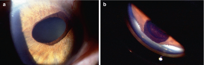
The three entities in their purest form may be easily recognized clinically; however, clear categorization can be difficult at times since patients may exhibit different clinical manifestations at various stages of the disease. In general, the extent of iris involvement is a differentiating factor among the three variants. Chandler’s syndrome usually presents with corneal edema and minimal iris alterations and is often not recognized initially by the clinician. Progressive iris atrophy is characterized by extensive iris stromal atrophy associated with corectopia and ectropion uveae. These latter findings are usually seen in the quadrant with peripheral anterior synechiae, with formation of stretch holes often in the opposite quadrant. In Cogan–Reese syndrome, any degree of iris atrophy may be present, but the predominant feature is the presence of multiple, pigmented, pedunculated iris nodules (Fig. 20.2a, b). These nodules are composed of tissue resembling iris stroma and are often surrounded by endothelial tissue.
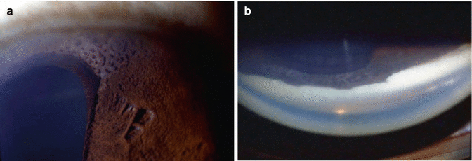
Fig. 20.2
A 39-year-old African American woman with Cogan–Reese syndrome. (a) Multiple, pigmented, pedunculated nodules and ectropion uveae (left) are located adjacent to relatively normal-appearing iris (right). (b) Peripheral anterior synechia observed by gonioscopy in the anterior chamber angle adjacent to the abnormal iris
The prevalence of glaucoma in ICE syndrome has been reported to be as high as 82 % [3]. Glaucoma appears to be more frequent and severe in patients with progressive iris atrophy and Cogan–Reese syndrome as compared with Chandler’s syndrome. The anterior chamber angle is usually open early in the disease process, and some patients may develop glaucoma at this stage. Often, broad peripheral anterior synechiae are present in the drainage angle, resulting from contraction of the abnormal basement membrane (Fig. 20.3a, b). These often extend to Schwalbe’s line, leading to aqueous outflow obstruction and culminating in secondary angle closure glaucoma [7].