Intraocular Lens Implantation in the Absence of Capsular Support


Intraocular Lens Implantation in the Absence of Capsular Support


M. Edward Wilson

Muralidhar Ramappa

Rupal H. Trivedi


When planning an intraocular lens (IOL) implantation in the absence of capsular support, key questions immediately come to mind. Are anterior chamber (AC) IOLs (angle-supported or iris claw), or posterior chamber IOLs (scleral or iris sutured or “glued”) safe and effective in children’s eyes with inadequate capsular support for in the capsular bag or ciliary sulcus fixation of IOLs? Is there an optimal choice of IOL design or material, method, and site of fixation in eyes with inadequate capsular support?

In this chapter, we explore these controversial issues and provide information that will help the pediatric cataract surgeon make decisions that provide the best possible visual outcome. By their very nature, pediatric surgeons are conservative while still being innovative. Risk must be minimized, but amblyopia must not be left untreated or undertreated. Nowhere is that balance more difficult than when there is inadequate capsular support in the face of contact lens and aphakic spectacle intolerance.

Some key points from the discussion that follows are included in Table 32.1.


BACKGROUND

IOL implantation in the absence of adequate capsular support is a challenging situation to manage, particularly in a pediatric eye. The long-term implications of these surgical interventions, when performed on children, are not well documented, and the optimal lens placement techniques for the pediatric eye are uncertain. Poor capsular support is often seen in pediatric aphakic eye, particularly after removal of congenital cataracts, traumatic cataract, or ectopia lentis. After extensive removal of the lens capsule, either by a surgeon at the primary lensectomy or at subsequent membranectomy in cases of reocclusion of the visual axis, there may not be enough of a capsular shelf to safely implant a secondary IOL in the ciliary sulcus. Also, in cases where extensive adhesion between capsular remnants and uveal tissues are broad and fibrotic, reestablishment of the ciliary sulcus may not be possible. In eyes with sufficient capsular rim, a posterior chamber IOL (PCIOL) can be safely implanted in the ciliary sulcus or in the re-opened capsular bag remnant. However, in eyes with poor capsular support, the possible locations to fixate the IOL include angle-supported anterior chamber intraocular lens (ACIOL), iris claw IOL, or transscleral or iris-fixated PCIOL. In eyes with either a partially subluxated or dislocated IOL, it can be repositioned by securing it to the sclera or iris in a minimally invasive or less traumatic procedure than IOL exchange. However, IOL fixation elsewhere poses several intraoperative and postoperative surgical challenges. ACIOL implantation is contraindicated in the presence of extensive damage to the iris and AC angle, preexisting glaucoma, peripheral anterior synechia (PAS), low endothelial cell count, and shallow AC.1 Iris claw IOLs, which are lenses placed in the AC and attached to the iris with small claw-like haptics, have been described to correct aphakia in adults,2 and children,3 with good visual results. These lenses have been available in most parts of the world for several years and are now being implanted in 20 sites in the United States under a compassionate-use FDA investigational device exemption. PCIOL offers several advantages, and many authors recommend them even in eyes lacking capsular or zonular support.4 Scleral fixation of PCIOL is a method to overcome the lack of capsular support but is technically more difficult and time consuming compared to ACIOL implantation. Due to its anatomic location in the eye, PCIOL is more appropriate for patients with glaucoma, low endothelial cell counts, PAS, and cystoid macular edema (CME).5


Table 32.1 KEY POINTS IN THE DISCUSSION OF IOL IMPLANTATION WHEN THERE IS AN ABSENCE OF CAPSULAR SUPPORT


  • Surgical aphakia in children is extremely difficult to handle; timely refractive correction is necessary in this vulnerable population to preserve monocular vision and binocularity and to protect against stimulus deprivational amblyopia


  • Conservative management may include correction with glasses or contact lenses, although these measures are not equally effective in all patients. Aphakic spectacles are an option for bilateral aphakic children but often undesirable for social and optical reasons


  • The possible locations to fixate the IOL in the absence of adequate capsular support include angle-supported AC, iris- (sutured or claw fixation) supported AC, or scleral (transscleral sutured or externalized haptics with glued flap)


  • Angle-supported ACIOL implantation has been linked in past literature with higher risks of iritis, pigment dispersion, corectopia, glaucoma escalation, and corneal endothelial attrition leading to corneal decompensation


  • A review of angle-supported ACIOLs and transscleral-sutured IOLs by the American Academy of Ophthalmology concluded that there was insufficient evidence to prove superiority of one lens type or fixation site.


Suitable candidates for IOL fixation elsewhere include those aphakic children who meet the following criteria: (1) unilateral or bilateral aphakia in children intolerant of or not suitable for contact lens wear; (2) extensive capsular deficiency precluding safety of stable in the bag or sulcus placement of an IOL; (3) absence of uncontrolled glaucoma, uveitis, or endothelial dysfunction; (4) absence of retinal hole/fluid, extensive lattice degeneration, or major retinal disease; and (5) motivated child’s parent(s), who acknowledges the risks/alternatives and the importance of follow-up examinations. Comprehensive preoperative assessments are needed, including examination under general anesthesia if complete office examination was not possible. This assessment includes age-appropriate testing of corrected distant visual acuity, refraction, sensorimotor examination of eye alignment/eye movement, measurement of intraocular pressure (IOP), slit-lamp examination, gonioscopy, and fundoscopy.


TRANSSCLERAL-SUTURED IOL FIXATION

Transscleral-sutured IOL fixation can be considered in cases with inadequate capsular support or in cases with compromised AC structure. The ciliary sulcus is located 0.83 mm posterior to the surgical limbus in the vertical meridian, whereas in the horizontal, it is 0.46 mm from surgical limbus.6,7 Duffey et al.8 passed the needle perpendicular to the sclera 1, 2, and 3 mm posterior to the surgical limbus and found that the needle exited internally at the ciliary sulcus, pars plicata, and pars plana, respectively. Scleral suturing of a PCIOL requires that the needle pass through vascular uveal tissue, with the attendant risk of bleeding. In many cases, this is minor and resolves spontaneously.9 Keeping the suture anteriorly (0.5-1 mm behind the surgical limbus) and avoiding the 3 o’clock and 9 o’clock positions may reduce the risk of bleeding by avoiding vascular ciliary body tissue and the long posterior ciliary arteries.10 In addition, taking care to maintain a pressurized globe and to minimize tissues traversed during transscleral needle passes will reduce the risk of intraoperative intraocular hemorrhage.11 Scleral fixation too close to either iris or pars plicata can have long-term protracted complications. Scleral fixation can be performed by either ab externo (passing needle from outside to inside) or ab interno approach (inside to out). The ab externo approach is most preferred. Adequate anterior vitrectomy is mandatory before placement of sutures and the IOL to avoid complications related to the vitreous base.


Surgical Technique


Ab Interno Approach

In this technique, two partial-thickness scleral flaps are made, 180 degrees apart. A right-handed surgeon should create the superior flap toward 1 o’clock and the inferior flap toward 7 o’clock for ease of maneuverability. The Ethicon CIF-4 long tapered needle can be used to pass a polypropylene suture through the inferior scleral flap, and the Ethicon TG 160-6 small curved needle can be used to pass a polypropylene suture through the superior scleral flap. The sutures can be looped through the eyelets of the haptics or tied to the haptic, or a girth hitch suture can be used. These sutures can be used for two-point or four-point fixation of the IOL. After passing out the polypropylene sutures through the scleral flaps, the ends of the sutures are tied in a 3-1-1 knot. The ends of the knot are cut 1 mm long. The scleral flaps are tied with 6-0 Vicryl. The corneoscleral wound is closed with 10-0 monofilament nylon or Vicryl, and the overlying conjunctival wound is closed with 8-0 Vicryl sutures or sealed with fibrin glue.


Ab Externo Approach

By this approach, the IOL can be placed more accurately in the ciliary sulcus. After designing the partial-thickness scleral flaps, the long straight needle is passed approximately 1 mm from the surgical limbus through one side; on the other side, a 27-gauge hypodermic needle should be passed. The straight needle is then negotiated through the hollow hypodermic needle. The hypodermic needle should be withdrawn with the straight solid needle inside it. The AC is
entered, and the suture is taken out with a hook. The suture is cut, and the end is tied to the eyelet of the IOL haptic. If the surgeon desires four-point fixation of the IOL, the above steps are repeated (Figs. 32.1 and 32.2). It should be noted that the main advantage of the ab externo approach is its greater precision in the location of scleral sutures.


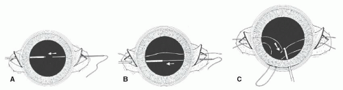
Figure 32.1. A: The double-suture variant of the Lewis ab externo technique begins similarly to the single-suture technique, except that the suture entry point under the scleral flap is displaced to one side. B: The second suture is passed parallel to the first, with 1 to 1.5 mm between the two sutures. C: Care must be taken to keep the sutures taut, to avoid crossing them or confusing which suture originates from each sclera site, while a Kuglen hook or similar instrument withdraws the suture loop through the previously prepared principal incision. (Reprinted from Steinert RF, Arkin MS. Secondary intraocular lenses. In: Stienert RF, ed. Cataract Surgery: Techniques, Complications, Management. Philadelphia, PA: Saunders, 2004:429-441, with permission.)

The polypropylene suture knot should not be kept exposed, with attendant risk of suture-related irritation or suture-wick endophthalmitis. Scleral flaps are recommended to cover it. If possible, the knot should be buried in the deep in the scleral pocket. Alternatively, a four-point fixation technique can be achieved where a loop of suture is created and the knot can be rotated into the sclera, thus avoiding the need for a scleral flap (Fig. 32.3). Another technique, known as a Hoffman pocket, creates a scleral pocket dissected from a corneal groove outward to the sclera. This is discussed more fully below. Some surgeons prefer leaving the polypropylene suture ends long and tucking them under the conjunctiva toward the fornix. It was believed that short ends of suture tend to extrude, whereas long ends tend to lie down against the sclera and are well tolerated. We have seen exposure and one infection from this tucking of the long suture ends method, and we no longer advocate it. The PCIOL used for scleral fixation should ideally have large diameter optics (6.5-7 mm), and haptics should ideally have large and well-polished eyelets.


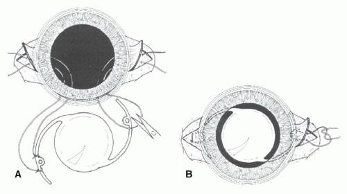
Figure 32.2. A: To achieve four-point stable fixation, the cut sutures are tied to the haptic on either side of the eyelet. B: The IOL is placed in the posterior chamber, keeping the sutures taut to avoid enlargement. The ends are then tied under the scleral flaps, and the conjunctiva is closed over the flaps. (Reprinted from Steinert RF, Arkin MS. Secondary intraocular lenses. In: Stienert RF, ed. Cataract Surgery: Techniques, Complications, Management. Philadelphia, PA: Saunders, 2004:429-441, with permission.)


Complications After Transscleral-Sutured Iols


Suture Breakage

A disturbing late complication of transscleral fixation of IOLs is the spontaneous breakage of the polypropylene suture leading to IOL displacement, especially in young patients.12,13 Vote et al.12 reported late breakage of polypropylene sutures in 16 of 61 eyes (26.2%) of mostly adult patients occurring about 50 ± 28 months after IOL fixation. Walter et al.14 had only 2.2% suture breakage in 89 eyes of adult patients during a mean follow-up of 24 months after penetrating keratoplasty and scleral-fixated PCIOL (SF-PCIOL) implantation. Buckley15 reviewed the literature on transscleral-sutured IOLs in children. He reported outcomes of 33 eyes with an average follow-up of 61 months (range 9-200 months). Twenty one of the eyes had >3 years of follow-up and 14 eyes (42%) had more than 5 years of follow-up. Four patients had spontaneous suture breakage at 38, 66, 96, and 107 months after implantation. An additional 13 cases of 10-0 Prolene suture breakage in children were uncovered by a survey of pediatric ophthalmologists. As
a conclusion of his Costenbader lecture, Buckley15 urged caution in the use of 10-0 polypropylene suture to fixate an IOL to the sclera in children. He recommended using an alternative material or an alternative size (9-0 polypropylene instead of 10-0). Appropriate ophthalmic needles are now available on the 9-0 Prolene suture. When necessary, however, the 9-0 Prolene suture can be tied to the end of a 10-0 Prolene and pulled through the eye using the 10-0 Prolene needle.


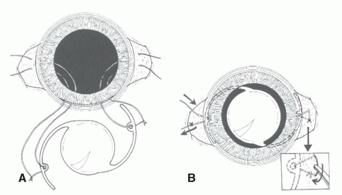
Figure 32.3. In this variant of the double-suture ab externo technique, the goal is to achieve a loop of suture where the knot can be rotated beneath the sclera, avoiding the necessity for a sclera flap. A: Cut ends of each suture are passed through the haptic positioning hole and tied. B: As the IOL is positioned in the posterior chamber, one end of the suture on each side is pulled, so it is cut off. The remaining suture ends are then tied together, and the knot is rotated beneath the sclera (inset), achieving the same result as illustrated in Figure 32.2. (Reprinted from Steinert RF, Arkin MS. Secondary intraocular lenses. In: Stienert RF, ed. Cataract Surgery: Techniques, Complications, Management. Philadelphia, PA: Saunders, 2004:429-441, with permission.)

May 24, 2016 | Posted by in OPHTHALMOLOGY | Comments Off on Intraocular Lens Implantation in the Absence of Capsular Support

Full access? Get Clinical Tree

Get Clinical Tree app for offline access