Intraocular Dyes for Vitreoretinal Surgery
Michel E. Farah
Eduardo B. Rodrigues
Mauricio Maia
Lihteh Wu
Fernando J. Arevalo
INTRODUCTION
The intraocular application of dyes to assist visualization of preretinal tissues during vitreoretinal surgery, “chromovitrectomy,” has become a very popular technique in the past few years (1). Chromovitrectomy was motivated by the difficulty in visualizing the several thin and transparent tissues in the vitreoretinal interface (2,3). Staining the internal limiting membrane (ILM), the epiretinal membrane (ERM), and the vitreous with a vital dye may assist the visualization of those fine transparent tissues, thereby leading to improvement in the postoperative visual acuity. The first vital dye used in chromovitrectomy, indocyanine green (ICG), facilitated the identification of the fine and transparent ILM (2). Following ICG, trypan blue (TB) has been introduced as an appropriate agent to identify the several ERMs, and triamcinolone acetonide (TA) has been found to stain the vitreous well. Recently, additional vital dyes such as infracyanine green (IfCG) and patent blue (PB) have been proposed for intraocular application during vitrectomy (4,5). In this chapter the current knowledge involving the use of dyes in chromovitrectomy will be discussed. In order to minimize the risk of toxic effects of dyes in chromovitrectomy, surgical recommendations are presented.
INDOCYANINE GREEN
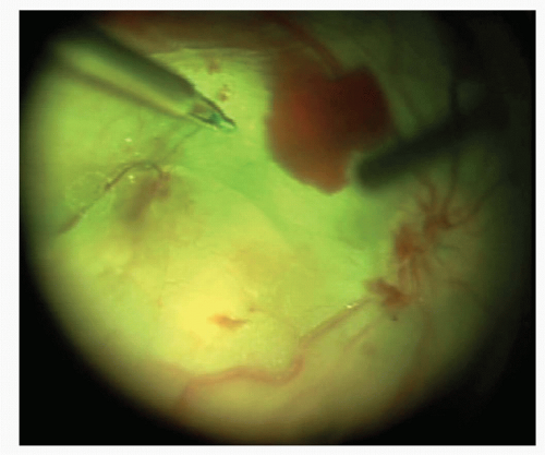
The application of intravitreal ICG was first reported in cadaver eyes to improve the visualization of the ILM. Since then, many articles have been released regarding the use of ICG as a surgical adjuvant to facilitate the identification and removal of the ILM in macular surgery (Figs. 29-1 and 29-2). ICG-assisted macular hole surgery demonstrated closure rates above 75% as well as visual improvements varying from 18% to 94% (3). However, soon after the introduction of ICG for the treatment of macular holes, caution among vitreoretinal surgeons took over the previous enthusiasm, as Gandorfer et al. in 2001 (6) postulated that intravitreous ICG-assisted ILM-peeling promoted worse visual outcomes in macular surgery. Histopathological analysis of tissue harvested during ICG-assisted vitreomacular surgery revealed various retinal elements adherent to the ILM. On electron microscopy, these retinal structures on the inner surface of the ILM were identified mainly as the plasma membrane of Müller cells (7,8). They are thought to originate from an altered cleavage plane induced by the ICG application. Most toxicity studies in animals have shown dose-dependent toxicity of ICG to various types of retinal cells. Interestingly, recent clinical reports have shown better surgical outcomes of ICGguided chromovitrectomy, which may be explained by a more selective and controlled ICG application (9,10).
 Get Clinical Tree app for offline access 
|


