Radiotherapy Treatment for Age-Related Macular Degeneration
Raul Velez-Montoya
Virgilio Morales-Canton
Hugo Quiroz-Mercado
INTRODUCTION
Age-related macular degeneration (AMD) is a complex disease and difficult to define. Since its first description by Holloway and Verhoeff in 1929 as a condition characterized by the loss of vision in older patients due to degenerative changes in the retina pigment epithelium (RPE) (1), the amount of knowledge about this disease, as well as the interest in finding an adequate therapy, has been increasing exponentially. Eighty years later, despite all the resources and efforts put forth to better understand the etiology and pathophysiology of AMD, many of its mechanisms remain elusive.
As life expectancy in developed countries and around the world has increased, AMD incidence has also risen to become the leading cause of vision loss among individuals aged 50 years or older (2, 3 and 4). It is believed that the annual incidence of some degree of AMD among patients 60 years or older reaches 40% to 50%. AMD-related disability and poor quality of life have become heavy socioeconomic burdens in industrialized countries (5, 6 and 7).
Although most AMD patients suffer the atrophic or “dry” form, the neovascular or “wet” form will be found in more than 10% of the cases. Wet AMD is characterized by deep choroidal neovascularization (CNV), subretinal fluid (SRF), subretinal hemorrhage (SRH), exudates, scarring, and severe visual loss (8, 9, 10 and 11). The natural history of the disease dictates that approximately 75% of the patients with subfoveal exudative lesions will become legally blind after 2 years (12,13). Population studies such as the Beaver Dam Eye Study demonstrated that approximately 10% to 20% of patients with the dry form of AMD will progress to the exudative form (especially if the patient’s retina exhibits large drusen [≥125 μm], soft indistinct drusen, retinal pigment epithelium abnormalities, or exudative and geographic lesions). This phenomenon of dry to wet progression was likely responsible for most of the estimated 1.2 million cases of visual loss due to AMD (14, 15 and 16) which occurred in the 1990s in the United States and may lead to 7 million by the year 2025, given that 4% of the wet AMD cases are classified as severe (17) (Fig. 32-1).
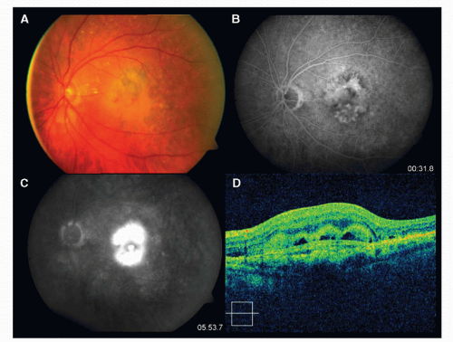 Figure 32-1. Color picture (A), fluorescein angiography (B and C), and spectral domain optical coherence tomography (D) of a 65-year-old female with clinical diagnosis of wet age-related macular degeneration. The color picture shows the presence of subretinal fibrosis and subretinal hemorrhage. B and C show middle and late fluorescein leakage, which indicates an active lesion. D shows the presence of a subretinal hyperreflective lesion (fibrosis) and hyperreflective spaces, which may indicate the presence of blood or subretinal fluid. |
Although the etiology of the AMD is still largely unknown, there has been an improvement in understanding the pathophysiology of the disease. In a normal eye, the byproducts of photoreceptor metabolism are usually removed by the RPE cells (18). It is believed that the aging process slows down RPE metabolism, diminishing its phagocytosis and lysosomal capacity, and resulting in accumulation of cell debris, clinically seen as drusen (5,18 and 19). Over time, these changes gradually thicken Bruch’s membrane and make it more hydrophobic, impairing oxygen delivery to photoreceptors (19, 20 and 21). It is not clear why this aging process ends in cell loss and atrophy in dry AMD, while in the wet form it stimulates a choroidal neovascular response. What we do know is that the hypoxia induces a low-grade inflammatory cascade which ends in the production of several growth factors, including vascular endothelial growth factor (VEGF), found to have a key role in CNV pathogenesis (2,22). VEGF is a dimeric lipoprotein whose principal biological activity is to produce an increase in the vascularization of hypoxic
tissues (23,24). This is accomplished through several mechanisms: increase in the mitotic activity of vascular endothelial cells; increase in vascular permeability via leukocyte-mediated endothelial cell injury, formation of fenestrae and dissolution of tight junctions; and induction of activated endothelial cells to produce enzymes and metalloproteinases which digest the capillaries’ basement membrane, the surrounding ground substance, and Bruch’s membrane, thus forming spaces and breaks through which newly formed vessels pass into the subretinal space (2, 18, 23, 24, 25 and 26).
tissues (23,24). This is accomplished through several mechanisms: increase in the mitotic activity of vascular endothelial cells; increase in vascular permeability via leukocyte-mediated endothelial cell injury, formation of fenestrae and dissolution of tight junctions; and induction of activated endothelial cells to produce enzymes and metalloproteinases which digest the capillaries’ basement membrane, the surrounding ground substance, and Bruch’s membrane, thus forming spaces and breaks through which newly formed vessels pass into the subretinal space (2, 18, 23, 24, 25 and 26).
It has also been demonstrated by histological studies that a granulomatous inflammatory response to the degenerated Bruch’s membrane is additionally responsible for the formation of breaks (23, 24, 25, 26, 27, 28, 29 and 30). This is supported by the observation of lymphocyte and macrophage infiltration of Bruch’s membrane and accumulation of macrophages at the site of neovascularization in some animal models of laser-induced CNV (23, 24, 25, 26, 27, 28, 29 and 30). However, it is still unknown whether this inflammatory response is part of the aging process of the eye or if the macrophage and lymphocyte activation is necessary for the CNV process (23).
Once the CNV reaches the subretinal space, the visual impairment will be closely related to the lesion size, the amount of exudates, and thickness of the disciform fibrosis. Any therapeutic approach that helps to control or reduce the three pathways of VEGF action described earlier may result in the stabilization or improvement of visual acuity in patients with wet AMD (31,32).
Basics About Radiation Therapy for AMD
The application of radiation to treat AMD is based on the concept that CNV formation is analogous to a wound-healing process, where the high rate of proliferating endothelial cells makes them more sensitive to the effects of radiation (33, 34 and 35). Reinhold described that a single dose of 8.7 Gy on normal capillaries leads within hours to swelling and vacuolation of the endothelium cells’ cytoplasm and local vasodilation (8,36). Reinhold also describes that the radiation effects continue through time, until the loss of endothelial nuclei occurs and a reduction is seen in the number and length of the capillaries and occlusive changes (8,36 and 37). The clinical effect of radiation on the eye vessels was first described in plaque-irradiated choroidal melanoma patients where, following plaque removal, a ring of choroidal atrophy with decreased or absent blood flow by fluorescein angiography (FA) was observed around the tumor’s base (18,38). A similar response has also been seen in choroidal hemangiomas and intracerebral arteriovenous malformations, where radiation is used to induce regression, and in ocular and cutaneous wounds, where radiation stunts the growth of newly formed vessels without thermal injury associated with lasers and cautery (8, 18, 34, 39, 40 and 41).
Although the biological justification to use radiation to treat CNV seems clear, there is controversy regarding the exact mechanism that drives the radio effect (42). The use of ionizing radiation induces the formation of free radicals (mainly from water molecules), which in turn cause irreparable damage to the DNA backbone and disrupt protein synthesis. The irradiated cell loses its ability to replicate and migrate but does not lose its cellular integrity nor undergoes necrosis (33, 34 and 35). On the other hand, there is evidence that radiation induces programmed cell death in vivo (33, 34, 35, 43 and 44). Miyamoto et al. studied the histologic appearance of rabbit eyes with experimental CNV, 4 weeks after a single fraction exposure to 20 Gy of focal X-ray irradiation (45,46). The degree of vascular formation and the number of endothelial cells in the CNV of irradiated eyes were less than in those of control eyes. And although the pathogenesis of experimental CNV and CNV due to AMD are not identical (45,46), the selective toxicity of radiation on rapidly dividing endothelial cells seems clear.
The use of sublethal radiation doses, low enough to spare cell life but high enough to induce DNA damage, can produce growth arrest by altering the genes of endothelial-growth-regulating cytokines and genes of inflammatory cytokines interactively affecting different cell populations (8,47 and 48). In vitro investigations of radiated endothelial cells showed an upregulation of basal intracellular adhesion molecule-1, which participates in the radiation-induced inflammatory reaction of the endothelium, facilitating inflammatory cells adhesion (49). All these actions decrease the CNV growth profile and the inflammatory response, which is thought by many to play a role in the formation of CNV (18,50 and 51).
Although radiation effects on endothelial cells are important in its ability to impact CNV, the power of radiation to affect inflammatory cells and fibroblasts is also a key factor, as it alters the formation and deposition of collagen, ultimately inhibiting scarring and fibrosis, which are important components of end-stage AMD (32,39,40 and 52). The results of the Belfast Study Group, showing that deterioration of visual acuity is less marked and scars are smaller in irradiated eyes than in untreated eyes at 6 to 24 months of follow-up, and that there is a positive correlation between scar size and better visual acuity, seem to support this assertion (53,54


Stay updated, free articles. Join our Telegram channel
Full access? Get Clinical Tree
