, Vincent Y. W. Lin2 and Joseph M. Chen2
(1)
Department of Otorhinolaryngology, Medical University of Vienna, Vienna, Austria
(2)
Department of Otolaryngology Head & Neck Surgery, Sunnybrook Health Sciences Center, Toronto, Ontario, Canada
Electronic supplementary material
Supplementary material is available in the online version of this chapter at 10.1007/978-3-7091-1490-2_12. Videos can also be accessed at http://www.springerimages.com/videos/978-3-7091-1489-6.
It must be understood that the medial wall of the vestibule forms the lateral wall of the internal auditory canal. Therefore, a small amount of bone removal is sufficient to unroof the internal auditory canal at its anterior (lateral) end, the fundus. Posteriorly, the route to the porus acusticus (the medial end of the canal) is much deeper because the canal is slanting away from the fundus to the porus. The IAC is in the same axis as the external auditory canal. It has a much more acute angle (more vertical) than most trainees expect.
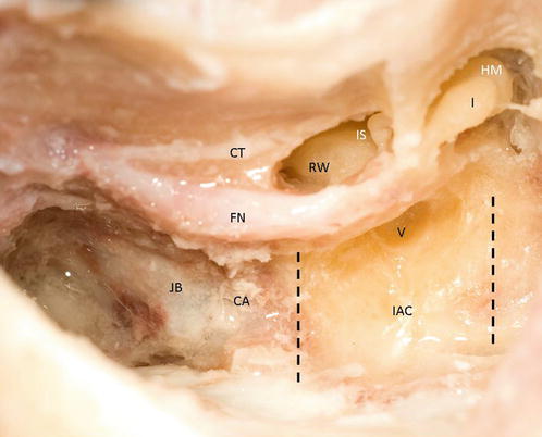
The superior limit of the IAC is defined by both the subarcuate artery and the ampullated end of the superior SCC. The inferior limit of the IAC is defined by the cochlear aqueduct medially and the P-SCC ampulla laterally. Of course, the real position of the IAC is subject to anatomic variations as well as underlying pathologies (e.g., meatal tumors). The cochlear aqueduct will appear during dissection between the jugular bulb and the internal auditory canal as a small white discoloration in the bone (Fig. 12.1). Cerebrospinal fluid will be released upon entry into the cochlear aqueduct. This can be done to intentionally release CSF.
Get Clinical Tree app for offline access
Fig. 12.1
The medial wall of the vestibule forms the lateral wall of the internal auditory canal. The superior (ampulla of superior-SCC and subarcuate artery) and inferior (cochlear aqueduct and ampulla of posterior-SCC) limits are plotted (CT chorda tympani, FN


Stay updated, free articles. Join our Telegram channel
Full access? Get Clinical Tree
