Infections of the Oropharynx
Vinidh Paleri
Introduction
Oropharyngeal infections are commonly caused by viruses, and the frequent pathogens are identified in Table 7.1. In adults, viral infection accounts for 80-90% of pharyngitis, with this figure reducing to 60% in children.
Table 7.1 Organisms causing oropharyngeal infections | |||
|---|---|---|---|
|
It is difficult to differentiate clinically viral infections from bacterial infections of the oropharynx, but the latter are more likely to present with severe local and systemic symptoms such as high-grade fever. Viral infections are more likely to be associated with nasal blockage and discharge. Throat swab cultures will identify bacterial pathogens, but this should be interpreted in the clinical context, as bacterial contaminants are common. Anaerobes such as Bacteroides species may have a greater role to play in peritonsillar abscesses but can be cultured from infected tonsils. Most uncomplicated infections of the oropharynx need symptomatic treatment only, and antibiotics are indicated in less than 20% of patients.
In acute pharyngitis (7.1) clinical symptoms include sore throat, halitosis, odynophagia, and tender cervical lymph nodes. The adenoid and tonsil hypertrophy associated with
the infection can cause mouth breathing, snoring, or even sleep apnoea. Systemic symptoms such as low-grade fever, lethargy, and malaise are common. Examination will reveal an inflamed pharynx with exudate or mucopurulent discharge running down from the pharynx. The tonsils may show mild inflammation.
the infection can cause mouth breathing, snoring, or even sleep apnoea. Systemic symptoms such as low-grade fever, lethargy, and malaise are common. Examination will reveal an inflamed pharynx with exudate or mucopurulent discharge running down from the pharynx. The tonsils may show mild inflammation.
 7.1 Acute pharyngitis showing diffuse erythema with prominent lymphoid follicles. |
When tonsillitis is present, the predominant findings can include all of the above with spots of exudates, or perhaps even a membrane covering the whole of the tonsils (7.2). Palatal petechiae can be present in both bacterial (typically Group A streptococci) and viral infections (typically with Epstein-Barr virus). Systemic examination is largely unproductive in the majority in identifying the pathogen. Presence of mucocutaneous ulceration, in the anus and genitalia, occurs during the acute seroconversion illness in HIV infection. Sometimes, the tonsillar hypertrophy is asymmetrical during an infectious episode (7.3). Follow-up is needed to ensure that the asymmetry settles down. Persistent enlargement of one tonsil should be investigated further to exclude neoplasm, and commonly tonsillectomy and histological examination is recommended. Lingual tonsillitis is a rare presentation (7.4A, B).
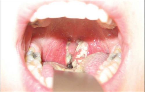 7.2 Tonsillitis showing tonsillar hypertrophy with greyish exudate on the surface. This patient had infectious mononucleosis. |
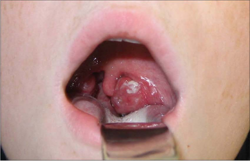 7.3 Asymmetrical hypertrophy of the left tonsil during acute tonsillitis. |
 7.4 (A) Prominent lingual tonsils in the noninflamed state. (B) Lingual tonsillitis in the same patient showing multiple follicular exudates. |
Viral infections
INFECTIOUS MONONUCLEOSIS
Infectious mononucleosis is caused by the Epstein-Barr virus (EBV), a double stranded DNA virus. This is one of the most common causes of viral pharyngotonsillitis in clinical practice. Transmission usually occurs through the saliva of infectious persons and subsequently the virus infects through lymphoid tissue in the pharynx.
Following a variable incubation period between 2-7 weeks, a prodrome of generalized myalgia, fever, and malaise occurs followed by the acute symptoms. These are constituted by fever, sore throat, loss of appetite, and cervical lymphadenopathy. Examination reveals any of the combination of symptoms and signs described above. Periorbital oedema, especially of the upper eyelids (Hoagland’s sign), can be found in nearly 30% of patients in the early days of the infection. Systemic examination will reveal splenomegaly in 60% of patients.
Blood tests are usually helpful in the diagnosis. The presence of atypical lymphocytes with a background picture of lymphocytosis usually suggests an EBV infection. It must be noted that other viral infections such as cytomegalovirus and HIV can also cause a similar atypical lymphocytosis. Other blood count abnormalities include thrombocytopenia and neutropenia. EBV infection also generates heterophile antibodies in the blood. The EBV shares similar epitopes with the cells of other animals. For example, treating the serum of affected humans with sheep red blood cells (RBC) causes an agglutination reaction. Using sheep RBC forms the basis for the Paul-Bunnell test and using horse RBCs forms the basis for the Monospot test. Other specific tests include looking for EBV-specific antibodies, like the viral capsid antigen.
Stay updated, free articles. Join our Telegram channel

Full access? Get Clinical Tree


