Infections of the Neck
Vinidh Paleri
Introduction
Each side of the neck houses approximately 200 lymph nodes, which drain the whole of the upper aerodigestive tract. The supraclavicular nodes on the left hand side also receive lymphatics from the abdominal viscera. These nodes are involved in any infectious or inflammatory process that may affect the aerodigestive tract and also in many systemic illnesses. In addition, there are many potential spaces in the neck through which infection can spread to other areas of the neck and outside the neck. Abscesses in these spaces can lead to serious and life-threatening complications if not recognized and treated expeditiously. Neck infections form a significant proportion of infectious problems seen in otolaryngologic practice.
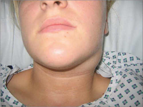 10.1 Reactive cervical lymphadenopathy in a patient with viral pharyngitis. |
Neck infections
CERVICAL LYMPHADENITIS
Reactive cervical lymphadenopathy is a common occurrence in children, presenting as neck lumps (10.1). These are usually secondary to a primary infection in the upper aerodigestive tract or a systemic illness. There are many organisms that can cause cervical lymphadenopathy and these are listed in Table 10.1. The history usually is of short duration, from a few days to 2 weeks. Clinical examination should include all relevant drainage sites, including the ears, nose, oral cavity, larynx, and pharynx. Table 10.2 sets out the drainage areas for different lymphatic groups in the neck. Cervical examination usually reveals small, soft cervical nodes, commonly in the jugulo-digastric region.
Table 10.1 Infections presenting with cervical lymphadenopathy | ||||||||||||||||||||||||||||
|---|---|---|---|---|---|---|---|---|---|---|---|---|---|---|---|---|---|---|---|---|---|---|---|---|---|---|---|---|
| ||||||||||||||||||||||||||||
The majority of these patients need no intervention and the lymphadenopathy settles down, although this can take 8-10 weeks. The challenge is to differentiate these reactive lumps from infections such as tuberculosis and neoplastic conditions. Symptoms and sign that should instigate further work-up are shown in Table 10.3.
Investigation for persistent lymphadenopathy should include a chest radiograph, serology for infectious agents (e.g. cytomegalovirus, Epstein-Barr virus, HIV, Toxoplasma, and Bartonella) and imaging of the neck. Rarely, pyogenic lymphadenitis can occur (10.2) and needs drainage (10.3). Fine needle cytology is not of great benefit in this setting. Inflammatory cervical lymphadenopathy is uncommon in adults and should always be investigated to rule out malignancy (see branchial cysts, below).
Monomicrobial infections
CAT SCRATCH DISEASE
Cat scratch disease is caused by a gram-negative bacillus, Bartonella henselae, and is so called as kittens are typically the vectors. It is a self-limiting illness characterized by papules or pustules at the inoculation site following an incubation period of 3-10 days. Regional painful lymphadenopathy then occurs, and can last up to 2 months. Suppuration occurs in 30% of patients. Systemic symptoms
like low-grade fever and malaise can be present. Rarely, serious neurological (transverse myelitis, encephalitis) and haematological (thrombocytopaenic purpura) complications ensue. Diagnosis is achieved by serological testing or PCR assay of lymph nodal tissue. For mild disease, symptomatic treatment is sufficient. Incision and drainage of suppurative lymphadenitis does not hasten recovery. In the immunosuppressed host, prolonged antibiotic treatment is needed.
like low-grade fever and malaise can be present. Rarely, serious neurological (transverse myelitis, encephalitis) and haematological (thrombocytopaenic purpura) complications ensue. Diagnosis is achieved by serological testing or PCR assay of lymph nodal tissue. For mild disease, symptomatic treatment is sufficient. Incision and drainage of suppurative lymphadenitis does not hasten recovery. In the immunosuppressed host, prolonged antibiotic treatment is needed.
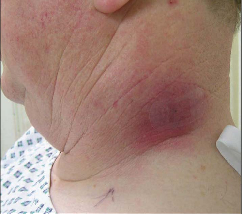 10.2 Suppurative bacterial cervical lymphadenitis in an otherwise well patient. |
Table 10.2 Cervical lymph nodal groups and their drainage areas | ||||||
|---|---|---|---|---|---|---|
|
Table 10.3 ‘Red-flag’ symptoms in patients with cervical lymphadenopathy that should instigate further work-up | |||||||
|---|---|---|---|---|---|---|---|
|
 10.3 Incision and drainage of the pus from the neck of the patient in 10.2. Note loculi being opened with blunt forceps. |
TOXOPLASMOSIS
Toxoplasmosis, caused by the protozoan Toxoplasma gondii, is an intracellular parasite. Cats are the definitive hosts for the organism and excrete oocysts. Infection is acquired by ingesting oocysts from faecal contamination or from tissue cysts in uncooked meat. Immunocompetent hosts present with sore throat, malaise, fever, and cervical lymphadenopathy, which is clinically indistinguishable from other viral infections, such as infectious mononucleosis. The lymphadenopathy can last several months. Immuno-fluorescent assay (IFA) for IgM antibody confirms acute infection. No treatment is required in the immuno-competent host. The infection can involve multiple systems in the presence of immunocompromise such as AIDS, manifesting as necrotizing encephalitis, pneumonitis, and myocarditis.
 10.4 Draining sinus from a tuberculous infection of a neck node. |
MYCOBACTERIAL INFECTIONS
Tuberculous and nontuberculous mycobacterial infections present with persistent cervical lymphadenopathy, with or without suppuration (10.4). This may be the sole presentation of nonrespiratory tuberculosis. Slow insidious growth and the examination findings of matted groups of nodes that feel rubbery on palpation should lead one to suspect the diagnosis of tuberculosis. In contrast, atypical mycobacterial infections involve a single nodal group and are characterized by relatively rapid growth and suppuration, usually in the submandibular or parotid region. Systemic symptoms such as fever, loss of weight, and malaise may not be present in the immunocompetent host. Diagnosis is established by identifying the organism on culture or on PCR assays.
Therapy for tuberculosis is medical and triple therapy has a high success rate in controlling the disease. Atypical mycobacterial infections, if localized, can be excised, which is curative. Disseminated disease in immunocompromised hosts will need chemotherapy.
ACTINOMYCOSIS
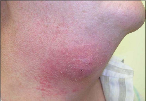
This infection is uncommon but important to recognize as it can mimic other diseases. Despite their fungal sounding name, these are gram-positive bacilli. Most members of the Actinomyces species are commensals in the oral cavity. The infection usually presents as a mass adjacent to the mandible, with surrounding induration, erythema, and abscess formation (10.5). The mass is caused by the organisms proliferating in the soft tissue rather than lymph node inflammation. The classical description is that of draining sinuses with bright yellow granules in the pus (sulphur granules) caused by the filaments of the organism. Diagnosis can be made only after microbiological confirmation. Prolonged antibiotic therapy is essential to effect a cure (10.6).
 10.5 Actinomycosis of the neck depicting a discharging sinus overlying a mass in the submandibular region. |
 10.6 Resolving actinomycosis 1 month after commencing treatment. |
 10.7 Cellulitis of the neck. |
Polymicrobial infections
CELLULITIS OF THE NECK
Superficial cellulitis of the neck occurs after minor trauma or spontaneously. Staphylococcus aureus and Streptococcus pyogenes are commonly implicated. Rarely, this can be the presentation of serious underlying disease in the upper aerodigestive tract. Clinical examination reveals superficial inflammation, with no symptoms of dysphagia or general systemic upset (10.7). Treatment with systemic antibiotics usually clears the vast majority of infections.
NECROTIZING CERVICAL FASCIITIS
This is a rapidly progressive infection of the soft tissues usually caused by group A haemolytic streptococci or Staphylococcus aureus. Other pathogens, including gramnegative organisms and anaerobes, have also been implicated. Minor trauma or surgery can trigger this, although it can begin spontaneously. Clinical signs are characterized by pain, rapidly increasing erythema, and crepitation in the affected area. The skin and subcutaneous tissue becomes necrotic, demonstrated by dark areas, which may not be contiguous. Blisters form over the affected skin, which soon sloughs off (10.8). Systemic symptoms of fever, hypotension, and respiratory failure indicating septicaemia are evident. Early intervention is essential as the condition can rapidly become fatal.
Stay updated, free articles. Join our Telegram channel

Full access? Get Clinical Tree


