Fig. 5.1
Hordeolum on the upper eyelid
The differential diagnosis of hordeolum includes acute chalazion, basal cell carcinoma, sebaceous gland carcinoma, and squamous cell carcinoma of the eyelid.
Hordeola usually have a self-limited course with a spontaneous rupture and drainage in 1–2 weeks. An internal hordeolum may evolve into a chalazion.
Medical therapy for hordeola includes eyelid hygiene, warm compresses and massages of the lesions for 10 min four times per day, and topical antibiotic ointment in the inferior fornix if the lesion is draining or if there is an accompanying blepharoconjunctivitis [7, 8]. Systemic antibiotics may be indicated if the hordeola is complicated by preseptal cellulitis. Oral doxycycline may also be added if there is a history of multiple or recurrent lesions or if there is significant and chronic meibomitis [9].
5.2.2 Chalazion
Chalazia appear most commonly as chronic subcutaneous nodules within the eyelid. A chalazion results from the obstruction of the meibomian gland leading to blockage of the gland’s duct at the eyelid margin and then release of the contents of the gland into the surrounding eyelid soft tissue [10, 11]. A chalazion arises as a mild to moderately tender red swelling of the upper or lower eyelid (Fig. 5.2). At various locations and stages, multiple lesions may appear. Clinical onset of a chalazion occurs over days, with the redness and tenderness subsiding while the lump remains [10]. Patients usually present when the lump becomes symptomatic, either due to cosmetic reasons or if the chalazion is of a considerable size, because it is causing ptosis, astigmatism, and/ or vision loss [12].
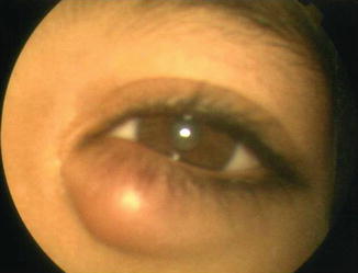
Fig. 5.2
Chalazion on the lower eyelid
Patients with underlying conditions such as rosacea, seborrheic dermatitis or blepharitis, and diabetes are more prone to multiple and recurrent chalazia.
If left untreated, chalazia may spontaneously resolve over many months. Management includes warm compresses application with topical corticosteroids and antibiotic, if signs of infection are present. If the lesion persists in spite of medical therapy, it may be incised and drained [11]. Steroid injections can be applied either intralesionally or subcutaneously and are considered to be a simple and effective treatment with reported success and resolution in 50–95 % of cases [12–17].
5.2.3 Bacterial Blepharitis
Anterior bacterial blepharitis affecting the base of the eyelashes is often caused by staphylococcal infection (Staphylococcus epidermidis, Staphylococcus aureus). Other causative organisms include Propionibacterium acnes, Corynebacteria, and Moraxella. Blepharitis predominantly involves young to middle-aged women and is associated with keratoconjunctivitis sicca in up to 50 % of patients [18]. There is a wide variety of ocular symptoms including sore eyelids, eyes feeling irritated, itchy, burning, or gritty, red eyes, dry or watery eyes, increased frequency of blinking, foreign body sensation, photophobia, contact lens intolerance, and eyelids sticking together, particularly in the morning [18]. Signs include swollen eyelids, inflamed lid margins with redness, thickening, and collarettes surrounding the eyelashes (Fig. 5.3), scaling, crusting, irregularity, and/or ulceration of the lid margins, altered eyelash appearance, individual lash poliosis, and lashes broken, misdirected, or crusted with fibrinous or sebaceous matter, as well as secondary alterations to the conjunctiva and cornea. Hordeolum and multiple and recurrent chalazia may occur in the course of bacterial blepharitis [18]. Staphylococcal blepharitis may be also associated to seborrheic blepharitis.
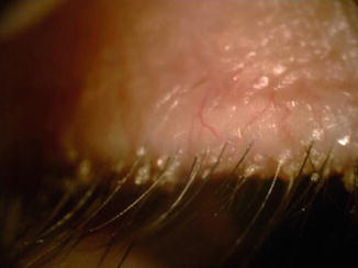
Fig. 5.3
Staphylococcal blepharitis with collarettes surrounding the eyelashes
The clinical diagnosis of staphylococcal blepharitis may be confirmed by culture, which usually reveals many S. aureus organisms on the lid margins.
A combination of lid hygiene with warm compresses, lid scrubs, and antibiotherapy is required. Erythromycin or bacitracin ointments and fusidic acid gel have been the most frequently used agents for blepharitis. Aminoglycosides such as gentamycin or tobramycin eyedrops or ointment are also useful but do not have the same broad coverage against gram-positive bacteria [18]. Topical vancomycin 50 mg/mL four times daily has been used to treat resistant Staphylococcus [18]. Fluoroquinolones such as ciprofloxacin ophthalmic solution 0.3 % and topical levofloxacin 0.5 % proved their efficacy in the management of bacterial blepharitis [18–20]. The fourth-generation fluoroquinolones are also used for the treatment of bacterial blepharitis [21]. Corticosteroid-antibiotic combinations are also available in either ointment or eyedrop form [22, 23]. Most experts recommend corticosteroids for short-term use only (less than 2 weeks) in an effort to “jump start” therapy for moderate-to-severe anterior blepharitis or blepharoconjunctivitis. One of the more recent additions to the management of blepharitis is topical azithromycin [24, 25]. Prevention is based on careful cleaning of the eyelids which is the most effective way to prevent bacterial blepharitis and its recurrence.
5.2.4 Erysipelas
Erysipelas is a bacterial skin infection most often caused by Group A Streptococcus. Patients typically develop symptoms including high fever, shaking, chills, fatigue, headaches, vomiting, and general illness within 48 h of the initial infection. Eyelid involvement appears as erythematous swollen, warm, hardened, and painful skin lesion with sharply demarcated raised edge that enlarges rapidly. More severe infections can result in vesicles, bullae, and petechiae, with possible skin necrosis [26, 27]. Lymph nodes may be swollen, and lymphedema may occur.
Penicillin administered orally or intramuscularly is sufficient for most cases of classic erysipelas and should be given for 10–20 days. A first-generation cephalosporin or macrolide, such as erythromycin or azithromycin, may be used if the patient has an allergy to penicillin [28].
5.2.5 Impetigo
Impetigo of the eyelid is caused by superinfection of skin disorder such as eczema, poison ivy, insect bites, and cuts or scrapes due to minor trauma by Staphylococcus aureus or Streptococcus pyogenes. The infection is more common in children younger than 6 years of age [29, 30]. Impetigo contagiosa of the eyelid is usually associated with infection of the face. Impetigo begins as tiny blisters, which eventually burst and leave small wet patches of red skin that may weep fluid. Gradually, a tan or yellowish-brown crust covers the affected area.
Cleansing of the affected area and local antibiotics ointments are generally effective. Systemic antibiotics are sometimes necessary.
5.2.6 Preseptal Cellulitis
The term preseptal cellulitis refers to an infectious process in the eyelids that is isolated to regions anterior to the orbital septum (Chap. 3).
5.3 Other Bacterial Infections of the Eyelids
5.3.1 Tuberculosis
The Mycobacterium tuberculosis can infect the eyelid as a primary infection or by spread of lupus vulgaris from the face. Lupus vulgaris is characterized by reddish-brown nodules that blanch to an “apple-jelly” color when pressure is applied and may appear on the skin of the eyelids. Tuberculosis can also manifest as a “cold abscess,” a soft, fluctuant mass without acute inflammation, or simulate a chalazion [31–35].
Diagnosis is based on epidemiological data, Mantoux test, and QuantiFERON and proved by histopathologic examination of lesion-exeresis biopsy showing granulomatous inflammation with areas of caseation, Langhans giant cells, and acid-fast bacilli [36].
5.3.2 Leprosy
Leprosy is a chronic disease caused by the bacteria Mycobacterium leprae and Mycobacterium lepromatosis. Thinning or loss of the eyebrows or eyelashes associated with a multiple upper eyelid crease pattern and trichiasis is common in leprosy [37]. Lagophthalmos or anesthetic, infiltrated macules or nodules can also be seen [38].
5.3.3 Syphilis
Syphilis is a sexually transmitted disease, caused by the spirochete bacterium Treponema pallidum. Eyelid involvement may occur by direct contact with an infected person. The chancre of primary acquired syphilis rarely occurs on the lid or lid margin. The skin of the eyelids can develop a macular or papular eruption in association with the generalized rash of secondary syphilis [41]. Pustules and ulcerative lesions can also occur, along with temporary syphilitic alopecia of the eyebrows and lashes. The skin eruption spontaneously fades as the secondary stage ends. In rare cases, benign tertiary syphilis involves the eyelid in the form of a gumma that begins as a deep swelling with minimal surrounding inflammation and eventually ulcerates to form exuberant granulation tissue. Congenital syphilis can involve the eyelid in the form of eruption, ulceration, or a gumma [42]. Temporary madarosis is also possible.
Penicillin G 12–24 million units per day for 6–21 days remains the treatment of choice in syphilis [42].
5.3.4 Anthrax
Anthrax is caused by Bacillus anthracis. Humans become infected as they come into contact with infected animals or animal products. Eyelid involvement present as a localized itchy erythematous papule of the eyelid that enlarges within 24–48 h, developing into an ulcer surrounded by vesicles that evolves into black, necrotic, central eschar characteristic of the disease [43, 44].
Treatment consists of ten million units of penicillin G given intravenously each day for a period of 7 days [43, 44]. With treatment, progressive healing of the skin occurs with or without complications that may include persistent cicatricial ectropion, lagophthalmos, palpebral symphysis, or restriction of upper eyelid movement.
5.4 Viral Infections of the Eyelids
5.4.1 Herpes Simplex Virus Blepharitis
Herpes simplex virus blepharitis is encountered primarily in children, although adults may also manifest this disorder. It usually presents in the form of small vesicles or pustules along the lid margin and/or periocular skin with associated regional lymphadenitis [45] (Fig. 5.4). Within the first week of infection, the vesicles may ulcerate or harden into crusts, although they will ultimately resolve without scarring [45–48].
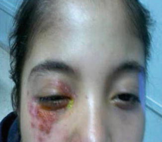
Fig. 5.4
Herpes simplex infection in a young patient involving the superior and inferior eyelids, with erythema-vesiculous lesions
The course of herpetic blepharitis is usually self-limiting within 2–3 weeks. The use of warm saline compresses with a topical drying agent is usually sufficient to palliate the patient. If the lesions are extensive, the concomitant use of topical antibiotic ointment is considered prudent to prevent a secondary bacterial infection [45]. The use of antivirals is advocated by some practitioners for severe cases.
5.4.2 Herpes Zoster Ophthalmicus
Herpes zoster ophthalmicus (HZO) is characterized by reactivation of the varicella-zoster virus infection. Early in the course of disease, the eyelids may become hyperemic and edematous [49–51]. HZO presents as an acute, painful, vesicular eruption distributed along a single dermatome. The rash evolves from an erythematous lesion, with macules, papules, vesicles, pustules, and crusts developing subsequently (Fig. 5.5a).
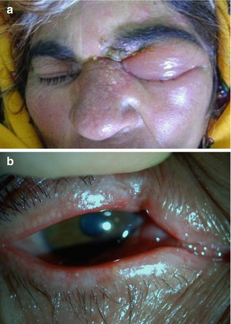
Fig. 5.5
(a) Acute herpes zoster ophthalmicus infection with eyelid edema and vesicles in a dermatomal pattern. (b) Cicatricial changes of the upper eyelid margin in a patient with history of herpes zoster ophthalmicus infection
The affected dermatome often heals within 2 weeks. As the inflammation resolves, there may be residual ptosis, lid scarring, deep scalp pitting, entropion, ectropion, loss of normal pigmentation, lid necrosis, loss of eyelid mobility (Fig. 5.5b), and lagophthalmos [52].
HZO is treated with acyclovir (800 mg five times daily for 7–10 days), valacyclovir (1 g three times daily for 7 days), or famciclovir (500 mg three times daily for 7 days). Palliative treatment of skin lesions with cool compresses and mechanical cleansing is useful [51]. Oral antiviral medication, if started within 72 h of the onset of the acute HZO rash, significantly shortens the periods of acute pain, virus shedding, rash, acute and reduce the rate of late-onset anterior segment complications and severity of postherpetic neuralgia [53].
5.5 Other Viral Infections of the Eyelids
5.5.1 Molluscum Contagiosum
Molluscum contagiosum infections are characterized by elevated, round, waxy, pearly white, noninflammatory lesions with umbilicated centers [54, 55].
Removal or expression of the nodule, allowing permeation of blood into its substance, is curative.
5.5.2 Kaposi’s Sarcoma
Kaposi’s sarcoma is a tumor caused by human herpesvirus 8 that usually occurs in patients with AIDS. Kaposi’s sarcoma of the eyelid appears as multiple purple to red nodules [54]. It can mimic a chalazion.
Therapeutic options include cryotherapy, surgical excision, radiation, and/or chemotherapy.
5.6 Fungal Infections of the Eyelids
5.6.1 Candidiasis
Candidal infections of the eyelid, usually associated with candidal infections elsewhere in the body, typically occur in immunocompromised patients. It may present as small ulcers, vesicles, or pustules at the bases of the eyelashes [56, 57]. Small granulomas are often found on the lid margin.
Candidiasis may be treated with fluconazole or itraconazole. Amphotericin B, voriconazole, or posaconazole are recommended in case of severe systemic-associated disease. Topical glucocorticoids and broad-spectrum antibiotics should be discontinued.
5.6.2 Eyelids Dermatophytosis
Dermatophytosis of the eyelids or tinea is an infection caused by a dermatophyte, which is most commonly of the Trichophyton genus. Tinea can affect the eyelid primarily or spread to the eyelid from other parts of the face. The early lesions begin as flattened, reddish papules that spread peripherally while the central area heals. The fully developed lesion has a ring-like appearance, with a reddish, scaly, sharply defined border and a central pinkish scaly area [58, 59]. Loss of the eyelashes is characteristic of these fungal infections, similar to the hair loss that occurs in kerions associated with tinea capitis [58].
Diagnosis is established by microscopic examination of edge scrapings with a drop of potassium hydroxide preparation and/or culture positive for fungal elements [58–60].
Treatment of eyelids dermatophytosis involves the use of topical imidazole preparations, including ketoconazole, bifonazole, miconazole, econazole, and clotrimazole. For cases that are widespread or recalcitrant to topical therapies, treatment with systemic antifungals including ketoconazole, itraconazole, or fluconazole can be considered [59, 61, 62].
5.6.3 Blastomycosis
Blastomycosis is caused by the dimorphic fungus Blastomyces dermatitidis. Blastomycosis of the eyelids is rare and occurs in approximately 25 % of patients with systemic blastomycosis. Eyelid involvement is characterized by granulomatous ulcers with thick crusts and an underlying purplish color [63–66]. Blastomycosis of the eyelid may be mistaken for a chalazion, basal cell, or squamous cell carcinoma [64].
5.6.4 Cryptococcosis
Cryptococcosis, caused by Cryptococcus neoformans, arises in immunosuppressed patients and can affect the skin of the eyebrow, forehead, and eyelids. Eyelid manifestations include papules, nodules, infiltrative plaques, pustules, ulcers, or subcutaneous abscesses [68, 69].
In patients with localized lesions, treatment is based on fluconazole 400 mg per os once/day for 3–6 months. For more severe disease, amphotericin B 0.5–1.0 mg/kg IV once/day with flucytosine 25 mg/kg per os q 6 h is given for several weeks.
5.7 Parasitic Infections of the Eyelids
5.7.1 Onchocerciasis
Onchocerciasis, also named river blindness or Robles disease, is a parasitic disease caused by the microfilariae Onchocerca volvulus. Onchocerciasis is endemic in many areas of sub-Saharan Africa and in isolated foci in Central and South America [70, 71]. Eyelids onchocerciasis is characterized by eyelid edema in the early phase of the infection. Later, severe pruritic dermatitis and intense photophobia can occur, followed by the development of subcutaneous nodules that occasionally involve the eyelid, especially in patients from Central and South America. Diagnosis of onchocerciasis is done by identification of worms from skin or subcutaneous nodules obtained by biopsy.
Treatment consists of ivermectin medical therapy given in a single oral dose of 150 μg/kg [72].
5.7.2 Leishmaniasis
Mucocutaneous leishmaniasis is caused by Leishmania braziliensis and Leishmania mexicana. The microorganism is transmitted by the bite of infected phlebotomine sand flies. Eyelid involvement presents as highly polymorphous lesions, such as swelling lesions, papules, plaques, nodules, and sometimes ulcerative or covered by crusts and scaling, erosions, ulcers, and blepharoconjunctivitis [73–77]. Ocular complications include palpebral scar, trichiasis, eyelash loss, lagophthalmos, epicanthus, trichiasis, and ectropion. If eyelid lesions remain untreated, the contiguous spread from the skin of the eyelid will extend to involve the conjunctiva, sclera, and even cornea, causing interstitial keratitis. Leishmania infection may simulate other conditions such as cysts, chalazion, dacryocystitis, tuberculosis, syphilis, sarcoidosis, and tumors [78, 79]. Diagnosis is primarily based on epidemiological data, history, and clinical findings. It can be confirmed by a positive leishmanin skin test, identification of the parasite by histopathologic sections, culture in NNN (Novy, McNeal, and Nicolle) medium, or by detection of the parasite DNA [73].
Pentavalent antimonial compounds such as Glucantime or sodium stibogluconate are the therapy of choice. The recommended dose is 20 mg/kg/day for at least 3 weeks [80].
5.7.3 Demodicidosis
Demodicidosis of the eyelids is caused by Demodex folliculorum, found in the eyelash follicle or Demodex brevis that burrows deep in sebaceous and meibomian glands [81]. Demodex is spread by direct contact and probably by dust containing eggs. Infection of Demodex often occurs in the course of chronic blepharitis, considered to be involved in its pathogenesis [82–84].
Stay updated, free articles. Join our Telegram channel

Full access? Get Clinical Tree


