Purpose
To report on the frequency of cysts and tumors of the pineal gland in patients with retinoblastoma.
Design
Observational retrospective case control study.
Methods
setting : Institutional. study population : Four hundred eight patients treated for retinoblastoma from January 2000 to January 2012 at Wills Eye Institute, Philadelphia, Pennsylvania, USA. observation procedure : Magnetic resonance imaging (MRI) features of the pineal gland were evaluated in all patients with retinoblastoma. Characteristics of patients with pineal cysts and pineoblastoma were reviewed. main outcome measures : Comparison of frequency of pineal gland cyst and pineoblastoma in children managed with systemic chemoreduction vs other methods.
Results
Of 408 patients, treatment included systemic chemoreduction in 252 (62%) and nonchemoreduction methods in 156 (38%). Overall, 34 patients (8%) manifested pineal gland cyst and 4 (1%) showed pineoblastoma. Of all 408 patients, comparison (chemoreduction vs nonchemoreduction) revealed pineal cyst (20/252 vs 14/156, P = .7) and pineoblastoma (1/252 vs 3/156, P = .1). The pineal cyst (n = 34) (mean diameter 4 mm) was asymptomatic (n = 34), followed conservatively (n = 34), and with minimal enlargement (n = 2, 9%) but without progression to pineoblastoma. The cyst was found in 22 germline and 12 nongermline patients ( P = .15). Among the 4 patients with pineoblastoma, all had germline mutation and 2 had family history of retinoblastoma. Among all patients with family history of retinoblastoma (n = 45), 2 (4%) developed pineoblastoma. The pineoblastoma was asymptomatic in 2 patients and symptomatic with vomiting and headache in 2 patients. The mean interval from date of retinoblastoma detection to pineal cyst was 2 months (median 2, range 0-8 months) and to pineoblastoma was 27 months (median 28, range 7-46 months). Management included aggressive chemotherapy and radiotherapy, with 2 survivors.
Conclusions
Pineal gland cyst was incidentally detected in 8% of retinoblastoma patients, causing no symptoms, and without progression to pineoblastoma. Pineoblastoma was detected in 1% of patients and fewer patients who received systemic chemotherapy developed pineoblastoma, possibly indicating a systemic protective effect.
In the pediatric population, the pineal gland can show alterations including cyst formation (pineal cyst) and solid tumor production (pineocytoma, pineoblastoma, germinoma, teratoma, astrocytoma, glial tumors). Pineal cyst is generally a coincidental finding in an asymptomatic child, as recognized by Karatza and associates in their study of pineal cysts in children with retinoblastoma. Pineoblastoma has historically been a highly fatal malignant tumor, found in children with the germline form of retinoblastoma, and preferentially in those with a positive family history. In the prechemoreduction era, pineoblastoma, also termed “trilateral retinoblastoma,” was found in 6% of bilateral retinoblastoma patients and 10% of patients with family history of retinoblastoma. Pineoblastoma is an important cause of mortality in retinoblastoma patients during in the first 5 years of life. Screening with neuroimaging can potentially improve survival if the tumor is detected early (<15 mm in size). However, with neuroimaging screening comes an increase in detection of coincidental pineal lesions. In this retrospective analysis, we explore the trends in the occurrence of pineal gland lesions in a consecutive series of children with retinoblastoma imaged during the chemoreduction era.
Methods
This was a retrospective review of medical records of patients treated for retinoblastoma from January 1, 2000 to January 1, 2012 at Wills Eye Institute, Philadelphia, Pennsylvania, USA. The study was approved by the Institutional Review Board of Wills Eye Institute and was in compliance with Health Insurance Portability and Accountability Act (HIPAA) regulations. Patients included in this study were those who had undergone brain imaging with magnetic resonance imaging (MRI) during the course of treatment. Each patient was evaluated for age at diagnosis, race, sex, and hereditary pattern (sporadic, familial). Eyes were assessed for laterality of involvement (unilateral, bilateral) and total number of retinal tumors per eye, and classified according to the International Classification of Retinoblastoma.
The MRI was evaluated by review of reports filed on the chart and specific information regarding abnormalities in the pineal region were documented. Those patients with follow-up MRI were documented for additional features. Statistical analysis was performed using χ 2 test, Fisher t test, and Mann-Whitney test. A P value of less than .05 was considered significant.
Results
There were 408 patients with retinoblastoma with adequate MRI information that were included in this analysis. The primary treatment modality was systemic chemoreduction in 252 patients (62%), enucleation in 143 (35%), external beam radiation therapy in 5 (1%), and plaque radiotherapy in 8 (2%). At initial MRI imaging, 34 patients (8%) were identified to have a pineal cyst and 4 patients (1%) were diagnosed to have pineoblastoma. There was no other structural brain abnormality in any case. The demographic features of all patients and an analysis of those with pineal cyst vs those with pineoblastoma are represented in the Table .
| Feature | All Patients (n = 408) | Patients With Pineal Cyst (n = 34) | Patients With Pineoblastoma (n = 4) |
|---|---|---|---|
| Age (months), mean (median, range) | 21 (13, 0-91) | 14 (9, 2-73) | 5 (1, 0-14) |
| Sex | |||
| Male | 202 (49%) | 15 (44%) | 2 (50%) |
| Female | 206 (51%) | 19 (56%) | 2 (50%) |
| Race | |||
| White | 287 (70%) | 26 (76%) | 4 (100%) |
| African American | 60 (15%) | 4 (12%) | 0 (0%) |
| Hispanic | 41 (10%) | 3 (9%) | 0 (0%) |
| Asian | 20 (5%) | 1 (3%) | 0 (0%) |
| Heredity | |||
| Sporadic | 363 (89%) | 27 (79%) | 2 (50%) |
| Familial | 45 (11%) | 7 (21%) | 2 (50%) |
| Laterality | |||
| Unilateral | 215 (53%) | 17 (50%) | 1 (25%) |
| Bilateral | 193 (47%) | 17 (50%) | 3 (75%) |
| Mutation | |||
| Germline | 215 (53%) | 22 (65%) | 4 (100%) |
| Nongermline | 193 (47%) | 12 (35%) | 0 (0%) |
Pineal Cyst Characteristics
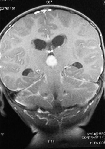
The pineal cyst was asymptomatic in all patients. The mean interval from diagnosis of retinoblastoma to detection of pineal cyst was 2 months (range 0-8 months). The mean size of pineal cyst was 4 mm (range 3-6 mm). The cyst was homogenous in 33 patients (97%) and heterogenous in 1 (3%). A ring of enhancement was seen in 18 cysts (53%). Follow-up imaging details were available for 23 patients and the cyst showed enlargement in 2 (9%), stable size in 17 (74%), and regression in 4 (17%). Among the 34 patients detected to have pineal cyst ( Figure 1 ), 20 patients (59%) were treated with systemic chemoreduction and 14 patients (41%) were treated with other modalities. This difference did not attain statistical significance ( P = .7). The incidence of pineal cyst was not increased in patients with germline mutation ( P = .15). There were 21 patients with follow-up MRI and a mean of 4 MRIs were performed during the course of treatment. None of the cysts showed evidence of compression on surrounding structures. None of the patients with pineal cyst developed second malignancies or metastatic disease during the period of follow-up (mean follow-up 59 months, range 6-138 months).
Pineoblastoma Characteristics
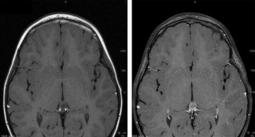
Of the total 408 patients, 4 patients were diagnosed to have pineoblastoma and 1 of these patients had received systemic chemotherapy prior to diagnosis of pineoblastoma ( Figure 2 ). Fewer patients who received systemic chemotherapy developed pineoblastoma (1/252 vs 3/156; chemoreduction vs nonchemoreduction), though this number did not attain statistical significance ( P = .1).