Immune Disease


Immune Disease


Anterior Uveitis

Richard F. Multack

Samuel J. Multack

Leonid Skorin Jr

ICD-9: 364.0


THE DISEASE


Pathophysiology

The uvea consists of the middle, pigmented, vascular structures of the eye and includes the iris, ciliary body, and choroid. Uveitis refers to inflammation of the uveal tract. Anterior uveitis can be divided into iritis, anterior cyclitis (ciliary body inflammation), and iridocyclitis. Anterior uveitis does not include involvement of the pars plana or the posterior segment. Adjacent, nonvascular structures such as the cornea (keratouveitis) and sclera (sclerouveitis) are often affected secondarily in the inflammatory process.

Anterior uveitis can also be divided by its clinical course. Acute uveitis refers to inflammation that lasts for weeks or a few months and resolves once the attack is over. Chronic uveitis may last for many months or years without clearing completely between exacerbations. Uveitis can be classified by its etiology, infectious or noninfectious. Most commonly anterior uveitis is a noninfectious/sterile process.

Acute and chronic uveitis can be further subdivided into granulomatous and nongranulomatous that is determined by clinical findings. Granulomatous uveitis presents with large, greasy, “mutton-fat” precipitates on the corneal endothelium with large clumps of inflammatory cells present in the anterior chamber because of exuberant macrophage activity. Nongranulomatous uveitis presents with fine corneal endothelial precipitates and anterior chamber activity.


Etiology

The causes of anterior uveitis are multiple and varied and commonly go undetected. Most types of anterior uveitis are sterile inflammatory reactions. Underlying associations include ankylosing spondylitis, reactive arthritis (Reiter’s syndrome), Adamantiades-Behçet’s disease (ABD), trauma, psoriatic arthritis, juvenile rheumatoid arthritis/juvenile idiopathic arthritis (JRA/JIA), postoperative iritis, glaucomatocyclitic crisis, intraocular lens-induced UGH syndrome (uveitis-glaucoma-hyphema), Fuchs heterochromic iridocyclitis (FHI), sarcoidosis, and idiopathic causes. Infectious causes of anterior uveitis may include Lyme disease, various viral etiologies, syphilis, and tuberculosis.


The Patient

The signs and symptoms can vary and depend on the anatomical involvement, onset, and duration of the uveitis and whether it is granulomatous or nongranulomatous.


Clinical Symptoms

The patient with an acute anterior uveitis will complain of ocular pain, red eye, photophobia, epiphora, and blurred vision. Pain from anterior uveitis will often be referred to the eyebrow area. These symptoms may be very mild or absent in chronic anterior uveitis. Characteristic
HLA-B27-associated uveitis is recurrent with alternating bilateral involvement.


Clinical Signs


  • Cells and flare in the anterior chamber


  • Circumcorneal ciliary flush


  • Nongranulomatous: fine keratic precipitates (KPs) on corneal endothelium


  • Granulomatous: “mutton-fat” precipitates on corneal endothelium


  • Iris nodules

    Koeppe: clusters of cells on the pupillary border

    Busacca: cells on the anterior iris surface

    Berlin: cells in the iris angle structure


  • Miotic pupil


  • Hypopyon


  • Hyperemia


  • Plasmoid iridocyclitis: fibrin with sluggish or no movement of cells in the anterior chamber


  • Spillover: cells in the anterior vitreous


  • Low intraocular pressure (IOP) secondary to cyclitis


  • Elevated IOP secondary to obstruction of the trabecular meshwork, trabeculitis, iris bombè causing pupillary block and angle closure


  • Cystoid macular edema (CME)


  • Posterior synechiae


  • Endothelial dysfunction with associated corneal edema


  • Fibrin pupillary membrane


Demographics

Anterior uveitis is the most common form of uveitis with an incidence of 9/100,000 people and increasing to 100/100,000 in patients over 65 years of age. Most patients affected are from 20 to 50 years of age. Some forms of anterior uveitis, such as acute nongranulomatous uveitis secondary to ankylosing spondylitis and reactive arthritis (Reiter’s syndrome), are more often found in men. Chronic anterior uveitis is more common in women. Immunologic and hormonal differences are thought to underlie some specific types of uveitis.

Seronegative spondyloarthropathies (rheumatoid factor [RF] negative) that are associated with acute anterior uveitis are often HLA-B27 positive (located on the short arm of chromosome 6). HLA-B27 is positive in 1.4% to 8% of the general population. In patients who present with acute iritis, up to 60% prove to be HLA-B27 positive. Ankylosing spondylitis, reactive arthritis, inflammatory bowel disease (IBD), and psoriatic arthritis are all included in the seronegative spondyloarthropathies.

ABD, is associated with HLA-B51. It is a perivascular inflammation of unknown cause that results in a generalized occlusive vasculitis. The Behçet’s triad includes a hypopyon with iritis, aphthous stomatitis, and genital ulceration. It is less common in the United States and is more common in Japan and Eastern Europe. Researchers in Japan have suggested a diagnostic system that includes major and minor criteria. Major criteria are recurrent oral ulcers, skin lesions, recurrent genital ulcers, and ocular inflammatory disease. Minor criteria include epididymitis, arthritis, vasculitis, gastrointestinal ulcerations, and psychiatric symptoms. Based on the number of criteria, different types have been developed including complete, incomplete, suspect, and possible. Eye findings may include a nongranulomatous anterior uveitis, retinal vasculitis, retinal hemorrhage, macular edema, retinal necrosis, ischemic optic neuropathy, and vitritis. Systemic signs may include erythema nodosum, nondestructive arthritis, IBD, mucous membrane ulcerations, pericarditis, myocarditis, and central nervous system (CNS) strokes.

Glaucomatocyclitic crisis (Posner-Schlossman’s syndrome) is a mild to moderate uveitis associated with mid to upper range elevated IOP, corneal
edema, fine KPs, and a mid dilated pupil. An association with HLA-B54 has been made with this entity.

Lens-associated uveitis or phacoantigenic endophthalmitis is a presumed immune response to lens protein after violation of the lens capsule.

Postoperative uveitis can include infection, lens-associated and intraocular lens (IOL)-associated inflammation. Infectious causes can include Staphylococcus epidermidis, Candida, and delayed endophthalmitis caused by Propionibacterium acnes. Manipulation of the anterior chamber results in some break down of the blood-aqueous barrier (BAB) leading to increased risk of uveitis.

Pseudophakic uveitis may be seen with closed loop anterior chamber intraocular lenses although it is much less common with newer flexible intraocular lens implants.

FHI or Fuchs uveitis syndrome is usually unilateral with varying symptoms of mild discomfort to none. The diagnosis is based on heterochromia, lack of synechiae, and diffusely distributed KPs.

Masquerade syndromes, such as syphilis, may present with an anterior uveitis. Syphilis accounts for 1% to 3% of all uveitis cases. Patients will develop anterior uveitis in 5% to 10% of secondary syphilis cases.


Significant History


  • History of ocular trauma or surgery


  • Recent viral or bacterial disease


  • History of underlying immunologic disease


Ancillary Tests

Ocular assessment includes visual acuity, pupil testing, IOP, slit-lamp examination, and dilated fundus evaluation.

Laboratory studies: complete blood count (CBC) with differential, erythrocyte sedimentation rate (ESR), chest and pelvis x-ray, Lyme titer, antinuclear-antibody, rapid plasma reagin (RPR) or VRDL, FTA-ABS, purified protein derivative (PPD), human leukocyte antigen (HLA) typing, angiotension converting enzyme, and serum lysozyme.


The Treatment

As the etiology of anterior uveitis is often unknown, treatment is directed to control the inflammation and to prevent damage to uveal vasculature that can result in chronic recalcitrant uveitis or secondary side effects, such as posterior synechiae, cataracts, and glaucoma.

Cycloplegic agents are used to relieve pain, decrease synechiae formation, and reduce the permeability of the iris vasculature. Cyclopentolate or homatropine can be used for mild to moderate inflammation, whereas scopolamine or atropine may be used for more severe inflammation.

Corticosteroids can be applied topically, by periocular injection, systemically, or by intravitreal implants such as fluocinolone acetonide/Retisert (Bausch and Lomb). The steroid therapy should never be stopped abruptly because this may lead to severe rebound in inflammation.

Patients who fail or are intolerant to steroids may require immunomodulatory agents, such as methotrexate or cyclosporine and azathioprine. Recently, tumor necrosis factor-alpha (TNF-a) inhibitors such as infliximab have shown promising results.


Ankylosing Spondylitis

Tammy P. Than

ICD-9: 720.0M


THE DISEASE


Pathophysiology

Ankylosing spondylitis belongs to a group of seronegative spondyloarthropathies in which the RF is absent. Ankylosing
spondylitis is an aggressive inflammatory arthropathy with a predilection for the central skeleton affecting the sacroiliac joint most severely. The joints between the spine and pelvis, and joints between the vertebrae of the spine, may eventually fuse.


Etiology

The etiology is unknown but is thought to be multifactorial. HLA-B27 has been found in 93% of patients with ankylosing spondylitis while present in only 6% of controls. In the absence of HLA-B27, genes for other inflammatory conditions may be important in predisposing individuals. Evidence for this is the greater association with other inflammatory conditions such as Crohn’s disease or psoriasis. Alternatively, bacterial and viral infections may trigger ankylosing spondylitis. In particular, Klebsiella pneumoniae, commonly found in the gastrointestinal tract, is more common in patients with the disease compared to controls. There appears to be a familial pattern as there is a higher incidence of ankylosing spondylitis if there is a positive history in a first-degree relative.


The Patient


Clinical Symptoms

Symptoms usually begin in the early 20s with an onset rarely after the age of 40. Systemic symptoms begin with intermittent hip and/or lower back pain that are worse at night or after inactivity. Bent posture eases the pain, which improves later in the day and with exercise. There may be pain or tenderness in the ribs, shoulder blades, hips, thighs, shins, and heels. The patient may also complain of fatigue, loss of appetite, and general discomfort. Ocular symptoms include pain, redness, and photophobia if uveitis is present.

The disease in women tends to be less severe than in men and may present with neck pain and breast pain in the absence of lower back pain.


Clinical Signs

Uveitis occurs in about 25% of patients with ankylosing spondylitis presenting as an acute, unilateral, nongranulomatous episode. The ocular manifestations may occur prior to joint and skeletal signs, and ankylosing spondylitis accounts for 10% to 33% of all cases of anterior uveitis.

Early on, limited flexibility of the lower spine is noted. Enthesopathy, new bone formation at the attachment of tendons and ligaments, may be noted. Over time, ankylosing spondylitis progresses to involve the entire spine—lordosis (forward curvature) of the lumbar spine, kyphosis (excessive curvature with convexity backward) of the thoracic spine, and hyperextension of the cervical spine. Other joints are involved in about 33% of cases, most frequently presenting with inflammation of the hips, knees, ankles, and shoulders. Osteoporosis is noted in early and late stages of the disease in 19% to 62% of cases and may result in vertebral fractures. Tendons and ligaments can also become inflamed. Cardiac complications include valvular disease (e.g., aortic valve stenosis) and aortitis. Ankylosing spondylitis can affect the bones of the rib cage, reducing lung capacity. Patients may have signs of psoriasis or IBD.


Demographics

The incidence of ankylosing spondylitis is 0.21% in Americans over the age of 15. The incidence is greatest among Native American Indians and Eskimos, and it is rare in African Americans. The peak age of onset is in the mid-20s, although a juvenile form develops around age 8 to 10 years. The male to female ratio is often overestimated because women are frequently underdiagnosed because
the disease manifests less severely in the female gender. The ratio is thought to be 2 to 3:1 (male:female).


Significant History


  • Lower back pain in the morning and after inactivity


  • Stooped posture


  • Sleeping in a fetal position to minimize pain


  • Awakening during the second half of the night because of back pain


  • Recurrent unilateral uveitis


Ancillary Tests


  • CBC—may reveal mild anemia


  • HLA-B27—positive


  • ESR—may be elevated during active stage


  • C-reactive protein (CRP)—may be elevated during active stage


  • Rheumatoid factor—negative


  • Antinuclear antibodies (ANAs)—negative


  • Spine and/or pelvis x-ray—look for Romanus lesion—early radiographic sign indicative of disc margin erosion


  • Technetium bone scan—more sensitive than plain film x-rays


  • Vitamin D levels—identify patients at risk for osteoporosis


  • Spine mobility measurements


  • Electrocardiogram


The Treatment

Exercises to strengthen the back muscles to maintain erect posture, to maintain chest expansion, and to maintain or improve spinal mobility should be part of the patient’s daily routine. Physical therapy is also useful. Heat applied to the joints helps reduce joint pain unless active inflammation is present in which case cold packs are preferred. The patient should be encouraged to sleep on a firm mattress with good neck support. A smoking cessation program, if pertinent, should be a priority. Oral nonsteroidal anti-inflammatory agents are the first line of therapeutic intervention. More aggressive management includes sulfasalazine and other disease-modifying antirheumatic drugs. TNF-a antagonists, such as etanercept, infliximab, and adalimumab, are useful if conventional treatment fails. Intraarticular corticosteroid injections may be beneficial. Topical corticosteroids and cycloplegics are used to manage the anterior uveitis. Approximately 90% of patients remain fully independent with little or no disability.


Behçet’s Disease

Tammy P. Than

ICD-9: 136.1


THE DISEASE


Pathophysiology

Behçet’s disease is a rare, nongranulomatous, obliterative vasculitis that is thought to be immune mediated. Antiendothelial cell antibodies are detected in an increased prevalence and neutrophils are hyperactive interacting with T cells. Activated T cells produce TNF-a which results in increased levels of proinflammatory cytokines. It affects arteries and veins resulting in thrombosis. The disease is chronic and multisystemic, characterized by oral and genital mucocutaneous ulcerations, skin rashes, arthritis, thrombophlebitis, uveitis, colitis, and neurologic symptoms.


Etiology

The exact etiology is unknown; however, immune regulation, immunogenetics, vascular abnormalities, or bacterial or viral infection may play a role in the development of Behçet’s disease. An infectious cause may be associated with herpes simplex virus I or Streptococcus sanguis in which there is autoimmunity
or cross-reactivity between the microbe and the oral mucosal antigens. In countries with a high prevalence of Behçet’s disease, HLA-B51 has been found in 72% of patients and appears to predispose patients (especially males) to a more severe disease. The association with HLA-B51 has not been demonstrated in the United States. Other trigger factors may include environmental toxins such as heavy metals or pesticides, English walnuts, Gingko nuts, chocolate, and tomatoes.


The Patient


Clinical Symptoms

Prior to the onset of Behçet’s disease, the patient may experience a prodrome and report malaise, weight loss, anorexia, general weakness, and headache. During the disease process, the patient will complain of a painful mouth and genital sores, photophobia with a red and painful eye, and joint pain.


Clinical Signs

Behçet’s disease waxes and wanes with periods of exacerbation and remission. As time passes, the recurrence frequency decreases and the severity diminishes.

Ocular signs are present in 75% of patients. Bilateral nongranulomatous uveitis, which may include a small hypopyon, usually follows the onset of oral ulcers by 3 to 4 years, but ocular disease is the initial presentation in 20% of patients. Late-stage ocular complications include neovascularization, glaucoma, cataracts, retinal detachment, vascular occlusive disease, and optic atrophy. Other potential ocular manifestations are sixth-nerve palsy, hemiparesis, visual field loss, idiopathic intracranial hypertension, internuclear ophthalmoplegia, and complications from intracranial artery aneurysms.

There are two common criteria for the diagnosis of Behçet’s disease. The International Study Group (ISG; 1990) criteria require the presence of oral ulceration, which may be a limitation. Therefore, some recommend applying the 1987 criteria from the Japanese group in conjunction with the ISG guidelines (Table 10-1).


Demographics

Behçet’s disease has a worldwide distribution and is most prevalent (and more virulent) in the Mediterranean, Middle East, and Far East with an estimated incidence of 14 to 380/100,000. The prevalence in the United States is 0.33/100,000. Those from high-risk areas who immigrate to low prevalence areas have an intermediate risk, which suggests that environmental factors play a role in the disease. The predominant age at diagnosis is in the third to fourth decades, with men affected more often and with a more severe course in some regions. Prognosis is poorer with neurological involvement, where there is a 20% mortality rate after 7 years. Chronic morbidity is usual, with ophthalmic involvement as the leading cause. Blindness occurs in up to 25% of eyes within 10 years of onset of ocular disease. The effects of the disease appear to be cumulative.


Significant History


  • Recurrent oral ulcerations


  • Genital sores


  • Recurrent uveitis


  • Hypopyon


Ancillary Tests

Ocular evaluation includes assessment of the anterior chamber and IOP. A dilated fundus examination should be performed to rule out occlusive disease and its sequelae, vasculitis, retinal detachment, and optic nerve disease.

There is no diagnostic laboratory test specific for Behçet’s disease, but the following may be helpful:


  • CBC with differential—anemia observed in some patients with chronic disease


  • ESR—may be elevated during active stage


  • CRP—may be elevated during active stage


  • Cerebral spinal fluid analysis—protein level elevated


  • Rheumatoid factor—negative


  • ANAs—negative


  • IgA—elevated


  • Complement 3 and 4 levels—elevated


  • Lipid levels—may be elevated, predisposing the patient to thromboses


  • Pathergy test (“skin prick”)—following an intradermal puncture to the forearm, a positive test results if the puncture site results in an inflamed and pustular area greater than 2 mm within 24 to 48 h; test is positive in up to 79% of patients with Behçet’s disease although positivity is quite low (˜5%) in white patients limiting the test’s use in many regions


TABLE 10-1 Two Criteria for the Diagnosis of Behçet’s Disease*


ISG Criteria for Diagnosis of Behçet’s Disease


Behçet’s Disease Research Committee of Japan Criteria


Major features


Recurrent oral ulceration (three occurrences within a 12-mo period)


Recurrent oral ulcers Skin lesions


Erythema nodosum Subcutaneous thrombophlebitis


Folliculitis Acneform lesions Cutaneous hypersensitivity


Ocular signs


Uveitis Chorioretinitis


Genital ulcers


Minor features


Recurrent genital ulceration


Arthritis without deformity


Ocular involvement (uveitis or vasculitis)


Gastrointestinal lesions Epididymitis


Skin lesions


Vascular lesions


Positive pathergy test (see ancillary tests under Behçet’s disease)


CNS symptoms


Diagnosis


1 major + 2 minor


Complete


4 major


Incomplete


3 major


2 major + 2 minor Typical ocular symptom + 1 major or 2 minor


Suspected


2 major OR 1 major + 2 minor


Data from Behçet’s Disease Research Committee of Japan. Behçet’s disease: Guide to diagnosis of Behçet’s disease. Jpn J Ophthalmol 1974:18:291-294; and International Study Group for Behçet’s Disease. Criteria for diagnosis of Behçet’s disease. Lancet 1990;335:1078-1080.


The Treatment

There is no established standard therapeutic regimen for Behçet’s disease, but treatment is guided by organ involvement. Prognosis has improved in the past decade with more aggressive treatment strategies.

Uveitis is managed with topical corticosteroids and cycloplegia. Laser photocoagulation may be used to manage retinal neovascularization. Ocular management may also include
cataract surgery and vitrectomy. Skin manifestations are treated with topical corticosteroids or antibiotic solutions. The systemic disease is successfully managed with immunosuppressants such as levamisole, colchicine, dapsone, tacrolimus, azathioprine, chlorambucil, cyclosporine A, and cyclophosphamide. Interferon a 2A and B are useful and are thought to have antiviral, immunomodulatory, and antiproliferative properties. TNF-a antagonists, such as infliximab, etanercept, and adalimumab, may be considered for refractory disease including uveitis. Acyclovir may be effective if the etiology is herpes simplex. Exercise, such as swimming or walking, is beneficial to keep joints strong and flexible.


Allergic Conjunctivitis

Salisa K. Williams

Daniel Ullmann

The ocular surface may present an immunologic response as an inflammation of the conjunctiva and cornea. According to the Gell and Coombs classification system for immunologic reactions, four types of hypersensitivity reactions are recognized:

Type I: Immunoglobulin E (IgE)-mediated. This is also known as immediate hypersensitivity that can manifest as an anaphylactic (systemic) or atopic (local) reaction. The antigen binds to the antigen-binding fragment (Fab) portion of the IgE on either mast cells or basophils. Mast cells produce the initial reaction, while basophils initiate the late phase reaction. The cross-linking causes degranulation and the release of histamine resulting in increased vascular permeability and smooth muscle contraction.

Type II: Antibody-mediated (cytotoxic). Circulating antibodies react with antigens on the surface of cells, which leads to cell damage by either phagocytosis or complement activation. These cytotoxic reactions appear to be the underlying etiology of cicatricial pemphigoid (CP) and Mooren ulcer.

Type III: Immune-complex mediated. Soluble antigen-antibody complexes form when the antigen is abundant. Complex deposition in tissues causes type III hypersensitivity. The Arthus reaction is a classic systemic type III reaction, while Wesley rings are an example of a corneal type III reaction.

Type IV: Cell-mediated (delayed type). Presentation of antigen to CD4+ T cells leads to tissue damage via the release of cytokines and the subsequent accumulation of neutrophils and macrophages. Ocular examples of type IV hypersensitivity include drug allergies, corneal allograft rejection, and contact dermatitis.

This section presents the major hypersensitivity conditions involving the conjunctiva, more commonly referred as allergic conjunctivitis.


Seasonal Allergic Conjunctivitis and Perennial Allergic Conjunctivitis

ICD-9: 477.9


THE DISEASE

The conjunctiva is a thin mucous membrane that lines the posterior surface of the eyelids and extends over the globe to the corneal limbus. It has a rich vascular supply and is an important, immunologically active defense against pathogens. Seasonal allergic conjunctivitis (SAC) is a mild to moderate manifestation of the immune response of the conjunctiva to a wide variety of antigens. Patients may experience symptoms on a seasonal basis when exposed to antigens or may exhibit symptoms year round,
thus presenting with perennial allergic conjunctivitis (PAC).


Pathophysiology

Once primary exposure and sensitization to antigens occur, repeat exposure to the antigen initiates the inflammatory response. Mast cells migrate into the conjunctival epithelium and are the primary cell type involved in the inflammatory response. Degranulation of mast cells and basophils releases histamine, bradykinins, and leukotrienes. These primary inflammatory mediators induce vasodilatation, tissue edema, and nerve stimulation.


Etiology

Among patients with allergic conjunctivitis, airborne allergens such as ragweed, pollens, dander, dust, or mold spores initiate the immune response resulting in symptoms of allergic conjunctivitis. Patients with SAC present with symptoms in the spring, summer, and fall due to the abundance of airborne allergens found in tree pollen, grass pollen, and weed pollen, respectively.

Patients with PAC may present symptoms throughout the year; thus, in addition to the seasonal allergens, other allergens such as dust mites, cockroaches, and pet dander play a role in the development of symptoms among these patients.


The Patient


Clinical Symptoms

Patients with SAC and PAC complain of conjunctival hyperemia, burning, tearing, and ocular itching. Symptoms are transient, recurrent, and often follow seasonal patterns. The timing of the symptoms differentiates between patients with SAC and patients with PAC.


Clinical Signs

Patients develop variable degrees of conjunctival hyperemia, chemosis (edema), and eyelid edema (Fig. 10-1). In some patients, a papillary conjunctival response may be observed. The conjunctiva often appears milky due to the edema.


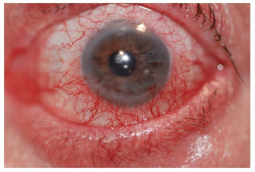
Figure 10-1. Allergic conjunctivitis. (Photo courtesy of Leonid Skorin Jr.)


Demographics

SAC and PAC are common conditions affecting all segments of the population.


Significant History


  • Exposure to allergens


  • Complaint of itching


  • Personal or family history (first-degree relatives) of atopic disease, that is, allergic rhinitis, bronchial asthma, and atopic dermatitis (AD)


Ancillary Tests


  • For most patients, the diagnosis of SAC and PAC is clinical and ancillary testing may not be required.


  • In severe cases of allergic conjunctivitis, superficial conjunctival scrapings may be obtained to evaluate for the presence of eosinophils.


  • Markers of allergic activity in tear samples may be obtained to assess for levels of IgE, histamine, and tryptase.


The Treatment


  • Conservative treatment includes the application of cold compresses, artificial tears, and topical ocular vasoconstrictors (Table 10-2). Topical vasoconstrictor/antihistamine combinations cause vascular constriction, decrease vascular permeability, and reduce ocular itching; however, these preparations have
    a short duration of action (Table 10-2). Topical ocular antihistamines reduce itching and vasodilation (Table 10-2).


  • Topical mast cell stabilizers are a safe and effective treatment for chronic allergy symptoms (Table 10-2). A therapeutic effect with these preparations may not be noted for 7 to 14 days; therefore, topical antihistamine/mast cell stabilizers may be indicated to provide rapid symptomatic relief (Table 10-2).


  • Topical ocular corticosteroids should be reserved for severe cases. However, use judiciously due to potential adverse effects, such as increasing IOP (Table 10-2). Loteprednol is a site-specific steroid with less potential to increase IOP.


  • Oral antihistamines may provide symptomatic relief from rhinocon-junctivitis. Of note, these drugs may exacerbate dry eye symptoms in some patients (Table 10-2).


  • Nasal steroid sprays may be a better option than oral antihistamines because they have less systemic effect (Table 10-2).


  • A topical ocular nonsteroidal anti-inflammatory drug (NSAID) is an alternative treatment option. Ketorolac tromethamine 0.5% is the only NSAID approved for the treatment of SAC (Table 10-2) but is less effective than the other classes of drugs.


TABLE 10-2 Commonly Used Medications to Treat Allergic Conjunctivitis


Topical ocular vasoconstrictors available over the counter


Drug


Recommended regimen


Use in children


Pregnancy risk


Phenylephrine 0.12% solution (Refresh Redness Relief)


One drop four times daily


Not recommended


Unknown


Naphazoline solution (Clear Eyes)


One drop four times daily


Not younger than 6 y.o. Category


C


Tetrahydrozoline solution (Visine)


One drop four times daily


Not younger than 6 y.o.


Unknown


Topical ocular vasoconstrictor and antihistamine combinations available over the counter


Naphazoline/antazoline solution


One drop four times daily


Not younger than 6 y.o.


Category C


Naphazoline/pheniramine solution (Opcon-A), (Naphcon-A), (Visine A)


One drop four times daily


Not younger than 6 y.o.


Category C


Topical ocular antihistamines


Emedastine difumarate 0.05% solution (Emadine)


One drop four times daily


Not younger than 3 y.o.


Category B*


Bepotastine besilate 1.5% solution (Bepreve)


One drop twice daily


Not younger than 2 y.o.


Category C


Topical ocular mast cell stabilizers


Lodoxamide tromethamine 0.1% solution (Alomide)


One drop four times daily


Not younger than 2 y.o.


Category C


Cromolyn sodium 4% solution (Crolom), (Opticrom)


One drop four times daily


Not younger than 4 y.o.


Category B*


Nedocromil sodium 2% solution (Alocril)


One drop twice daily


Not younger than 3 y.o.


Category B*


Pemirolast potassium solution (Alamast)


One drop four times daily


Not younger than 3 y.o.


Category C


Topical ocular antihistamine and mast cell stabilizer combinations available over the counter


Ketotifen fumarate 0.025% solution (Zaditor), (Alaway), (Refresh), (Zyrtec Itchy Eyes)


One drop twice daily


Not younger than 3 y.o.


Category C


Topical ocular combinations of antihistamine and mast cell stabilizer


Olopatadine hydrochloride 0.1% solution (Patanol)


One drop twice daily


Not younger than 3 y.o.


Category C


Olopatadine hydrochloride 0.2% solution (Pataday)


One drop once daily


Not younger than 3 y.o.


Category C


Azelastine hydrochloride 0.05% solution (Optivar)


One drop twice daily


Not younger than 3 y.o.


Category C


Epinastine hydrochloride 0.05% solution (Elestat)


One drop twice daily


Not younger than 3 y.o.


Category C


Topical ocular corticosteroids


Loteprednol etabonate 0.2% suspension (Alrex)


One drop four times daily


Not recommended


Category C


Fluorometholone 0.1% suspension (FML), (Flarex)


One drop four to six times daily


Not recommended


Category C


Loteprednol etabonate 0.5% suspension (Lotemax)


One drop four times daily


Not recommended


Category C


Prenisolone acetate 1% suspension (Pred Forte)


As often as 1 drop once every 1-2 h for 7 d, then rapid taper


Not recommended


Category C


Oral antihistamines available over the counter


Diphenhydramine liquigels or tablets (Benadryl)


25-50 mg orally every 4-6 h


Children’s formulation available


Category B*


Cetirizine hydrochloride tablets (Zyrtec)


10 mg orally once daily


Not younger than 2 years old


Category B*


Fexofenadine hydrochloride tablets (Allegra)


Up to 180 mg orally daily


Not younger than 6 months old


Category C


Loratadine tablets (Claritin), (Alavert)


10 mg orally once daily


Not younger than 2 years old


Category B*


Chlorpheniramine tablets (Chlor-Trimeton)


4 mg orally every 4-6 h


Not younger than 6 years old; sustained release form not younger than 12


Category C


Oral antihistamine


Desloratadine tablets (Clarinex)


5 mg orally once daily


Not younger than 12 years old


Category C


Nasal corticosteroids


Fluticasone propionate (Flonase)


Two sprays in each nostril once daily


Not younger than 4 y.o.


Category C


Mometasone (Nasonex)


Two sprays in each nostril once daily


Not younger than 2 y.o.


Category C


Budesonide (Rhinocort Aqua)


One spray in each nostril once daily


Not younger than 6 y.o.


Category B*


Triamcinolone (Nasacort AQ)


Two sprays in each nostril once daily


Not younger than 2 y.o.


Category C


Beclomethasone (Beconase AQ)


One to two sprays in each nostril twice daily


Not younger than 6 y.o.


Category C


Fluticasone furoate (Veramyst)


Two sprays in each nostril once daily


Not younger than 2 y.o.


Category C


Topical ocular NSAIDs


Ketorolac tromethamine 0.5% solution (Acular)


One drop four times daily


Not younger than 3 y.o.


Category C


Topical corticosteroid


Hydrocortisone 1% ointment or cream


Two or three times daily for 5-10 d


Use with caution


Category C


Triamcinolone 0.025%-0.5% ointment or cream


Two or three times daily for 5-10 d


Use with caution


Category C


Topical immunosuppressives/calcineurin inhibitors


Pimecrolimus cream (Elidel)


Twice daily


Not younger than 2 years old


Category C


Tacrolimus ointment (Protopic)


Twice daily


Not younger than 2 years old


Category C


Oral immunosuppressives


Cyclosporine (Neoral), (Sandimmune)


5 mg/kg orally daily


Use with caution


Category C


Oral corticosteroids


Prednisone


20 mg orally daily for 7 days for acute cases; 5-10 mg orally daily for chronic cases


Use with caution


Category C


Topical ocular antibiotics


Bacitracin zinc/polymyxin B sulfate ointment


Every 3-4 h for 7-10 d


Safe


Category C


Trimethoprim 0.1% with polymyxin B solution (Polytrim)


One drop four times daily


Not younger than 2 mo


Category C


Azithromycin 1% solution (AzaSite)


One to two drops rubbed into lashes twice daily for 2 d, then once daily for 2-4 wk


Not younger than 1 y


Category B*


Topical ocular corticosteroid with antibiotics


Dexamethasone 0.1% and Tobramycin 0.3% suspension (TobraDex)


One drop four times daily; as often as one drop every 2 h


Not younger than 2 y.o.


Category C


Loteprednol 0.5% and Tobramycin 0.3% suspension (Zylet)


One to two drops every 4-6 h


Safety not established


Category C


Prednisolone acetate 1% and Gentamicin 0.4% suspension (Pred-G)


One drop four times daily; as often as one drop every two hours


Safety not established


Category C


Oral antibiotic


Doxycycline capsules or tablets


50-100 mg orally, daily for 3 mo


Not younger than 8 y.o.


Category D


Topical ocular mucolytic preparation


Acetylcysteine 10% solution (compounded by a pharmacist) (Mucomyst)


One drop four times daily


Use with caution


Category B*


Topical ocular cyclosporine


Cyclosporine 0.05% emulsion (Restasis)


One drop four times daily


Not younger than 16 y.o.


Category C


Cyclosporine 1%-2% emulsion in olive or castor oil (compounded by a pharmacist)


1 drop four times daily for 2 weeks


Not younger than 16 y.o.


Category C


Oral NSAID


Aspirin


25 mg/kg orally daily


Contraindicated if weight <24 kg or if concurrent fever


Category D


Topical ocular antineoplastic preparation


Mitomycin-C 0.01% (compounded by a pharmacist)


One drop three times daily for 2 wk


Safety not established


Category D


* Category B denotes: presumed safe, animal studies.

Category C denotes: uncertain safety, animal studies show an adverse effect.

Category D denotes: unsafe, use may be justifiable in certain circumstances.


Atopic Dermatitis

ICD-9: 691.8


THE DISEASE

AD is a chronic, relapsing inflammatory condition that often presents with ocular involvement. This disorder emanates from a complex interaction among various susceptible genes, defects in skin barrier function, immunological responses, host and environmental factors, and infectious agents. The atopic triad consists of AD in association with allergic rhinitis and asthma.


Pathophysiology

Approximately 80% of patients with AD have elevated serum levels of IgE. This increase in the concentration of IgE is more pronounced when other atopic disorders are present. It appears that patients with AD have an overactive type 2 T-helper (Th2) cell cytokine immune response, which stimulates B-cells to produce IgE. In addition, patients with AD have defective cell-mediated immunity as manifested by an increased susceptibility to viral and fungal infections, which are also present in atopy (AD, allergic rhinitis and asthma). Decreased numbers and diminished function of T lymphocytes usually correlate with disease activity. Defective chemotactic activity of neutrophils and monocytes is also found among patients with AD.


Etiology

Atopy (AD, allergic rhinitis, and asthma) is a form of hypersensitivity reaction whose immunologic factors relate to potential genetically mediated defects in metabolism or biochemical response to exogenous substances.


The Patient

Most patients develop AD before the age of 5 years. Symptoms and signs of atopy include pruritic, eczematous lesions, which typically present on the face, scalp, antecubital and popliteal areas, eyelids, neck, outer canthi, and behind the ear lobes.


Clinical Symptoms

The patient with atopy will complain of intense pruritus (itching) of the skin and the eyes with partial relief from rubbing.


Clinical Signs


  • Prominent signs


    • Infantile phase (birth to 2 years): these patients present with facial erythema and crusting; extensor extremity lichenification (leathery induration and thickening); exudative papules on the forehead.


    • Childhood phase (2 to 12 years): patients in this age group present with xerosis (dryness) of skin and lichenification of the flexural extremities.


    • Adolescent or adult phase (after 12 years): these patients present with chronic relapsing dermatitis, immediate skin test reactivity and oftentimes dermographism.


  • Ocular signs


    • Eyelid lichenification


    • Weeping eczematous lesions


    • Eversion or stenosis of lacrimal puncta


    • Dennie-Morgan fold—double lower lid fold


    • Bilateral keratoconjunctivitis


    • Papillary conjunctival hypertrophy


    • Superior corneal shield ulcer


    • Corneal neovascularization


    • Symblepharon


    • Entropion


    • Trichiasis


    • Keratoconus


    • Anterior and posterior subcapsular cataracts in up to 25% of patients


    • Blepharitis


      • Atopic keratoconjunctivitis (AKC)


      • Scarring of the palpebral conjunctiva


      • Limbal deposits of eosinophils (Trantas’ dots)


      • Atopic cataracts


  • Dermatological Evaluation of eczematous lesions to assess the extent of dermatitis and lichenification on the face, scalp, antecubital and popliteal areas, eyelids, neck, outer canthi, and behind the ear lobes. Consultation with a dermatologist is advisable.


Demographics

The prevalence of AD has been estimated from 10% to 20% in children and from 1% to 3% among adults. It typically develops before 5 years of age (90% of cases) and most often by 1 year of age (60%). There is a family history of AD, asthma, or hay fever in 70% of patients. About 40% of patients experience a spontaneous remission by age 5 years.


Significant History


  • Chronic, pruritic, erythematous inflammation of the skin


  • Asthma


  • Allergic rhinitis (hay fever)


Ancillary Tests


  • Serum IgE level and evaluation of skin test reactivity may aid in confirming the diagnosis.


  • Culture of the eyes with conjunctivitis associated with AD usually grow out Staphylococcus aureus.


The Treatment


  • Moisturizers, cold compresses, and preventive measures: Treatment should be directed at decreasing xerosis and pruritis with frequent moisturization of the skin with creams (i.e., Eucerin, Cetaphil) or ointments (i.e., white petrolatum jelly or Vaseline). Additionally, cold compresses applied directly to the skin, as needed, decrease itching. Avoidance of sudden changes in temperature or humidity help to decrease symptoms, as does avoidance of sweating or overheating. Scratchy materials (i.e., wool or other irritants) and harsh soaps, detergents, and solvents should also be avoided. Environmental factors that trigger allergies (i.e., pollens, molds, dust mites, and animal dander) should be identified and avoided. For ocular symptoms, artificial tears, instilled four to six times per day, or cold compresses applied to the eyelids may provide relief.


  • Corticosteroids have been shown to be effective for both skin and ocular symptoms. Relief can be provided by topical corticosteroids during periods of exacerbation when applied sparingly to the affected area, including the periorbital region (Table 10-2). One of the adverse effects of topical corticosteroids is atrophy or thinning of the skin, so use judiciously around the eyes. For severe cases, the addition of topical ocular corticosteroids may be warranted (Table 10-2). Oral corticosteroids are not recommended for treatment of AD, except in very severe cases. Consultation with a dermatologist is recommended.


  • The topical immunosuppressive/calcineurin inhibitors, pimecrolimus and tacrolimus, have shown excellent results for refractory eczema (Table 10-2). A black box warning, however, has been issued for this class of drugs due to reports of an increased risk of skin malignancy and lymphoma. Consultation with a dermatologist is recommended.


  • Oral immunosuppressives are reserved for treatment of patients with severe disease in whom conventional therapy is ineffective (Table 10-2). Consultation with a dermatologist is strongly recommended.


  • Acute ocular symptoms may respond well to topical ocular antihistamines (Table 10-2) or topical ocular antihistamine/mast cell stabilizers (Table 10-2). In addition, topical ocular NSAID can be used as an alternative treatment (Table 10-2).


  • Chronic ocular symptoms may be managed with topical ocular mast cell stabilizers (Table 10-2) or with the antihistamine/mast cell stabilizer preparations (Table 10-2).


  • Oral antihistamines: There is little evidence that oral antihistamines are effective in the treatment of AD. Clinically, it may be difficult to distinguish the antipruritic effect of oral antihistamines from the sedative effect since reported improvements in disease severity and quality of life may be due primarily to promotion of restful sleep rather than a reduction in symptoms.


  • Phototherapy is the supervised use of ultraviolet (UV) light A, B, or a combination of both, by a dermatologist. UV is usually used for mild to moderate cases of AD in adults. It is used only for severe symptoms in children.


Atopic Keratoconjunctivitis

ICD-9: 372.05


THE DISEASE

AKC is a rare but potentially blinding condition characterized by bilateral and symmetric conjunctivitis more prevalent in men than in women. In addition, patients with AKC may present with affected eyelid skin, lid margins, as well as affected cornea and lens. Among patients with AKC, 95% have a long standing history of AD, while 87% have a history of asthma. Decreased vision and blindness are the results of superficial punctate keratitis, epithelial defects, corneal scarring/thinning, keratoconus, cataracts, and symblepharon formation.


Pathophysiology

AKC is a perennial disorder characterized by both type I and type IV hypersensitivity reactions; thus, the inflammatory response of AKC has features of immediate and delayed changes of the conjunctiva and cornea. The serum and tear IgE levels are elevated during exacerbated periods of AKC. Among these patients, the prevalence of AD is approximately 3%, with 25% of them having ocular disease.


Etiology

The etiology of AKC remains unknown. Approximately, 5% of patients present with childhood vernal keratoconjunctivitis (VKC). In addition, patients with AKC have a family history of allergic disease, hence atopy. Patients with atopy often present with environmental allergies, asthma (allergic), rhinitis, and AD.


The Patient


Clinical Symptoms

Patients present with bilateral, symmetric itching and tearing. While some patients experience seasonal exacerbations, others are able to identify allergens associated with an increase in symptoms. In cases of corneal ulceration or erosion, pain and photophobia may occur. Late symptoms may include reduced vision from corneal scarring.


Clinical Signs

The conjunctiva develops hyperemia and can become noticeably thickened.
Some patients develop a ropy mucous discharge. A papillary conjunctival reaction is common on the tarsal conjunctiva. The lower eyelids are typically more involved than the upper eyelids. Horner-Trantas’ dots may appear as multiple, small, discrete, white spots that form under the epithelial surface at the limbus. These represent accumulations of eosinophils. Chronic tissue changes associated with AKC include conjunctival scarring, fornix foreshortening, and symblepharon formation. Up to 75% of patients develop corneal complications associated with AKC, and these are commonly associated with loss of vision.

Corneal findings include persistent keratopathy, corneal neovascularization, corneal scarring, and the development of keratoconus. AKC has also been associated with a higher incidence of cortical cataract formation (10% of cases). Patients with AKC have depressed T-cell function and may develop staphylococcal blepharitis, meibomianitis, and herpetic keratitis as secondary complications. Rhegmatogenous retinal detachment (equivalent to traumatic detachment) has been linked to AKC attributed to vitreal degeneration secondary to excessive eye rubbing practice by patients to alleviate symptoms of itching.


Demographics

Patients with AKC range from late teens to 50 years of age, with peak incidence between the ages of 30 and 50. This condition occurs more often in men than in women. Among patients with AD, the prevalence of AKC is from 25% to 40%.


Significant History


  • Medical history of atopic disease (dermatitis, asthma, rhinitis)


  • Medical history of multiple allergies including those to food


  • Ocular history of symptoms of itching, tearing, ropy discharge, burning, photophobia, and decreased vision


Ancillary Tests

Ancillary tests are generally not useful in diagnosing AKC. Conjunctival biopsy can help differentiate AKC from CP. Conjunctival biopsy results reveal excessive eosinophils, mast cells, and goblet cells. Conjunctival biopsy can also help to histologically differentiate AKC from CP by the presence of basement membrane antibodies or complement components in CP.


The Treatment


  • Symptomatic relief may be obtained with cold compresses, applied to the eyelids for 10 minutes, four to six times daily, and artificial tears instilled four to six times daily. Maintenance of a cool, moist environment is helpful. In the case of a patient with blepharitis or meibomianitis, the application of warm compresses to the eyelids for 10 minutes, four times daily, and eyelid scrubs, twice daily, provide reduction of symptoms.


  • Acute ocular symptoms may be controlled with topical ocular antihistamines (Table 10-2), antihistamine/mast cell stabilizers (Table 10-2), or an ocular NSAID (Table 10-2). For patients with AKC, oral antihistamines (Table 10-2) may be needed to control intense itching.


  • Chronic ocular symptoms can be managed with topical ocular mast cell stabilizers (Table 10-2) or combination of antihistamine/mast cell stabilizers (Table 10-2).


  • Eyelid eczema may be treated judiciously with a topical corticosteroid ointment or cream (Table 10-2). Topical ocular corticosteroids should also be used judiciously for acute and severe cases (Table 10-2).
    Oral corticosteroids are not recommended for the treatment of eyelid eczema, except in very severe cases (Table 10-2). Referral to an allergist or dermatologist is recommended.


  • Blepharitis or meibomianitis may be treated with topical ocular antibiotics (Table 10-2) or with a topical ocular corticosteroid/antibiotic preparations (Table 10-2). An oral antibiotic, such as doxycycline, may be of clinical utility for chronic posterior blepharitis (Table 10-2).


  • Excessive mucous production may be treated with a mucolytic agent, such as acetylcysteine (Table 10-2), compounded to a preparation of 10%, formulated by a pharmacist, for topical ocular use.


  • Topical ocular cyclosporine has been shown to be effective in cases that are refractory to other treatments (Table 10-2). For most severe cases, oral cyclosporine may be used (Table 10-2). However, consultation with an allergist or dermatologist is warranted.


Vernal Keratoconjunctivitis

ICD-9: 372.13—VERNAL CONJUNCTIVITIS

ICD-9: 370.32—LIMBAL AND CORNEAL INVOLVEMENT IN VERNAL CONJUNCTIVITIS


THE DISEASE

VKC is a severe bilateral conjunctival inflammation that affects the pediatric population, usually boys, presenting with symptoms in the first decade of life. In most cases, this condition subsides by the late teens. Unresolved cases, however, develop AKC. VKC is characterized by a peak incidence in the spring, hence the term vernal. Although cases may present year around, symptoms may be mild. More than 90% of patients with VKC have associated atopy (allergic rhinitis, AD, or asthma). VKC may be classified as palpebral disease, limbal disease, or mixed disease.


Pathophysiology

VKC is an immunologic disorder that involves both IgE-mediated and cell-mediated immune mechanisms; thus, type I (immediate) and type IV (delayed) hypersensitivity reactions are involved. Elevated levels of IgE, immunoglobulin G, and mediators of the inflammatory response (histamine, tryptase) have been isolated from patients with VKC. Mast cells, eosinophils, and their mediators play major roles in the development of clinical manifestation of VKC. In addition, Th-2 cells-derived cytokines, chemokines, growth factors, and enzymes are over expressed in the conjunctiva of VKC patients; thus, these factors play a role in the pathophysiology of VKC. The proliferation of fibroblasts within the substantia propia leads to conjunctival thickening. In addition, structural cells such as epithelial cells and fibroblasts are involved both in the inflammatory process and in the tissue remodeling phase, ultimately resulting in the formation of giant papillae (diameter > 1 mm). Corneal changes are likely the result from the effect of inflammatory cells and the release of mediators that are found in high concentrations in the conjunctival epithelium and substantia propria.


Etiology

The etiology of VKC remains unknown.


The Patient


Clinical Symptoms

Patients with VKC complain of intense bilateral itching. In addition, a stringy, mucus discharge is often present. Visual acuity can be affected, especially if patients experience corneal complications or if mucus strands transiently cross the
visual axis. Patients may also report foreign body sensation from large papillae or mucus in the tear film. Symptoms typically wax and wane with periods of exacerbation occurring during warmer spring and summer months and decreased symptoms during fall and winter months.


Clinical Signs

Three forms of VKC are generally recognized, palpebral, limbal, and mixed. In the palpebral form, large papillae (cobblestones) develop on the superior tarsal conjunctiva. The papillae are diffuse and may have flattened tops. In the limbal form, gelatinous nodules form in a thickened limbal conjunctiva. These nodules may have small white dots (Horner-Trantas’ dots), which are accumulations of eosinophils. Many patients may present with signs of both limbal and palpebral forms, and hence patients are classified as having mixed VKC. Prominent mucus discharge is often present and can adhere to the giant papillae. In some cases, the discharge can precede the development of giant papillae. The most common corneal finding is punctate keratitis, which can lead to further epithelial breakdown and the development of corneal ulceration or shield ulcer. Shield ulcers are typically located in the superior one third of the cornea, and they tend to have a horizontally oval shape with sharp borders. Shield ulcers are rarely painful. These ulcers are likely the result of epithelial breakdown from exposure to inflammatory mediators or from direct trauma from the giant papillae. Corneal lesions are found in only 3% of severe forms and up to 50% of palpebral forms of VKC. About 30% of patients who develop corneal involvement will have decreased visual acuity. Patients with VKC also have an increased incidence of keratoconus, pellucid marginal degeneration, and cataracts.


Demographics

VKC is a condition that affects younger patients with peak incidence occurring between the ages of 8 and 12. Younger patients tend to be predominately male; however, this sex predilection may be reduced with increasing age of onset. The majority of affected patients are under the age of 20 years. The average duration of VKC is 4 years. In most patients, this condition is resolved by age 30. The disease is more common in dry, warm climates or in areas with polluted air. In more temperate climates, VKC tends to be seasonal with symptoms increasing in the spring and decreasing in the fall. In the sub-Saharan region of Africa, VKC is a significant global health issue.


Significant History


  • Personal or family history of atopy (allergic rhinitis, AD, or asthma)


  • Medical history of intense itching, lacrimation, foreign body sensation, blepharospasm, and photophobia


Ancillary Tests

VKC remains a clinical diagnosis; therefore, ancillary testing is not done.


The Treatment

Jul 21, 2016 | Posted by in OPHTHALMOLOGY | Comments Off on Immune Disease

Full access? Get Clinical Tree

Get Clinical Tree app for offline access