Head and Neck Imaging
Vishvesh M. Mehta
Deborah Reede
Gady Har-El
Imaging plays a profound role in the head and neck diagnostic process. Head and neck imaging is continuously undergoing refinement through improvement in computed tomography (CT) and magnetic resonance imaging (MRI). This chapter discusses the various radiologic modalities available for the diagnosis of common otorhinolaryngologic diseases.
EAR
Temporal Bone Imaging
State-of-the-art imaging of the temporal bone includes three modalities: CT, MRI, and digital subtraction angiography. The natural contrast between air in the middle ear and mastoid and the surrounding bony framework makes CT the imaging modality of choice for temporal bone imaging. Imaging planes include both axial and coronal. Evaluation of the middle and inner ear requires use of sections of 1 to 1.5 mm in thickness. The axial scan should extend from the base of the skull inferiorly to the apex of the superior semicircular canal superiorly. Coronal sections should include the cochlea anteriorly to the posterior extent of the semicircular canal. In most cases, a noncontrast study is adequate. Intravenous contrast material is used to evaluate adjacent vascular structures and mass lesions.
MRI can be performed in three planes (axial, coronal, and sagittal) without changing the patient’s position in the gantry. It is used primarily for the diagnosis of tumors in the internal auditory canal, cerebellopontine angle (Fig. 48-1), and to assess the integrity of the semicircular canal in the preoperative evaluation of patients for cochlear implants. MRI can complement the CT in displaying soft tissue structures in and around the temporal bones, including brain, and cerebral spinal fluid (CSF). MR angiography (MRA) and venography (MRV) can highlight normal blood vessels and vascular lesions.
Inflammatory Disease
Acute Mastoiditis
CT is an excellent study for the evaluation of inflammatory disease of the ear. Normal mastoid air cells appear as a myriad of air-filled spaces (cells) with adjacent intact bony septa. These cells become opacified when acute mastoiditis develops. The bony septa, however, are preserved. Untreated mastoiditis leads to coalescence of the mastoid cells (breakdown of septa between cells). Extramastoid extension may occur when the bony cortex that surrounds the mastoid air cells is destroyed. If the medial or superior mastoid cortex is disrupted, complications such as meningitis, sigmoid sinus thrombosis, or abscess (epidural or brain) may develop. The complications of mastoiditis are often better visualized by MRI.
 FIG. 48-1. T1-weighted magnetic resonance image with gadolinium enhancement of a patient with bilateral acoustic neuromas shows bilateral tumor in the internal auditory canals. |
Cholesteatoma
A cholesteatoma is a sac lined by keratinizing stratified squamous epithelium that gradually enlarges in the air spaces inside the middle ear and mastoid cavity. Erosion of the lateral semicircular canal, the most common complication of chronic otitis media with cholesteatoma, as well as erosion of the tegmen may be identified with CT. CT is also the imaging study of choice in the evaluation of the middle ear and mastoid after surgery.
Trauma
Temporal bone injuries occur with approximately 22% of all skull fractures. The three major complications of temporal bone trauma are hearing loss, facial nerve paralysis, and cerebrospinal fluid leak. A temporal bone fracture should be excluded when clinically there is evidence of any of the following: hemorrhage (hemotympanum), cerebrospinal fluid leak (otorrhea), facial nerve injury, hearing loss, or evidence of vestibular injuries (vertigo). A CT scan of the temporal bone in the axial and coronal planes is the most effective means of locating a fracture.
Tumors
Tumors of the cerebellopontine angle are best assessed with MRI. The main advantages of MRI are that it provides images in
multiple planes, does not require ionizing radiation, and precisely depicts the intra- and extra-canalicular portions of the seventh and eighth cranial nerves. Evaluation of temporal bone tumors may include both MRI and CT, which can be supplemented with angiography or MRA. A combination of MRI and CT is useful in both diagnosis and follow-up evaluation of glomus tumors (glomus jugulare, glomus tympanicum).
multiple planes, does not require ionizing radiation, and precisely depicts the intra- and extra-canalicular portions of the seventh and eighth cranial nerves. Evaluation of temporal bone tumors may include both MRI and CT, which can be supplemented with angiography or MRA. A combination of MRI and CT is useful in both diagnosis and follow-up evaluation of glomus tumors (glomus jugulare, glomus tympanicum).
MRA is useful to evaluate vascular compression and vessel patency and to characterize vascular masses and malformations. It is often helpful in delineating the vascular anatomy before resection of a glomus tumor. Embolization of feeding vessels by means of interventional angiography may be performed to minimize intraoperative blood loss (Figs. 48-2, 48-3).
Congenital Anomalies
CT allows excellent evaluation of congenital anomalies of the outer, middle, and inner ear. Evaluation of congenital sensorineural hearing loss includes CT because approximately 20% of these patients have radiographic anomalies of the inner ear.
Cochlear Implantation
Computed tomographic (CT) scanning is essential before cochlear implantation. Assessment of the size and anatomic features of the hypotympanum and round window niche is necessary
to perform the procedure successfully. MRI is useful in determining whether there is fibrous tissue in the semicircular canals. This diagnosis cannot be made on CT.
to perform the procedure successfully. MRI is useful in determining whether there is fibrous tissue in the semicircular canals. This diagnosis cannot be made on CT.
 FIG. 48-2. T1-weighted magnetic resonance image of a patient with bilateral glomus tumors—glomus jugulare on the right and glomus vagale on the left. The flow voids in the right neck mass characterize this mass as a vascular tumor. |
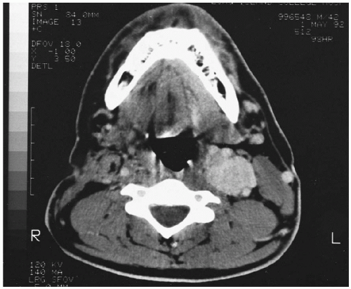 FIG. 48-3. Computed tomographic scan with contrast enhancement of a patient with a left carotid body tumor shows splaying of external and internal carotid arteries. |
Necrotizing (Malignant) Otitis Externa
The initial radiographic evaluation of necrotizing (malignant) otitis externa should include CT and a radionuclide examination. CT is used to evaluate the temporal bone for the presence of bone destruction and inflammatory changes in the adjacent soft tissues. A radionuclide bone scan may show inflammatory changes not detected at CT. If an intracranial abnormality is suspected, MRI is the study of choice.
Stay updated, free articles. Join our Telegram channel

Full access? Get Clinical Tree


