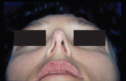
10 Functional Rhinoplasty: Principles and Techniques Nose plays the central role in respiration, olfaction, humidification of inspired air, and filtration. However, the importance of these factors on quality of life is frequently overlooked until these functions are compromised.1 Nasal obstruction is a problem frequently encountered by otolaryngologists. The etiologies are legion, yet the nasal valve is an important site of obstruction. Understanding the anatomy and physiology of the nasal valve is critical in evaluation, treatment, and planning for functional rhinoplasty. The nasal valve was first described over 100 years ago by Mink, referring to the narrow section of the nasal cavity at the border of the upper lateral cartilage (ULC) and lower lateral cartilage (LLC).2 Since then, the definition has evolved to divide the valve into internal and external components. The internal valve consists of the lateral nasal soft tissue and is bordered by the ULC, nasal septum, and head of the inferior turbinate. The internal valve angle is 10 to 15 degrees in the leptorrhine (European) nose and wider in the platyrrhine (Asian and African) nose,3 with an average cross-sectional area of 55 to 83 mm2.3–5 The external valve represents the alar vestibule and is defined by the nasal sill, alar rim, medial crus, and nasal spine. The external valve has little in the way of structure and is comprised mostly of fibroareolar tissue. A third and critically important, yet ironically less frequently discussed, site of the nasal sidewall is the intervalve area. This area has no rigid support except for occasional sesamoid cartilage, and is located at the inferolateral aspect of the lateral crura of the LLC. The corresponding superficial landmark is the supra-alar crease, and a deep supra-alar crease implies that the intervalve is the epicenter of collapse. Pathology there can be due to recurvature of the lateral crus, a paradoxically concave lateral crus, or simply a weak and collapsing soft tissue. Understanding the nasal valve is crucial because it is the narrowest and flow limiting segment of airway, which accounts for approximately half of the total nasal resistance. Obstruction at the valve can be static or dynamic, and can also be affected by the nasal muscles. Cole and Roithmann described the nasal dilators (nasalis anterior and nasalis posterior) and compressors (procerus, levator labii, superioris alaeque nasi, nasalis, and depressor septi).5 Additionally, external changes in nasal shape can impact function. For example, ptosis of the nasal tip is frequently seen in the aging population and can alter the intranasal space and create valve narrowing. Further, tip ptosis is not infrequently seen after primary rhinoplasty when tip support mechanisms are compromised. For this reason, the rhinoplasty surgeon must be mindful when destabilizing these structures. There are major and minor support mechanisms for the nasal tip, although the degree of contribution of each definite structure varies among individuals. The major tip support mechanisms include the inherent strength of the LLC, the attachment of the medial crura of the LLC to the caudal septum (pods), and the attachment of the ULC to the LLC (scroll). The cartilaginous septum can also be considered a major support mechanism because it functions to support much of the lower two-thirds of the nose. Minor tip support mechanisms include attachments of the alar cartilage to the skin, interdomal ligament, and nasal spine. Small decreases in diameter of the nasal passage result in large increases in resistance and therefore nasal obstruction. Resistance to flow is inversely proportional to the radius raised to the fourth power (Poiseuille law). Further, airflow velocity increases when the diameter/cross-sectional area decreases. The higher velocity results in amplified turbulence and resistance, which leads to increasing obstruction in a self-propagating cycle. As per the Bernoulli principle pressure decreases with the increase in rate of flow. Applying this principle to nasal airflow explains the lower pressure at the nasal walls exacerbating collapse. To counteract Bernoulli principle the lateral nasal wall needs to be rigid enough to withstand inward forces, though some degree of collapse is expected at high rates of flow. The differential diagnosis for nasal obstruction is extensive, and the diagnoses and treatments are beyond the scope of this chapter. Clearly, one must rule out masses (polyp, encephalocele, tumor, etc.). Additional possibilities include rhinitis, granulomatous disease, empty nose/paradoxical obstruction, scar bands, and foreign body. When faced with nasal anatomic abnormalities, many otolaryngologists first consider the septum as the site of pathology. While septoplasty is a widely performed procedure, its role in nasal obstruction has been challenged. Constantian and Clardy6 evaluated patients before and after surgery with rhinomanometry. They found that septal surgery alone did not show any improvement in mean nasal airflow, while both internal and external valve reconstruction improved flow significantly. Obviously, the exact etiology and degree of improvement varies with each patient. Some nasal valve obstruction results from etiologies such as trauma, Mohs surgery, facial paralysis, and commonly, iatrogenic collapse after rhinoplasty.7–10 A reduction rhinoplasty, whether to the dorsum or tip, is a common culprit of creating iatrogenic valve collapse and nasal obstruction. Postrhinoplasty obstruction can be manifest as tip ptosis, destabilization of the vestibule, narrowing of the bony piriform or open roof, or destabilized ULC after dorsal reduction. Middle vault collapse and tip ptosis can also occur from disease processes that compromise the bony framework and produce a subsequent saddle nose deformity. Wegener granulomatosis, septal hematomas with necrosis, and iatrogenic causes can all result in saddle nose deformities. Anatomic variation in nasal anatomy is a comprehensive topic that can fill an entire textbook. However, it bears mentioning briefly variants in anatomy that predispose patients to functional obstruction in addition to pitfalls that can occur after rhinoplasty. Dorsal hump reduction in patients with short nasal bones is challenging, especially when osteotomies are also performed. These patients typically also have long ULCs that lack the same support as nasal bones. Dorsal hump reduction weakens the support between the ULC and dorsal septum. The destabilized ULCs fall into the middle vault with associated contracture of the nasal mucosa and soft tissue envelope, which together obstructs the nasal passage and yields a poor cosmetic appearance. Further, the internal valve is narrowed because the cut edge of the dorsal septum lacks the natural cartilaginous widening present in the native dorsum. In addition to a constricted nasal passage, patients may suffer with cosmetic defects such as the “inverted-V deformity” or the related “hour glass deformity.” Osteotomies with medicalization of the bone, by their very nature, narrow the internal nasal valve. When malformations of the LLC are present, tip-narrowing procedures, such as dome-binding sutures, can create nasal obstruction. Intrinsic weakness of the lateral crura of the LLCs or vertically oriented LLCs each results in weak nasal sidewalls. The poor structure is unable to withstand the negative inspiratory pressure and leads to dynamic collapse of the nasal sidewall and/or external valve (Fig. 10.1). Additionally, certain tip procedures cause LLC buckling at the intermediate crura leading to unsightly bossae. Any maneuver, such as additional resection (former complete strip operation), which further compromises the cartilage must be avoided. The LLCs can also have a paradoxical curvature with the concavity facing intra-nasally and creating a static obstruction. Tip maneuvers frequently medialize the lateral crura and must be considered at the time of surgery. Figure 10.1 Base view demonstrating severe external valve collapse with inspiration. The obstruction is greater on the right, but still severe on the left. Although not an anatomic issue per se, the surgeon must be cautious when making intercartilaginous incisions during rhinoplasty. The intercartilaginous cuts can destabilize the scroll area. When they are made too close to the nostril margin, scarring of the external valve occurs, and the destabilized junction of the ULCs and LLCs cannot withstand the forces of contracture. Further blunting at the internal valve angle aggravates obstruction. Tension nose is a naturally occurring deformity resulting from overgrowth of the quadrangular septal cartilage. This leads to an appearance of a “big nose,” with a high nasal dorsum and an anterior septal angle that sits cephalad to the tip-defining point.11 This combination stretches nasal skin and soft tissue, resulting in alar flattening and a very narrow static internal valve. A deviated septum is one of the most common causes of nasal obstruction. The caudal septum is more easily addressed with standard septoplasty approaches. However, a more challenging situation is present when the septum is deviated at the dorsal aspect that creates a static narrowing at the internal valve. As is the rule in functional rhinoplasty, identifying not only the location of obstruction but also the anatomic etiology is the key. A thorough history will uncover previous surgery and trauma. History additionally helps delineate reversibility, seasonal differences, laterality, pain, and epistaxis. It is also important to inquire about impact on daily life.
Anatomy
Physics of Resistance and Airflow
Etiology
Anatomic Considerations in Rhinoplasty
Evaluation
Stay updated, free articles. Join our Telegram channel

Full access? Get Clinical Tree


