3 Flow Charts and Checklists
Initial Management
 See p. 2.
Objective | Measure |
Establish and maintain airways (intubation, cricothyrotomy, tracheotomy) | Clearing the upper airways of discharge or prolapsed tongue (combination fractures of mandible and maxilla), dentures |
Stabilize circulation | Establishing intravenous access, monitoring, fluid resuscitation |
Exclude intracerebral pressure, intracranial hematoma, brain swelling | Neurologic status, CT, possibly intracranial pressure (ICP) catheter, decompression, hematoma evacuation |
Exclude internal bleeding | Thoracic trauma: CT, drainage if necessary |
Abdominal trauma: ultrasonography, CT, or peritoneal lavage, laparotomy if necessary | |
Pericardial packing: ECG, echo-cardiogram, pericardiocentesis | |
Exclude paraplegia and compartment syndrome | Neurologic status, CT, decompression and stabilization of the spine if necessary |
Clinical signs, sensation, motor response, fasciotomy if necessary, fracture reduction, osteosynthesis | |
Soft tissue injuries | Wound management |
Initial Evaluation
Patient history | Loss of consciousness, vomiting |
Impaired nasal breathing, loss of olfactory/taste sensation, impaired vision, sensory disturbance | |
Inspection and palpation | Hemorrhage, open wounds, foreign bodies |
Cerebrospinal fluid or brain matter leak | |
Swelling, hematoma, air crepitus | |
Osseous step-offs, osseous gaps, abnormal mobility | |
Functional testing | Visual function |
Taste sensation/olfactory function | |
Sensory assessment | |
Diagnostic imaging | CT, a.-p. cranial views, lateral cranial views |
Immediate care | Extruding brain matter, intracerebral hemorrhage, intracerebral pressure, vision loss |
Interval treatment | Cerebrospinal fluid leak, pneumocephalus |
– Orbit and globe | Pupil width and reaction, gross evaluation of vision, globe position, motility, diplopia |
Diagnostic imaging | Radiologic evaluation of the paranasal sinuses, compare zygomatic arches, OPG, Clementschitsch, CT |
Immediate care | Replantation of teeth |
Management of open fractures | |
Interval treatment | Craniofacial reduction and reconstruction |
Injuries of the Neurocranium
 Diagnosing Injuries of the Neurocranium, Chapter 5, p. 38.
 Treatment of Injuries of the Neurocranium, Chapter 16, p. 137.
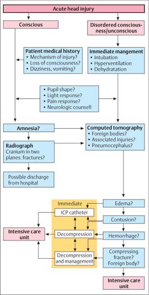
Fig. 3.1 Flow chart for acute head injury.
Injuries of the Skull Base
 Diagnosing Injuries of the Skull Base, Chapter 6, p. 45.
 Treatment of Injuries of the Skull Base, Chapter 17, p. 140.
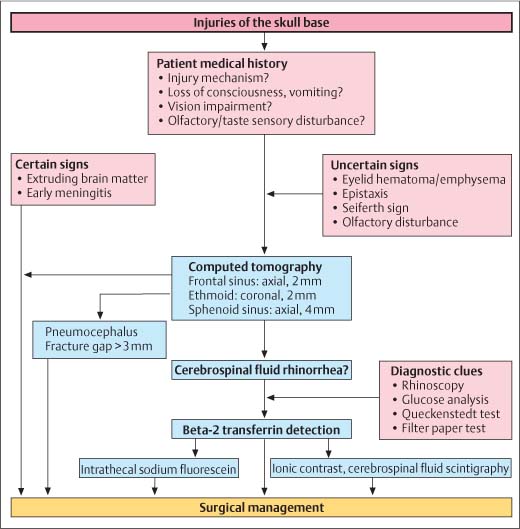
Fig. 3.2 Flow chart for injuries of the skull base.
Injures of the Ear and Lateral Skull Base
 Diagnosing Injuries of the Ear, Chapter 7, p. 53.
 Treatment of Injuries of the Ear, Chapter 18, p. 147.
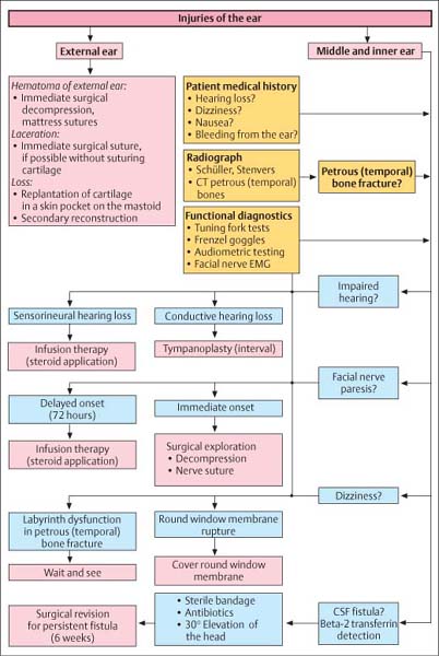
Fig. 3.3 Flow chart for injuries of the ear.
Patient medical history |


