Purpose
To quantify the vascular density of the choroid of normal eyes and to identify the influencing factors using en face images obtained with swept-source optical coherence tomography (SS OCT).
Design
Prospective cross-sectional study.
Methods
One hundred and sixty-three eyes of 163 healthy volunteers (83 female; mean age 42.2 ± 22.6 years) with a corrected visual acuity of ≥1.0 were investigated. En face SS OCT images of the choroid were used for quantitative assessment of the vascular density in the large choroid vessel layer. Relationships between vascular density of the choroid and age, sex, refractive error (RE), axial length (AL), and subfoveal choroidal thickness (SCT) were also investigated.
Results
There was a significant negative relationship between vascular density of the choroid and subject age ( P < .001). Analysis according to age showed a significant correlation in the group aged >30 years ( P < .001), but not in the group aged ≤30 years ( P = .225). SCT had a significant positive relationship with vascular density of the choroid ( P < .001). However, a significant correlation was not observed between sex, RE, or AL and vascular density of the choroid ( P = .981, P = .292, and P = .216, respectively). Multivariable regression analysis with vascular density of the choroid as the dependent variable and age, sex, RE, AL, and SCT as independent variables showed that age and SCT are important determinants of vascular density of the choroid ( P < .001).
Conclusion
Age and SCT affect vascular density of the choroid.
The choroid is the layer of blood vessels and connective tissue between the sclera and the retina, and plays an important role in maintaining the structure and function of the retinal pigment epithelium (RPE) and the sensory retina. It is well known that neovascularization and/or hyperpermeability of the choroid lead to many chorioretinal diseases, including age-related macular degeneration, polypoidal choroidal vasculopathy, central serous chorioretinopathy, and high myopia–related chorioretinal degeneration. Furthermore, the concept of “pachychoroid” has recently been proposed to cover choroidal anatomic manifestations that may include an increase in absolute thickness, a diminution in the relative thickness of the choriocapillaris layer, or an alteration in the caliber of the large choroidal vessels, indicating the increasing importance of morphologic evaluation of the choroid.
In general, the morphology of vascular tissue is affected by a number of factors, such as age and sex. Therefore, it is important to exclude the effects of these factors on the normal structure of the choroid in order to detect when a pathologic change has occurred. Among several parameters that contribute to the structure of the choroid, choroidal thickness has been evaluated extensively by histologic analysis and enhanced-depth imaging optical coherence tomography (EDI OCT). Factors affecting the thickness of the normal choroid are age, axial length (AL), and sex. Recently, Sonoda and associates quantified the luminal and stromal areas of the choroid (ie, the vascular density of the choroid) by binarization of EDI OCT images, and concluded that aging and elongation of the AL are factors that decrease the luminal area. However, these EDI OCT evaluations are for only 1 cross-sectional image of the choroid below the fovea, so the overall vascular density of the choroid and its relationships with these factors are still unclear.
One possible method to understand the vascular structure of the choroid with wide observation range is to assess the choroid not only with 1 cross-sectional image but also with coronal view. The recent advent of swept-source optical coherence tomography (SS OCT), which is highly penetrating and has a high scan speed, has made it possible to capture 3-dimensional images of the structure of the choroid. In particular, en face images (coronal-view images generated after computerized flattening along a specific retinal layer boundary) enable layer-by-layer analysis over a wide observation range.
To the best of our knowledge, there are currently no published studies investigating en face images of the normal choroid, and the stratified vascular structure of the choroid is not known. Therefore, in the present study we captured en face images of the choroid in normal eyes of Japanese people, and sought to identify the factors that affect the vascular density of the choroid.
Methods
Study Design
This was a prospective cross-sectional study.
Subjects
One hundred and sixty-three healthy volunteers were examined by SS OCT (DRI OCT-1 Atlantis; Topcon Corporation, Tokyo, Japan), autorefractometry (RC 5000; Tomey Corporation, Nagoya, Japan), and optical biometry (OA 1000; Tomey Corporation). Inclusion criteria were no prior history of eye disease and a corrected visual acuity of ≥1.0, and exclusion criteria were a spherical equivalent refraction of ±6.0 diopters (D) or more and a history of diabetes or cardiovascular disease. The right eye of each subject was examined. All examinations were performed under nonmydriatic conditions. Analysis of en face images and measurements of subfoveal choroidal thickness (SCT) were done by one of the authors in a masked fashion without knowledge of subject information. All of the investigative procedures conformed to the tenets of the Declaration of Helsinki. The study was approved by the Ethics Committee of Okayama University Hospital, Okayama, Japan.
Constructing En Face Images of the Large Choroidal Vessel Layer and Quantifying Vascular Density of the Choroid
Three-dimensional OCT volume data of the choroid were obtained using the DRI OCT-1 Atlantis, which is based on SS OCT technology and has a scanning speed of 100 000 A-scans per second. The scanning protocol was 12 × 9 mm oblong and contained 512 × 256 A-scans.
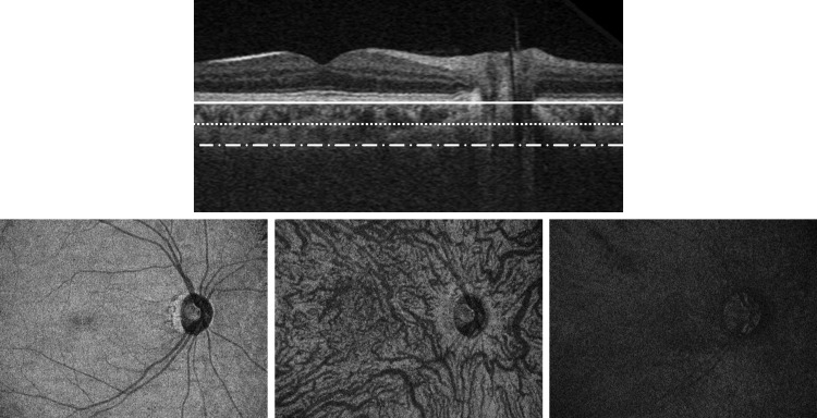
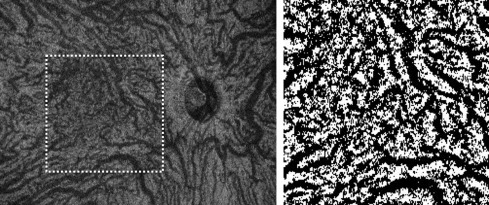
To construct en face image and quantify vascular density of the choroid, 2 software tools were applied: EnView version 1.0.1 software (Topcon Corporation) and ImageJ version 1.48 software (National Institutes of Health, Bethesda, Maryland, USA). Based on retinal layer boundary information, EnView can align the 3-dimensional OCT volume data along a specific retinal layer boundary and generate an en face image at the arbitrary depth. In this study, 3-dimensional OCT images were flattened relative to the Bruch membrane, and en face images were extracted at the level where choroidal thickness was 50% of the total choroidal thickness in order to examine the large choroidal vessel layer that has been reported to account for 70% of total choroidal thickness ( Figure 1 ). From the extracted en face image, a 5 × 5-mm-square image centered on the fovea was binarized according to Niblack’s method ( Figure 2 ). The black region was considered as vascular area and its number of pixels was quantified with ImageJ software. The vascular density of the choroid (%) was calculated by dividing the number of pixels of the vascular area by that of the 5 × 5-mm region of interest (44 521 pixels). The main outcome measures were the relationships between age, sex, refractive error (RE), AL, or SCT and the vascular density of the choroid.
Subfoveal Choroidal Thickness Measurements
The SCT was measured using OCT to capture cross-sectional images of the choroid. It was measured from the outer portion of the hyperreflective line corresponding to the RPE to the inner surface of the sclera.
Statistical Analysis
The statistical analysis was performed using Statistical Package for the Social Sciences software (SPSS, version 22.0.0.0; IBM Corporation, Armonk, New York, USA). Techniques used for the analysis are described in the results. A P value of <.05 was considered to be statistically significant.
Results
Demographic Data
One hundred and sixty-three volunteers (163 eyes) participated in the study. Their demographic characteristics are shown in Table 1 . The mean age of the subjects was 42.2 ± 22.6 years (range 6–80 years), and 83 subjects (50.6%) were female. Mean RE was −1.7 ± 2.6 D (range +5.8 to −5.9 D), mean AL was 24.2 ± 1.5 mm (range 20.8–27.9 mm), and mean SCT was 270.3 ± 94.5 μm (range 56.0–599.0 μm).
| Age Group | Age (y) | N (Eyes) | Sex (F/M) | Refractive Error (Diopters) | Axial Length (mm) | Subfoveal Choroidal Thickness (μm) |
|---|---|---|---|---|---|---|
| 1–10 | 7.9 ± 1.1 | 18 | 9/9 | −0.1 ± 2.2 | 23.0 ± 1.2 | 305.4 ± 70.9 |
| 11–20 | 13.8 ± 2.2 | 20 | 12/8 | −1.4 ± 3.3 | 24.2 ± 1.6 | 288.3 ± 71.9 |
| 21–30 | 24.8 ± 2.9 | 16 | 6/10 | −3.5 ± 1.7 | 25.2 ± 1.0 | 254.2 ± 70.8 |
| 31–40 | 34.8 ± 2.8 | 20 | 9/11 | −2.4 ± 2.8 | 24.6 ± 1.2 | 326.2 ± 118.2 |
| 41–50 | 46.2 ± 2.6 | 20 | 12/8 | −3.4 ± 1.7 | 25.3 ± 1.3 | 234.7 ± 83.2 |
| 51–60 | 54.8 ± 2.8 | 21 | 10/11 | −1.3 ± 1.8 | 24.1 ± 1.4 | 275.3 ± 103.3 |
| 61–70 | 65.2 ± 2.7 | 28 | 15/13 | −0.8 ± 2.4 | 23.9 ± 1.2 | 259.2 ± 78.5 |
| 71–80 | 74.3 ± 3.3 | 20 | 9/11 | −0.8 ± 2.4 | 23.8 ± 1.7 | 222.3 ± 99.4 |
Relationship Between Age and Vascular Density of the Choroid
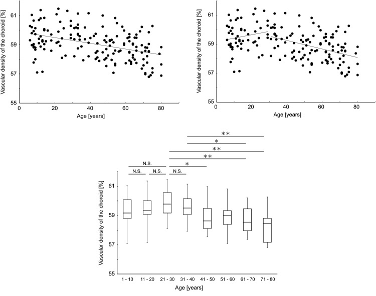
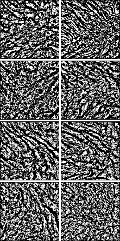
There was a significant negative relationship between vascular density of the choroid and subject age ( P < .001, R 2 = 0.3, regression analysis; Figure 3 , Top left). Because of a previous report indicating that development of the eye stops in the third decade of life, the results were re-examined with an age cutoff of 30 years. There was no significant correlation between age and vascular density of the choroid in subjects aged 30 years or younger ( P = .225, R 2 = 0.2, regression analysis), but there was a significant correlation in those aged older than 30 years ( P < .001, R 2 = 0.3, regression analysis; Figure 3 , Top right). These trends were supported by the results of the 10-year breakdown analysis. Although there was no significant difference in vascular density of the choroid among groups of subjects aged 40 years or younger, the vascular densities of the choroid of subjects aged 21–30 years and 31–40 years were significantly higher than that of groups of subjects over 61 years old ( P = .040, 1-way analysis of variance [ANOVA] with Bonferroni test; Figure 3 , Bottom). Representative images of the choroidal vessels in each age group are shown in Figure 4 .
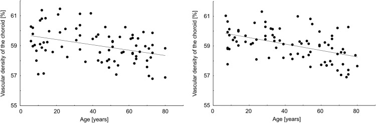
There was a significant negative relationship between subject age and vascular density of the choroid in female subjects ( P < .001, R 2 = 0.3, regression analysis; Figure 5 , Left). There was also a significant negative relationship between subject age and vascular density of the choroid in male subjects ( P < .001, R 2 = 0.5, regression analysis; Figure 5 , Right). There was no statistically significant difference in vascular density of the choroid between male and female ( P = .981, unpaired t test).