Purpose
Previously published literatures of acute studies on few subjects have shown contradictory evidence on the reproducibility and characteristics of the elicited phosphenes, despite using the same stimulating parameters with epiretinal electrode arrays. In this study, we set out to investigate the long-term repeatilibity and reproducibility of phosphenes in subjects chronically implanted with the Argus II retinal prosthesis (Second Sight Medical Products, Inc., Sylmar, CA, USA).
Design
Retrospective interventional case series and reliability study.
Methods
Six Argus II subjects of >5 years implantation from a single site participated. The 4-electrode cluster (“quad”) closest to fovea was stimulated in each subject with a fixed biphasic current. Perceived phosphenes were depicted relative to subjective visual field center. The stimulus was applied at reducing time intervals from 20 minutes to 1 second. Two sets of stimulations were performed on the same day and 2 further sets repeated on a separate visit >1 week apart.
Results
Each subject depicted phosphenes of consistent shapes and sizes, and reported seeing the same colors with the fixed stimulating parameters, irrespective of the interstimuli intervals. However, there is a wide intersubject variation in the phosphene characteristics. Four subjects drew phosphenes in the same visual field quadrant, as predicted by the quad-fovea location. Two subjects depicted phosphenes in the same hemifield as the expected locations.
Conclusion
Phosphenes for each subject were consistently reproducible in all our chronically implanted subjects. This has important implications in the development of long-term pixelated prosthetic vision for future devices.
Since entering the commercial market as a retinal prosthetic device for the treatment for end-stage retinitis pigmentosa (RP), the Argus® II (Second Sight Medical Products Inc., Sylmar, CA, USA) retinal prosthesis system has been implanted in an increasing number of patients. However, despite this growing clinical use, data describing the features of artificial vision perceived by the users remain scarce in the published literature. The idea of developing useful vision by epiretinal electrical stimulation hinges on the premise that stimulation with a single electrode gives rise to a discrete focal percept in a retinotopic manner. Simultaneous stimulation with multiple electrodes therefore theoretically leads to perception of a pattern in concordance with the pattern defined by the stimulating electrodes.
In earlier studies, Rizzo and associates have called into question the consistency and reproducibility of phosphenes elicited by patterned epiretinal microelectrode stimulation. In a study involving 5 end-stage RP patients and 1 patient with normal retina, only 48% of the single-electrode stimulations and 32% of the multielectrode stimulations elicited visual percepts that matched the electrical stimulation patterns. Of the single-electrode stimulations, 3 subjects reported “a line” on some occasions, while “clusters of 2 or 3 images” were seen on other occasions. In particular, the authors reported that only 66% (out of 99 stimulations) of the elicited visual percepts were reproducible in 3 RP patients on 2 separate trials, despite using the same stimulating parameters to activate the same electrodes. Such inconsistencies in the form and reproducibility of phosphenes would seriously undermine the formation of pixelated vision.
In this study, we set out to investigate the consistency and reproducibility of phosphenes elicited in a cohort of subjects chronically implanted with the Argus II system at a single site. All of the subjects described have had the device implanted and functioning for more than 5 years.
Methods
Subject Inclusion/Exclusion Criteria
This is a single-center prospective study. All but 1 subject from our center implanted with the Argus II retinal prosthesis system as part of the phase I/II clinical trial ( clinicaltrials.gov identifier: NCT00407602 ) took part in the study (n = 6). One subject was excluded, as his device ceased to function after he developed retinal detachment and thick macular pucker as a result of a fall. The participating subjects’ demographics and operation dates are shown in Table 1 . The study was approved by the institutional review boards and ethics committee, and adhered to the tenets of the Declaration of Helsinki.
| Subject ID | Diagnosis | Year of Operation | Age at Time of Operation (y) |
|---|---|---|---|
| 001 | Retinitis pigmentosa | 2008 | 70 |
| 003 | Retinitis pigmentosa | 2008 | 72 |
| 005 | Retinitis pigmentosa | 2009 | 55 |
| 006 | Choroideremia | 2009 | 66 |
| 007 | Retinitis pigmentosa | 2009 | 63 |
| 009 | Retinitis pigmentosa | 2009 | 45 |
Selection of Stimulating Electrodes and Parameters
For each subject, a cluster of 4 electrodes (hereinafter referred to as a “quad”) closest to the fovea, which were functioning with thresholds within the safety charge density limit, was selected for stimulation, and the elicited phosphenes characterized for the purpose of this study ( Figure 1 ).
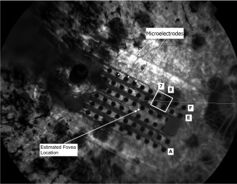
An estimated location of the fovea was made on the fundus photograph (taken at the outset of the study) and was used for each subject as a reference point, measuring 15.5 ± 1.1 degrees from the center of the optic disc horizontally and −1.5 ± 0.9 degrees vertically. The foveal position was estimated, as there were no remaining features of the fovea on color photographs, fluorescein angiograms, or optical coherence tomography (OCT) scans owing to severe end-stage RP.
Once the designated quad was chosen, the stimulating current for each subject was arbitrarily set to be 100 μA above the threshold (measured within the last 6 months) initially, and then adjusted according to the strength of response and comfort level reported by the subject. We aimed to elicit a clear, definite visual percept without causing any discomfort or physical “tingling” sensation for each subject. Default settings for the Argus II retinal prosthesis system Clinical Fitting System (CFS) employed for device fitting and standard testing were likewise used for this study, which generate cathodic-first, charge-balanced biphasic square waves to avoid tissue damage from charge build-up. These default waveform parameters were as follows: phase width of 0.46 ms, interphase duration of 0 seconds, and total stimulation duration of 250 ms at the frequency of 20 Hz. Swept-source OCT (DRI OCT-1 Atlantis; TOPCON, Topcon Medical Systems, Tokyo, Japan) imaging through the chosen quad for each subject was performed, to assess the contact between the stimulating electrodes and the retinal surface. The selected quad, quad-retina relation, quad threshold, and stimulating current for each subject were as shown in Table 2 .
| Subject ID | Quad | Quad-Retina Relation (on OCT) | Threshold (μA) | Stimulating Current (μA) | Phosphene Features | Phosphene Duration, t (s) |
|---|---|---|---|---|---|---|
| 001 | C07C08 D07D08 | In contact no significant ERM a | 137 | 277 | White filled-in circle | 0.5 < t < 1 |
| 003 | A07A08 B07B08 | In contact no significant ERM a | 250 | 350 | Electric blue filled-in circle | t < 0.5 |
| 005 | E05E06 F05F06 | In contact no significant ERM a | 137 | 237 | Bluish-gray vertical line, with fizzy vertical edges | t < 0.5 |
| 006 | A07A08 B07B08 | Quad-retina separation = 377 μm; no ERM visible | 371 | 552 | Yellow “7” shape | 0.5 < t < 1 |
| 007 | E07E08 F07F08 | In contact no significant ERM a | 24 | 124 | Orange filled-in ring that ripples out | 0.5 < t < 1 |
| 009 | E07E08 F07F08 | In contact no significant ERM a | 97 | 124 | Orange horizontal lines x2, with fizzy brightness in between the lines | 0.5 < t < 1 |
a In images where the electrode array is in direct contact with the retinal surface, owing to the artefact caused by the acoustic shadow of the array, it is difficult to ascertain the presence, if any, of mild epiretinal membrane.
Phosphenes Depiction
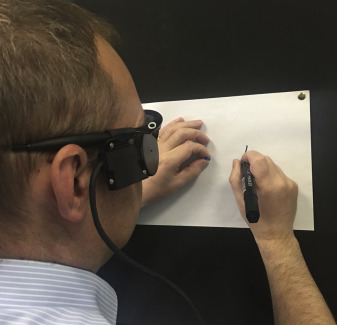
To record the phosphenes perceived by each subject, we constructed a wall covered with smooth-surface black mats. The subjects were first asked to stand up and stretch out both arms fully to touch the black wall, so that their shoulders were square, facing the wall. The standing position of each subject was then adjusted so that the distance between the front of their eyes and the wall equaled 30 cm. Next, the subjects were asked to point with the index finger of both hands simultaneously on the black wall, to where they believed the center of their visual field was, while keeping their head and eyes pointing straight ahead. A stack of white A4-size papers (in landscape layout) was then placed underneath the index fingers of each subject and pinned to the wall, so that the index fingers were pointing at the center of the top sheet of paper (ie, the center of the paper was approximating the proclaimed center of each subject’s visual field). A drawing pin with a protruding cylindrical head was then inserted at the point where their index fingers contacted with the wall, so as to mark the location of the proclaimed visual field center.
During the experiment, the subjects were asked to position themselves according to the setup above, hold onto the preplaced drawing pin head, and adjust their head and eye position until they felt the center of their visual field was in alignment with the drawing pin. With each quad stimulation, the subjects were instructed to keep their nondominant hand on the drawing pin as a point of reference, while drawing on the paper with a marker pen the outline of the phosphene they perceived in relation to their visual field center. A fresh sheet of paper was used for each phosphene depiction ( Figure 2 ).