, Vincent Y. W. Lin2 and Joseph M. Chen2
(1)
Department of Otorhinolaryngology, Medical University of Vienna, Vienna, Austria
(2)
Department of Otolaryngology Head & Neck Surgery, Sunnybrook Health Sciences Center, Toronto, Ontario, Canada
Electronic supplementary material
Supplementary material is available in the online version of this chapter at 10.1007/978-3-7091-1490-2_4. Videos can also be accessed at http://www.springerimages.com/videos/978-3-7091-1489-6.
The facial recess (posterior tympanotomy) is a triangular region delineated by the fossa incudis superiorly, the facial nerve posteriorly, and the chorda tympani anteriorly (Figs. 4.1 and 4.2). Anterior to the chorda tympani lies the annulus fibrosus of the tympanic membrane.
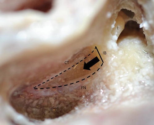
Fig. 4.1
After the external auditory canal is thinned out, drilling is evenly advanced medially (arrow). The posterior tympanotomy is opened between the facial nerve and chorda tympani. The superior border is formed by a bony strut (incus buttress) which protects the incus (FN facial nerve, CT chorda tympani, IB incus buttress, I incus in fossa incudis)
Fig. 4.2
The facial recess is defined by the line drawn through the body of the incus, across the posterior bony canal wall. Be faithful to this line during the initial dissection. Use a 2 mm cutting burr to start. Look for small air cells to guide you
The safest and the most effective identification of the facial recess was best described by Ugo Fisch. He used an imaginary line drawn through the profile of the incus in the “slot” position to delineate the FR. This is when the temporal bone is rotated away from the surgeon in such a way to create the maximal space medial to the ossicles (Figs. 4.3 and 4.4). In this view, the fallopian canal in the tympanic segment can be visualized.
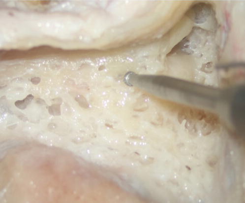
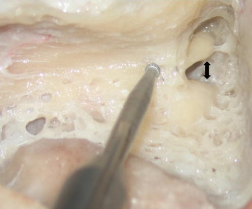
Fig. 4.3
Incorrect position of the patient (no slot position) and direction of drill towards the facial nerve during drilling of the facial recess
Fig. 4.4
Correct position of the patient and drill during drilling of the facial recess: patient is tilted away to delineate the slot position between ossicles and fallopian canal (arrow). This slot also delineates the facial recess
Once the slot position is obtained by rotating the temporal bone, imagine a line drawn through the body of the incus in its profile (Figs. 4.1 and 4.2


Stay updated, free articles. Join our Telegram channel
Full access? Get Clinical Tree
