CHAPTER 46 Eyelid malpositions
Entropion and ectropion
Introduction
Intact eyelids cover the eye completely while closed and the lid margins stay in direct contact with the globe while opened1. The lid margin’s contact line to the ocular surface is of vital importance to the integrity of the ocular surface. Dysfunction of lid motility and position can lead to symptoms varying from mild discomfort and dry eye to severe corneal ulceration and sight-threatening ocular surface disease. It is therefore most important to address lid malposition with the appropriate surgical therapy2–4. (Further suggested reading).
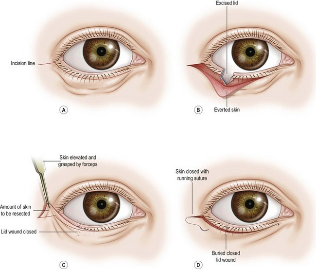
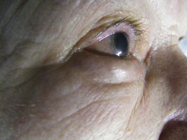
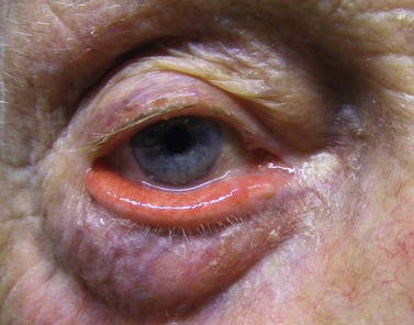
Entropion is a condition in which the lid margin is turned inward onto the ocular surface, while ectropion by definition is the contrary, describing a lid margin that turns outward, leading to exposure of tarsal conjunctiva (Figs 46.1 and 46.2).
Epidemiologic consideration and terminology
Congenital eyelid malpositions are rare in Caucasians, but can be potentially sight threatening if lash abrasion of the corneal surface leads to ulceration and infection (e.g. tarsal kink). Continuous and profuse secretion, pain, photophobia, and blepharospasm in a baby should always raise the suspicion and lead to immediate investigation under general anesthesia. In the Asian population, congenital epiblepharon – an inward positioning of the lashes in the medial part of the lid due to a fold of orbicularis muscle and skin – is a common diagnosis. If it is more severe, all of the lid margin is turned towards the eye, causing a true entropion. True and chronic congenital ectropion is also rare and most often an associated condition of a genetic disorder, e.g. trisomy 21.
Lid malposition can be the result of cicatrizing changes of either conjunctiva or skin after trauma or surgery, or in destructive skin and mucous membrane diseases (e.g. scleroderma, ocular pemphigoid). Ectropion might also be the result of paralysis of the orbicularis muscle, as in facial palsy.
The majority of cases of entropion and ectropion, however, are caused by involutional changes that alter the homeostasis of skin tension, muscle action, and stabilizing forces of the lid retractors.
Anatomical considerations
Anatomically, both upper and lower eyelids can be separated into an anterior and a posterior lamella. The anterior lamella consists of skin and orbicularis muscle. The posterior lamella consists of the tarsal plate, containing the meibomian glands and the conjunctiva covering the posterior surface. Medially and laterally, the canthal tendons fixate the lids to the medial and lateral orbital wall.
It is essential for any surgical procedure at the canthal region to understand that, in order to hold the lids correctly against the ocular surface, the vector of the deep canthal fixation has to be as posterior into the orbit as possible, at least as posterior as the equator of the globe.
Lid posture and vertical movement are dependent on the levator and tarsal muscle in the upper eyelid and on the complex system of lid retractors in the lower lid. This distinct difference makes it impossible to transfer surgical principles from lower to upper lid one to one.
Medially, the lacrimal punctum and canaliculi are embedded in the lid margin and canthal fixation system. Any surgical procedure aiming to reconstruct or tighten the canthal tendon has to address this delicate anatomical situation.
Fundamental principles and goals for surgery
In repairing ectropion and entropion, all the general principles of oculoplastic surgery need to be applied. With the exception of tumor removal, incisions around the lids should follow the relaxed tension lines in order to avoid visible scars. It is necessary to understand the complex anatomy, especially of the medial canthal area. All procedures addressing eyelid malpositions aim to restore the lid function while maintaining or restoring an adequate esthetic result: complete closure, blinking, tear distribution, tear pump, position of lashes, and texture of lid skin.
Indications for surgery
In principle, surgical correction of eyelid malposition is indicated whenever patient discomfort can be repaired with sufficient safety and predictability.
While minor entropion or ectropion may be mainly disturbing for esthetic reasons and the indication for repair considered only optional, malpositions causing ocular surface infection and ulceration should lead to immediate repair.
Preoperative assessment
In addressing the patient with lid margin malposition, it is most important to first decide whether the condition is disturbing normal lid function and surgery is needed to prevent further damage to the ocular surface or if it is mainly an esthetic problem for which surgery can be delayed.
The choice of procedure depends on the clinical assessment of the anatomical changes, which follows a catalog of questions:
Anesthesia
The majority of surgical procedures can be done under local anesthesia. Claustrophobic or psychologically unstable patients can also usually be operated on with intravenous or preoperative peroral sedation. It is much more comfortable for the patients if the whole face is surgically cleaned and then left open. Critically ill and cardiovascular unstable adults should always be supervised by an anesthetist during surgery and children under the age of 16 should always be considered for general anesthesia.
As local anesthetic 2% Xylocaine (lidocaine, lignocaine) with 1 : 200 000 epinephrine (adrenaline) is a very good choice for local infiltration. If prolonged anesthesia is required, bubivacaine can be added in equal volume. Some surgeons add hyaluronidase to the solution to obtain a faster effect.
Operation techniques
Both entropion and ectropion are more common in the lower eyelid; upper and lower eyelids are addressed separately in this section.
Lower lid ectropion
Ectropion occurs either as a result of aging changes to skin, muscle, or connective fibers, loss of muscle tone due to the lack of innervation as in seventh nerve palsy, or as a result of scar formation in the anterior lamella of the lid. Surgical repair aims either at restoring horizontal tightness at the medial or lateral canthal tendon and the lid margin itself or at reconstructing the anterior lamella. If the underlying cause of a neurological deficit cannot be addressed, the lid must be either elevated or horizontally tightened or both.
Tarsoconjunctival excision
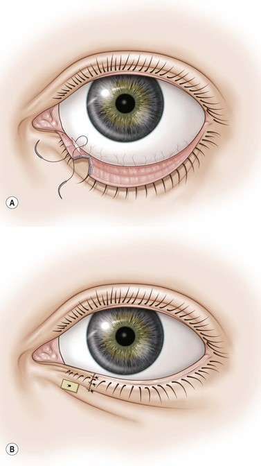
If the canthal tendon is tight and ectropion of the medial third of the lid has led to eversion of the lower punctum, an excision of the posterior lamella with or without plication of the lower lid retractors is sufficient to correct the problem5.
Technique
The conjunctiva is grasped with a forceps and a diamond-shaped excision of the tarsoconjunctiva is made below the punctum using straight scissors held parallel to the lid margin (a scalpel can be used if preferred by the surgeon), leaving enough conjunctiva below the punctum to place a needle. The canaliculus is protected by a probe. The horizontal extent of the excision is about 7 mm. A double armed 6-0 absorbable suture is passed through the upper edge of the wound just below the punctum; the other arm of the suture is passed through the lower edge of the wound, picking up the lower lid retractors. The knot is tied and buried in the wound and the suture left for spontaneous absorption.
Alternatively – if the eversion is more extensive – a non-absorbable suture can be used, passed through the skin and tied over a bolster. This will produce a stronger inverting effect. (See ‘lazy-T’ procedure below). The suture can be removed after 10–14 days.
(Tarso)conjunctival excision, inverting sutures, and horizontal lid shortening (lazy T procedure)
If the canthal tendons are tight and the eversion is mainly in the medial third of the lid, inverting the lid margin by excising a part of the conjunctiva and shortening the lid horizontally will correct the malposition. The resulting incision lines after closure forms a T that lies lazily on its side, hence the name6.
Technique (Fig. 46.3 A,B)

Fig. 46.3 (A&B) Lazy-T procedure. A diamond-shaped tarsconjunctival excision, pentagonal full-thickness lid resection.
The inverting suture is tied to just invert the lid margin to the globe. A little overcorrection is acceptable. Sutures can be removed at 10 days, the inverting suture at 14 days.
Horizontal lid shortening and skin excision (Kuhnt–Szimanowski)
If horizontal lower lid laxity and an abundance of skin is present while the medial and lateral canthal tendons are firm, the lid can be tightened by a pentagonal excision and the skin excess reduced via a subciliary incision.
Technique (Fig. 46.4 A–D)

Fig. 46.4 (A–D) Horizontal lid shortening and skin excision. (A) Incision. (B) Everted skin flap and excised lid. (C) Skin resection. (D) Wound closure.
A full-thickness pentagonal excision of the lid is performed with straight scissors or a number 11 blade scalpel. The pentagonal wound is closed in the same fashion as after a tumor resection: two marginal sutures (6-0 silk) are placed to adjust the gray line. The end of these sutures are left long to be later buried under the skin suture. Two to three 6-0 Vicryl sutures that pick up the tarsus are used to close the wound.
With the patient opening his or her mouth and looking upwards, the amount of resectable skin is assessed and cut. The skin is then closed with an intracutaneous or regular continuous 6-0 nylon suture.

Full access? Get Clinical Tree

