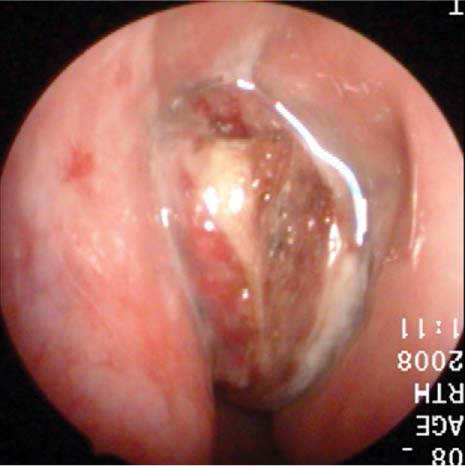
49 A 37-year-old man, a nonsmoker, presents with a 3-month history of left nasal congestion and intermittent epistaxis. The bleeding has been fairly slight and has not required trips to the emergency department or placement of nasal packing. He has a history of mild allergic rhinitis but denies chronic sinusitis. He does not ordinarily take any medication for his allergies, and he is not on any medicine that might affect his bleeding time or coagulation. When questioned, he does admit to mild hyposmia. He denies weight loss, fevers, or night sweats. Examination reveals anterior deviation of the nasal septum to the left with slight crusting, making the left posterior and superior nasal cavity somewhat difficult to visualize. However, no other lesions are noted on anterior rhinoscopy. His basic head and neck examination is otherwise unremarkable. Visual acuity and extraocular movements are intact. Pupils are equal, round, and reactive. Nasopharyngoscopy reveals no polyps or evidence of ostiomeatal disease within the right nasal cavity, but does suggest some mild allergic inflammation of the inferior and middle turbinates. Examination of the left side does reveal a polypoid, slightly fibrous irregular mass in the superior nasal vault, medial to the middle turbinate (Fig. 49.1). It appears slightly friable. Maxillofacial computed tomography (CT) confirms a soft tissue mass filling the left nasal vault, with no apparent erosion of the cribriform plate, but involvement of the left ethmoid sinus and possible erosion of the left lamina papyracea. A subsequent magnetic resonance imaging (MRI) scan of the head and neck with contrast also reveals a large homogeneously enhancing mass filling the left nasal vault, with no intracranial extension, and sparing of the soft tissues of the orbit. Because of the possible vascular nature of the mass, he is scheduled for nasal endoscopy and nasopharyngoscopy with biopsy in the operating room. Fig. 49.1 Fibrous, irregular mass left upper nasal cavity. 1. The etiology of this patient’s symptoms could easily have been attributed to his deviated septum. However, a 3-month history of recurrent epistaxis warrants a more thorough examination of the nose. A unilateral nasal mass should immediately raise concerns that this might be something other than a benign respiratory polyp. Benign neoplasms to consider include inverting papilloma, hemangioma, schwannoma, antrochoanal polyp, and meningocele or meningoencephalocele. 2. Malignant nasal masses that may present in the nose include squamous cell carcinoma, both within the setting of a preexisting inverting papilloma and as a new, isolated neoplasm. Other malignancies include adenocarcinoma, adenoid cystic carcinoma, mucoepidermoid carcinoma, hemangiopericytoma, blue cell tumors such as esthesioneuroblastoma, rhabdomyosarcoma, plasmacytoma, malignant mucosal melanoma, lymphoma, sinonasal undifferentiated carcinoma, and nasopharyngeal carcinoma. 3.
Esthesioneuroblastoma
History
Differential Diagnosis—Key Points
![]()
Stay updated, free articles. Join our Telegram channel

Full access? Get Clinical Tree


