In the past decade, we have introduced various techniques of endothelial keratoplasty in the management of corneal endothelial disorders, including Descemet stripping (automated) endothelial keratoplasty (DSEK/DSAEK) and Descemet membrane endothelial keratoplasty (DMEK); that is, isolated transplantation of a donor Descemet membrane and its endothelium. With these techniques, long-term decrease in endothelial cell density (ECD) may be the main indicator in predicting long-term graft survival. After DSEK/DSAEK, a 30% to 40% decrease in ECD has been observed and attributed to donor tissue manipulation during surgery. In contrast, graft preparation and implantation in DMEK can be performed as completely “no-touch” procedures, so that better ECD may be anticipated.
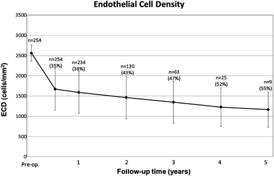
In the current study, for which we can now present a fifth year of follow-up results, 300 consecutive eyes that underwent DMEK for Fuchs endothelial dystrophy or pseudophakic bullous keratopathy were evaluated. ECD measurements were available in 254 eyes with 6 months of follow-up; 234 had 12 months, 130 had 24 months, 63 had 36 months, 25 had 48 months, and 9 had 60 months of follow-up ( Figure ). Compared with preoperative values, ECD decreased by 35% at 6 months, 38% at 12 months, 43% at 24 months, 47% at 36 months, 52% at 48 months, and 55% at 60 months—a sharp decrease in the first 6 months, followed by a yearly decrease of approximately 7% compared with the previous follow-up, up to 5 years after DMEK ( Figure ).