Endoscopy is advantageous in many aspects of pediatric ear surgery, especially for tympanoplasty and cholesteatoma. The narrower pediatric meatus can restrict access for a totally endoscopic approach, but when possible, avoidance of an external incision is greatly appreciated by children. In cholesteatoma surgery, less residual disease and better hearing from ossicular preservation are achievable. Factors influencing selection of a totally endoscopic approach, or use of endoscopy as an adjunct are reviewed, along with tips to facilitate successful endoscopic surgery in children. The optimum approach will depend upon the extent of the disease, morphology of the child’s ear and surgeon’s experience.
Key points
- •
Permeatal access to middle ear is restricted by the narrower width, but facilitated by the shorter length in younger children.
- •
In many cases it is the curvature of the ear canal instead of the age of the child that governs endoscopic access. Totally endoscopic middle ear surgery can be completed successfully, even in infancy.
- •
Endoscopic visualization of the recesses of the middle ear allows a less invasive canal wall up approach that is ideally suited to children.
- •
The retrotympanum is a common location for cholesteatoma in children and benefits from endoscopic removal.
- •
Residual cholesteatoma rates can be reduced with endoscopic inspection and dissection.
- •
Removal of cholesteatoma from the medial epitympanum can be completed endoscopically without disarticulating an intact ossicular chain and so improve hearing outcome.
Videos of Endoscopic Techniques in Children accompany this article
Introduction
The wide-angle view from the tip of a rigid endoscope provides advantages to the otologist in many aspects of pediatric care, ranging from assessment of the ear drum in clinic to minimally invasive access to the hidden recesses of the temporal bone during surgery. In clinic, the recording of endoscopic images improves accuracy in monitoring changes in tympanic membrane retraction. In surgery, endoscopes are used for inspection (eg, to reveal occult remnants of cholesteatoma) but, more importantly, they can be used with angled instruments and laser to remove disease from under hidden recesses without destructive bone removal. The child benefits from the consequent reduction in residual cholesteatoma and gain in hearing from more frequent ossicular preservation. An intact canal wall approach is generally favored for pediatric cholesteatoma surgery and the endoscope is an invaluable asset in the combined approach through ear canal and mastoid. Although access is more challenging in the smaller pediatric ear canal, totally endoscopic middle ear surgery through the canal is still appropriate in many cases, avoiding the disadvantage to the child of an external incision. The opportunities and methods for application of endoscopes to pediatric middle ear surgery are outlined in this article.
The principle advantage of a totally endoscopic surgical approach for the child is avoidance of an external incision. For the surgeon, the avoidance of a small endaural incision or of a cosmetically placed incision just behind the postauricular sulcus might seem a small advantage. However, the universal expression of relief and pleasure on the faces of parents and children on learning that it has been possible to avoid an external incision indicates that this is perceived as a significant benefit by the patient. In addition to the psychological and cosmetic benefits, more tangible advantages include the possibility of a shorter hospital stay (same day discharge can be anticipated) and faster return to the physical sports in which many children participate. These benefits are outweighed by the principles of achieving a safe dry ear, and the functional importance of an intact tympanic membrane and ossicular chain. Therefore, selection of a totally endoscopic permeatal approach has to be considered carefully and not allowed to compromise the primary objectives of surgery.
The advantages to the otologist of endoscopy are clearly demonstrated throughout this issue and center around the panoramic view of the middle ear cleft that endoscopy provides. The extent to which an endoscope is used in the ear of a child depends not just on the condition of that child’s ear but also on the availability of resource and experience of the otologist. With appropriate circumstances, a full range of otologic procedures can be completed in part or totally with endoscopy. The tools required are, in the main, no different from those used in adults because the middle ear and tympanic membrane approximate to adult size at birth. By starting with sinonasal endoscopes and conventional middle ear instruments, the surgeon can gradually develop the skills and experience that may then justify procurement of more specialized instruments. These in turn will facilitate development of greater expertise. So, although in the first instance the endoscope may simply be used for inspection in clinic or intraoperatively, with time it will supplement, and in some cases ultimately supplant, the microscope for pediatric middle ear surgery. This article is based on experience using the endoscope increasingly in pediatric middle ear surgery over the last decade and outlines the potential scope of pediatric otoendoscopic practice, steps for appropriate case selection, procedural tips for children’s ears, and discussion of the place of endoscopy in comparison with microscopy in pediatric ear surgery.
Range of endoscopic interventions
It is conceivable that the scope of endoscopic applications in pediatric otologic practice will increase in the future. A wide range of interventions is currently available.
Preoperative and Postoperative Assessment
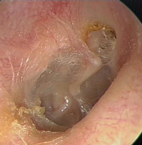
Short (6 cm) rigid endoscopes are very useful for image-capture for patient and parental education, monitoring tympanic membrane retraction, and for careful preoperative planning. Insufflation adapters allow endoscopic assessment of adherence of retracted areas ( Fig. 1 , [CR] ). The clearer optics of the 4 mm scope are preferable for most children, though the 2.7 mm scope is occasionally necessary for the narrower meatus of young children. Although angled endoscopes can help to visualize the depths of a retraction pocket, in practice it can be difficult to get the endoscope sufficiently close to a child’s tympanic membrane to be able to see deeply into a pocket. Any skin contact of the endoscope within the bony meatus is painful and likely to preclude further cooperation.
Intraoperative Inspection
The rigid endoscope is used by many otologists to inspect the retrotympanum or other hidden areas after microscope-guided dissection of cholesteatoma. This intervention has the potential to lower residual disease rates significantly. For thorough inspection, curved suckers must be used to clear blood from these areas.
Tube Insertion
Endoscopy provides excellent visualization for tube insertion, which is helpful for teaching and in settings in which a microscope is not available ( [CR] ). It is not practical in the youngest or syndromic children with narrow ear canals.
Tympanotomy
Elevation of a tympanomeatal flap and insertion of an angled endoscope provides a good view of the middle ear cleft and under the ossicular chain for assessment of conductive hearing loss and residual cholesteatoma. The availability of thin, angled endoscopes (eg, 1.7 mm 30°) makes it possible to achieve limited inspection of the middle ear cleft though a myringotomy.
Tympanoplasty and Cholesteatoma
The full range of tympanoplasty techniques is used in pediatric tympanoplasty. A simple patch myringoplasty or a butterfly graft is ideal for tympanostomy tube site perforations. Underlay or lateral onlay techniques can both be completed endoscopically for larger pediatric perforations. The endoscope is especially beneficial in surgery for tympanic membrane atelectasis because direct visualization for elevation of the retraction from the retrotympanum is provided.
As foreseen by Tarabichi, the endoscopic approach remains most valuable in tympanoplasty and cholesteatoma surgery (see later discussion).
Other
Petrous apex lesions and perilymph leak are rare in children but can be approached endoscopically. Endoscopic visualization of the Eustachian tube and cochlea may prove valuable in future.
Case selection
As in all aspects of surgery, good results can only be anticipated from appropriate case selection. It can be hard to determine preoperatively whether an entirely endoscopic approach will be possible, or even appropriate, in any given pediatric patient. Therefore, it is important to obtain consent for an external incision in case it is needed. Several variables influence the likelihood of a successful, totally endoscopic approach.
Morphology of the External Auditory Meatus
The width, length, and tortuosity of the ear canal, particularly the bony portion, determine the feasibility of endoscopic access. A meatus narrower than the 4.5 mm speculum is likely to resist an endoscopic permeatal approach. The bony meatus is narrower in young children, restricting access for instruments alongside the endoscope; however, it is also shorter, which increases the angulation and range of movement achievable. A deep anterior recess or prominent hump on floor of meatus can significantly impede access, though bone can be removed with curettage to improve access if necessary. Piezoelectric bone removal may prove effective (see the article by Badr-El-Dine and colleagues elsewhere in this issue), but drilling of bone is limited by the challenges of clearing irrigation fluid and spray. As shown in ( Fig. 2 ), individual configuration of these proportions is more important than age in determining access and totally endoscopic middle ear surgery can be completed successfully even in infancy. The author’s personal experience includes, for example, the identification and successful patching of a traumatic round window perilymph leak in a two-and-one-half year old child totally endoscopically ( [CR] ).


