Fig. 7.1
The fourth-week embryo
Fig. 7.2
The pharyngeal arches and clefts
Fig. 7.3
Frontal view of the embryo at the beginning of fourth week
Fig. 7.4
Frontal view of the embryo at the end of fourth week
7.1.3.2 Fifth Week
In the fifth embryonic week, the nasal placodes become hollow and form nasal pits. The inner and outer edges of the nasal pit are called the medial and lateral nasal prominences, respectively. As these two prominences grow, the nasal pits become deeper (Fig. 7.5) and develop into the nasal sac, which ultimately becomes the nasal cavity (Fig. 7.6). Subsequently, the maxillary prominences grow medially and compress the medial nasal prominences. The cleft between the medial nasal prominence and the maxillary prominence disappears, and the medial nasal prominences fuse at the midline to form the intermaxillary segment.
Fig. 7.5
Frontal view of the embryo in the fifth week
Fig. 7.6
Sagittal section of the embryo in the fifth week
7.1.3.3 Sixth Week
At the beginning of the sixth week, the maxillary prominences and the lateral nasal prominences are separated by the nasolacrimal groove. The surface ectodermal layers of these two prominences fuse, forming a groove, and subsequently, a canal known as the nasolacrimal duct. The upper end of the nasolacrimal duct widens to form the lacrimal sac. At the deeper layer, the primary palate is derived from the intermaxillary segment (Fig. 7.7). The dorsal region of the frontonasal prominence grows longitudinally, later becoming the nasal septum. Also, the lateral palatine process starts to expand (Fig. 7.8).
Fig. 7.7
Upward view of the palate at the beginning of sixth week
Fig. 7.8
Upward view of the palate at the end of sixth week
7.1.3.4 Seventh Week
During the fifth and sixth embryonic weeks, the nasal pits invaginate deeper into the underlying mesenchyme. The oronasal membrane initially separates the pits from the oral cavity (Fig. 7.9). Subsequently, in the seventh week, the membrane is penetrated and the primitive choanae are formed. At the end of seventh week, the primitive choanae divide the nasal cavity and the pharynx, and the nasal meatus plug closes the external nares. Distinct areas of the face develop from each of the facial prominences: the forehead and dorsum, apex, and nose root from the frontonasal prominence, the nasal wing from the lateral nasal prominence, the nasal septum and philtrum from the medial nasal prominence, the top half of the cheek and upper lip from the maxillary prominence, and the bottom half of the cheek, lower lip, and chin from the mandibular prominence. At this time, the nasal cavity is covered by undifferentiated stratified cuboidal epithelium (Fig. 7.10).
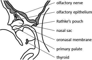
Fig. 7.9
Sagittal section of the embryo in the seventh week
Fig. 7.10
Frontal view of the embryo in the eighth week
7.1.4 Fetal Period
7.1.4.1 Eighth Week
The nasal sac, a cartilaginous tissue that spans from the nasal septal area and the ethmoid bone to the inferior nasal conchae, is the origin of the supportive tissues of the nasal and paranasal sinuses. The nasal sac extends to the sphenoid bone at posterior part of the nasal cavity (Fig. 7.11). From the lateral walls, three pairs of nasal conchae form the preturbinates. During this time period, the majority of the nasal cavity epithelium consists of undifferentiated or nonciliated cells. However, on the roof of the nasal cavity, ectodermal epithelium differentiates into olfactory epithelium. Subsequently, the olfactory epithelium differentiates into olfactory cells and supporting cells, and the olfactory nerve fiber is formed.
Fig. 7.11
Coronal section of the embryo in the eighth week
7.1.4.2 Ninth to Tenth Week
During the ninth week, the bilateral maxillary prominences form shelf-like growths called lateral palatine processes, which fuse at the midline and produce the secondary palate (Figs. 7.12 and 7.13). The secondary palate fuses with the primary palate, and the formation of the palate is finally completed. The nasal septum fuses with the palate (Figs. 7.14 and 7.15). Ciliated epithelial cells appear in the nasal septum and the inferior nasal conchae, and angiogenesis occurs in lamina propria. Thus, the formation of the fetal face is almost complete by the end of the tenth embryonic week (Fig. 7.16).
Fig. 7.12
Sagittal section of the embryo in the ninth week
Fig. 7.13
Upward view of palate at the end of tenth week
Fig. 7.14
Upward view of palate at the end of the twentieth week
Fig. 7.15
Coronal section of embryo in the twentieth week
Fig. 7.16
Frontal view of the embryo in the ninth week
7.1.4.3 Eleventh to Fourteenth Week
In the eleventh and twelfth embryonic weeks, the lateral nasal wall, which is anterior to the middle nasal concha, retracts and forms a space known as the ethmoidal infundibulum. The epithelium at the lower part of the ethmoidal infundibulum migrates toward the maxillary bone and initiates construction of the maxillary sinus. In the thirteenth and fourteenth weeks, the bony structure of the maxilla expands and replaces the cartilaginous tissue of the nasal sac, and the inferior part of the lateral wall is formed. Meanwhile, in the eleventh and twelfth weeks, the process of nasal bone ossification commences and the superior part of the lateral wall is formed. Also, posterior ethmoidal air cells are generated behind the superior nasal meatus. Ossification of the vomer starts in the twelfth week with continued growth until adulthood.
By the thirteenth week, the nasal septum is abundantly covered in ciliary epithelium cells. Goblet cells are present between the ciliary cells, and nasal glands are detectable in the submucosal layer. However, the respiratory epithelial cells of the lateral wall are not yet differentiated, with only a small number of immature cells detectable. Ethmoidal air cells are also covered by undifferentiated respiratory epithelium.
7.1.4.4 Fifteenth to Nineteenth Week
Before the start of the fifteenth week, formation of the superior, middle and inferior nasal conchae is complete, and ossification has started from the inferior nasal conchae. In addition, completion of the nasolacrimal duct occurs in the fifteenth week. In the seventeenth to eighteenth weeks, the anlage of the sphenoid sinus appears at the end of the nasal cavity.
The lining of the nasal cavity contains many ciliated epithelial cells and goblet cells. The lamina propria becomes thicker and more highly vascularized. At the roof of the nasal cavity, formation of the cribriform plate and bundles of olfactory nerve fibers is evident. At this time, all nasal mucosal tissue above the superior nasal concha is olfactory epithelium. At the anterior part of the nasal cavity, the nasal vestibular develops. The border between respiratory epithelial cells, and stratified squamous epithelium is defined, and nasal hair follicles are observed.
7.1.4.5 After the Twentieth Week
From the twentieth to the twenty-second week, ossification takes place around the nasolacrimal duct, and the border with the ethmoidal infundibulum becomes clear. Ossification of the nasal conchae rapidly progresses from the lateral wall side. The superior and middle nasal conchae develop as part of the ethmoidal bone, while the inferior nasal conchae form an independent bone. Development of the uncinate process and ethmoidal bulla result in formation of the hiatus semilunaris. Ossification of the maxilla also progresses from the twentieth week onward. Formation of the characteristic structures of the maxillary bone, including the alveolar process, palatine process, inferior orbit wall, zygomatic process, and frontal process, occurs before the twentieth week. Substantial development of the maxillary sinus results in sinus aeration after the twentieth week. Furthermore, ossification of the lateral part of the nasal sac starts and form orbital plate of ethmoid and walls of ethmoidal cells. Histologically, with the exception of the posterior ethmoidal cells, epithelial differentiation of all the paranasal sinuses is complete. The bony structure of the external nose consists of the nasal bone, the frontal process of the maxillary bone, and the nasal part of frontal bone. Ossification of the maxillary bone starts from the canine fossa in the sixth week, extending to the edge of the nasal capsule, and delineating the border zones of the external nose, the lateral wall of the nasal cavity, and the ethmoidal cells. Differentiation of the respiratory epithelium is near completion before the twenty-fourth week.
7.1.4.6 Sixth Month
By the sixth month, the structures of the nose are almost established. After the closure of the external nares by the nasal meatal plug during the eighth embryonic week, the external nares are now recanalized. Formation of the greater alar cartilage and the lateral nasal cartilages are completed; therefore, the appearance of the nose is quite similar to that of an infant (Fig. 7.17).
Fig. 7.17
Frontal view of the embryo in the fourteenth week
7.1.5 Postnatal Growth
7.1.5.1 External Nose
At birth, the lateral and longitudinal diameters of the external nares are 7–8 mm and 5–7 mm, respectively, and they double in size within 6 months. The elliptical shape of each external naris changes from being laterally longer at birth to being longitudinally longer at the time of puberty (Fig. 7.18).
Fig. 7.18
Bony structure of the external nose
7.1.5.2 Paranasal Sinuses
The maxillary sinus has a lateral diameter of 7–8 mm at birth, and it is located slightly lateral to the inferior nasal concha. Initially, the maxillary sinus expands laterally and reaches the position of the infraorbital nerve within a year. It then expands downward, extending to the middle level of the inferior nasal meatus at 7 years, the level of the nasal base at 12 years, with cessation of growth at 17 years of age.
Ethmoid cellulae are detectable as mucosal invaginations at the sixteenth embryonic week. Each ethmoid cell progressively swells, resulting in a compact structure at 2 years of age. Rapid growth continues until the age of 6 years, and the basic structure of the maxillary sinus is mature at this time point.
Development of the frontal sinus is slower than that of the other sinuses. Aeration is detectable at 1 year of age, extending to the level of superior orbit wall at the age of 8, with growth completed during adolescence. The size of the frontal sinus shows greater interindividual variation compared with the other sinuses.
Stay updated, free articles. Join our Telegram channel

Full access? Get Clinical Tree


