Deep neck infections present significant morbidity and mortality, particularly when associated with predisposing factors that impair a functional immunologic response. Familiarity with deep neck spaces and fascial planes is critical, because these form the basis for the emergent nature of the disease process. Common and potentially life-threatening complications include airway obstruction, jugular vein thrombosis, descending mediastinitis, sepsis, acute respiratory distress syndrome, and disseminated intravascular coagulation. The most common primary sources of deep neck infection are odontogenic, tonsillar, salivary gland, foreign body, and malignancy. Microbiology typically reveals mixed bacterial flora, including anaerobic species, that can rapidly progress to a fulminating necrotizing fasciitis. The treatment cornerstone remains securing the airway, providing efficient drainage and appropriate antibiotics, and improving immunologic status. A prolonged hospital stay should be anticipated.
Deep neck infections (DNIs) are unique among infectious diseases for their versatility and potential for severe complications. Complex head and neck anatomy often makes early recognition of DNIs challenging, and a high index of suspicion is necessary to avoid any delay in treatment. Aggressive monitoring and management of the airway is the most urgent and critical aspect of care, followed by appropriate antibiotic coverage and surgical drainage, when needed.
Since the discovery of penicillin, the widespread use of antibiotics has dramatically reduced the overall incidence of DNIs . More recent trends include the increasing prevalence of resistant bacterial strains, a decline in the number of DNIs caused by pharyngitis or tonsillitis, and a relative increase in cases related to odontogenic infection . In addition, a growing number of patients who have immune dysfunction, as in diabetes mellitus and HIV infection, are at risk for atypical and more complicated cases of DNI. Low socioeconomic status and poor oral hygiene have been associated with higher rates of odontogenic infections, including Ludwig’s angina .
Relevant anatomy
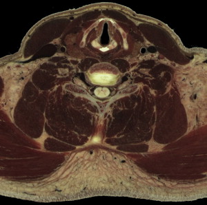
Multiple layers of cervical fascia encase the contents of the neck and form the potential head and neck spaces. These fascial planes constitute important anatomic limitations for the spread of infection and serve to direct infectious spread once their natural resistance is overcome. Therefore, anatomic considerations are paramount in dealing with DNIs, especially when planning treatment strategies and guarding against potential complications. Figs. 1 and 2 present fascial plane anatomic features.

Cervical fascial planes
The superficial cervical fascia underlies the skin of the head and neck in a continuous plane and covers adipose tissue, sensory nerves, superficial vessels (including the external jugular vein), lymphatics, the platysma muscle, and the muscles of facial expression ( Fig. 3 A). It extends from the superior aspect of the head down to the shoulders, axilla, and thorax, and includes the superficial musculoaponeurotic system. Although the area contained within this fascial plane is not considered a deep neck space, it may serve as an additional barrier for containing edema and pressure caused by infections in the underlying muscular and visceral compartments of the neck. Superficial space infections usually take the form of cellulitis, but can cause an obvious abscess with localized fluctuance, erythema, warmth, and tenderness beneath the skin. A superficial abscess can be drained with a transverse incision along Langer’s lines over the area of prominence, and cellulitis and abscess should be managed with appropriate antibiotic therapy .

The deep cervical fascia is divided into three layers (superficial, middle, and deep) that envelop the contents of the head and neck and form the potential deep neck spaces. The superficial layer of deep cervical fascia surrounds the neck as it courses from the posterior spinous processes of the vertebrae to its anterior insertions into the sternum, hyoid, mandible, and zygomatic arches ( Fig. 3 B). It follows a “rule of two’s,” splitting to envelop two muscles that cross the neck (trapezius and sternocleidomastoid [SCM]), two muscles above the hyoid bone (anterior belly of the digastric and masseter), two salivary glands (submandibular and parotid), and two fascial compartments (parotid and masticator spaces). Just deep to the SCM, it contributes to the lateral aspect of the carotid sheath. The anterior-superior aspect of the superficial layer of deep cervical fascia forms the floor of the submandibular space as it covers the anterior belly of the digastric and mylohyoid muscles .
The middle layer of deep cervical fascia encloses the anterior contents of the neck and has two divisions. The muscular division surrounds the infrahyoid strap muscles (sternothyroid, sternohyoid, and thyrohyoid) and extends from the hyoid bone down to the sternum, clavicle, and scapulae ( Fig. 3 C). The visceral division envelops the trachea, larynx, pharynx, esophagus, and thyroid gland that lie posterior to the strap muscles, and extends from the pharyngeal constrictor muscles and hyoid bone down into the anterior mediastinum overlying the fibrous pericardium and great vessels ( Fig. 3 D). The posterior-superior aspect of the middle layer of deep cervical fascia, also known as the buccopharyngeal fascia, courses around the posterior pharynx and forms the anterior wall of the retropharyngeal space. The muscular and visceral divisions of the middle layer of deep cervical fascia contribute to the anteromedial aspect of the carotid sheath .
The deep layer of deep cervical fascia encloses the posterior contents of the neck. It originates posteriorly along the vertebral spinous processes and courses anteriorly beneath the trapezius muscle while surrounding the deep neck musculature and vertebral column ( Fig. 3 E). The deep layer of deep cervical fascia divides as it reaches the longus colli muscle and anterior aspect of the vertebral bodies to form the prevertebral fascia and the alar fascia. Anteriorly, the alar fascia forms the posterior wall of the retropharyngeal space that extends from the base of the skull down to the level of the second thoracic vertebra. The alar fascia also serves as the anterior boundary of the danger space, which extends downward into the posterior mediastinum to the level of the diaphragm. Posteriorly, the prevertebral fascia adheres directly to the vertebral bodies and cervical muscles and serves as the posterior wall of the danger space. The prevertebral space lies between the vertebral bodies and prevertebral fascia and extends from the skull base down to the coccyx. In addition to surrounding the deep neck musculature, the prevertebral fascia envelops the brachial plexus, phrenic nerve, vertebral vessels, and subclavian vessels inferiorly and eventually gives rise to the axillary sheath. The alar and prevertebral divisions of the deep layer of deep cervical fascia contribute to the posterior aspect of the carotid sheath .
The carotid sheath receives connective tissue contributions from all three layers of deep cervical fascia, and yet it remains anatomically independent as a barrier against the spread of infection into the carotid space from the adjacent deep neck compartments, of which the parapharyngeal space is the most intimately associated ( Fig. 3 F). The carotid artery, internal jugular vein, cervical sympathetic chain, and cranial nerves IX, X, XI, and XII are all protected by the carotid sheath.
Cervical fascial planes
The superficial cervical fascia underlies the skin of the head and neck in a continuous plane and covers adipose tissue, sensory nerves, superficial vessels (including the external jugular vein), lymphatics, the platysma muscle, and the muscles of facial expression ( Fig. 3 A). It extends from the superior aspect of the head down to the shoulders, axilla, and thorax, and includes the superficial musculoaponeurotic system. Although the area contained within this fascial plane is not considered a deep neck space, it may serve as an additional barrier for containing edema and pressure caused by infections in the underlying muscular and visceral compartments of the neck. Superficial space infections usually take the form of cellulitis, but can cause an obvious abscess with localized fluctuance, erythema, warmth, and tenderness beneath the skin. A superficial abscess can be drained with a transverse incision along Langer’s lines over the area of prominence, and cellulitis and abscess should be managed with appropriate antibiotic therapy .
The deep cervical fascia is divided into three layers (superficial, middle, and deep) that envelop the contents of the head and neck and form the potential deep neck spaces. The superficial layer of deep cervical fascia surrounds the neck as it courses from the posterior spinous processes of the vertebrae to its anterior insertions into the sternum, hyoid, mandible, and zygomatic arches ( Fig. 3 B). It follows a “rule of two’s,” splitting to envelop two muscles that cross the neck (trapezius and sternocleidomastoid [SCM]), two muscles above the hyoid bone (anterior belly of the digastric and masseter), two salivary glands (submandibular and parotid), and two fascial compartments (parotid and masticator spaces). Just deep to the SCM, it contributes to the lateral aspect of the carotid sheath. The anterior-superior aspect of the superficial layer of deep cervical fascia forms the floor of the submandibular space as it covers the anterior belly of the digastric and mylohyoid muscles .
The middle layer of deep cervical fascia encloses the anterior contents of the neck and has two divisions. The muscular division surrounds the infrahyoid strap muscles (sternothyroid, sternohyoid, and thyrohyoid) and extends from the hyoid bone down to the sternum, clavicle, and scapulae ( Fig. 3 C). The visceral division envelops the trachea, larynx, pharynx, esophagus, and thyroid gland that lie posterior to the strap muscles, and extends from the pharyngeal constrictor muscles and hyoid bone down into the anterior mediastinum overlying the fibrous pericardium and great vessels ( Fig. 3 D). The posterior-superior aspect of the middle layer of deep cervical fascia, also known as the buccopharyngeal fascia, courses around the posterior pharynx and forms the anterior wall of the retropharyngeal space. The muscular and visceral divisions of the middle layer of deep cervical fascia contribute to the anteromedial aspect of the carotid sheath .
The deep layer of deep cervical fascia encloses the posterior contents of the neck. It originates posteriorly along the vertebral spinous processes and courses anteriorly beneath the trapezius muscle while surrounding the deep neck musculature and vertebral column ( Fig. 3 E). The deep layer of deep cervical fascia divides as it reaches the longus colli muscle and anterior aspect of the vertebral bodies to form the prevertebral fascia and the alar fascia. Anteriorly, the alar fascia forms the posterior wall of the retropharyngeal space that extends from the base of the skull down to the level of the second thoracic vertebra. The alar fascia also serves as the anterior boundary of the danger space, which extends downward into the posterior mediastinum to the level of the diaphragm. Posteriorly, the prevertebral fascia adheres directly to the vertebral bodies and cervical muscles and serves as the posterior wall of the danger space. The prevertebral space lies between the vertebral bodies and prevertebral fascia and extends from the skull base down to the coccyx. In addition to surrounding the deep neck musculature, the prevertebral fascia envelops the brachial plexus, phrenic nerve, vertebral vessels, and subclavian vessels inferiorly and eventually gives rise to the axillary sheath. The alar and prevertebral divisions of the deep layer of deep cervical fascia contribute to the posterior aspect of the carotid sheath .
The carotid sheath receives connective tissue contributions from all three layers of deep cervical fascia, and yet it remains anatomically independent as a barrier against the spread of infection into the carotid space from the adjacent deep neck compartments, of which the parapharyngeal space is the most intimately associated ( Fig. 3 F). The carotid artery, internal jugular vein, cervical sympathetic chain, and cranial nerves IX, X, XI, and XII are all protected by the carotid sheath.
Deep neck spaces
At least 11 deep neck spaces lie within the complex framework formed by the cervical fascial planes and they functionally contain DNIs as long as their natural points of resistance along communicating fascial boundaries are not overcome. Anteriorly, strong fascial attachments to the hyoid bone are an important barrier to downward infectious spread. As a result, the deep neck spaces are often classified into three anatomic groups, based on their relation to the hyoid:
- 1.
Those located above the level of the hyoid (peritonsillar, submandibular, parapharyngeal, masticator/temporal, buccal, and parotid spaces)
- 2.
Those that involve the entire length of the neck (retropharyngeal, danger, prevertebral, and carotid spaces)
- 3.
The anterior visceral, or pretracheal, space located below the hyoid
Spaces above the level of the hyoid
Peritonsillar space
The peritonsillar space lies between the palatine tonsil and the adjacent superior pharyngeal constrictor muscle and is bounded anteriorly and posteriorly by the tonsillar pillars. It contains loose connective tissue, and is not generally considered a “deep” neck space. However, the peritonsillar space is anatomically contiguous with several deeper spaces, and peritonsillar infections can potentially involve the parapharyngeal and retropharyngeal spaces. The presence of severe trismus suggests parapharyngeal space and medial pterygoid muscle involvement. Infections limited to the peritonsillar space often present with fever, sore throat, dysphagia, odynophagia, a muffled “hot potato” voice, and cervical adenopathy. Examination of the oropharynx may reveal tonsillar edema, visible exudates, bulging of the superior pole of the tonsillar pillar, and deviation of the uvula away from the side of the infection .
Infections of the peritonsillar space arise from tonsillitis and are usually seen in older children . Diagnosis of a peritonsillar infection is often based on clinical presentation and careful examination of the oropharynx. Less complicated infections without an obvious abscess or airway compromise are best treated with an initial course of intravenous antibiotics for 12 to 24 hours. However, it is important to distinguish cellulitis from a drainable abscess, which can be accomplished by attempting intraoral needle aspiration in cooperative patients or by using intraoral ultrasound. A contrast-enhanced computed tomography (CECT) scan is required when attempts fail to obtain an aspirate, in the presence of trismus, in patients who do not improve with initial antibiotic therapy, and when extension into the deep neck spaces is suspected, to evaluate the need for more extensive drainage procedures. Because of a higher recurrence rate, adults with peritonsillar infections generally should be treated subsequently with tonsillectomy .
Submandibular space
The submandibular space, as it is often collectively referred to, is composed of two potential spaces that span from the mucosal covering of the floor of the mouth down to the superficial layer of deep cervical fascia as it encloses the space between the mandible and the hyoid bone. The mylohyoid muscle traverses the space horizontally and divides it into a supramylohyoid compartment, also known as the sublingual space, and an inframylohyoid compartment, also known as either the anatomically correct submandibular or submaxillary space. In order to avoid confusion, the term “submandibular space” will henceforth refer to the combined inframylohyoid and supramylohyoid compartments. These two compartments communicate freely along the posterior aspect of the mylohyoid muscle , and infections have been noted to spread to the parapharyngeal space posteriorly and the anterior visceral space inferiorly . The roots of the second and third mandibular molars lie below the attachment of the mylohyoid to the mandible, and posterior dental infections can spread directly into the inframylohyoid compartment. In contrast, the remaining mandibular dental roots lie above the mylohyoid line, and more anterior dental infections spread directly into the supramylohyoid compartment. Other sources of infection include sialadenitis, suppurative lymphadenitis, oral trauma, and upper respiratory infections . Aspirates from submandibular space infections reveal a prevalence of odontogenic pathogens, including aerobic Streptococcus viridans and Staphylococci and anaerobic Prevotella and Peptostreptococcus spp. .
The supramylohyoid compartment (sublingual space) contains loose areolar tissue, the sublingual glands, the submandibular (Wharton’s) duct, geniohyoid muscles and the lingual and hypoglossal nerves. Supramylohyoid infections often present with induration, swelling, and tenderness in the floor of the mouth that often begins laterally. Protrusion and elevation of the tongue can occur as the swelling proceeds medially.
The inframylohyoid compartment (submaxillary space) contains the anterior bellies of the digastric muscles, submandibular glands, and lymph nodes. The “mental space” refers to the portion of the inframylohyoid compartment confined between the anterior bellies of the digastric muscles. Inframylohyoid infections cause induration, swelling, and tenderness below the mandible and can progress to elevation and protrusion of the tongue. Infection within this compartment requires an extraoral surgical approach .
Ludwig’s angina
The term Ludwig’s angina is derived from the Latin term “angere,” or “to strangle” . Ludwig’s angina is diagnosed clinically based on the classic description of a “rapidly spreading, firmly indurated cellulitis that originates intraorally and involves [supramylohyoid and inframylohyoid compartments] bilaterally, but without abscess or lymphadenopathy,” resulting in rapidly progressing upper airway obstruction . Most cases are of odontogenic origin; less common causes include peritonsillar or parapharyngeal abscesses, epiglottitis, and penetrating injuries to the floor of the mouth. Patients such as diabetics with impaired immune function are at increased risk for developing Ludwig’s angina secondary to submandibular space infections . The infectious process typically involves cellulitis within the submandibular space, which can progress into fasciitis and eventually form a true abscess . Swelling within the enclosed submandibular space displaces the tongue superiorly and posteriorly and causes odynophagia, dysphagia, and drooling. Tongue protrusion can potentially culminate in rapidly progressive airway obstruction and asphyxiation if the airway is not urgently addressed. Fiber optic nasotracheal intubation may afford the best initial approach to airway management and should be attempted with the patient sitting up or in the semi-Fowler position, rather than supine . Tracheotomy is the standard approach in Ludwig’s angina patients who fail intubation.
The definitive treatment of Ludwig’s angina involves early airway protection and a combination of broad-spectrum antibiotics and surgical decompression of a firmly indurated submandibular space. In addition to airway compromise, Ludwig’s angina may lead to complications such as mediastinitis, osteomyelitis of the mandible, pleural effusion, empyema, and infection of carotid sheath structures . With aggressive airway management, intravenous antibiotics, and surgical decompression, the mortality of Ludwig’s angina has been reduced to less than 10% .
Parapharyngeal space
The parapharyngeal space, also known as the lateral pharyngeal or pharyngomaxillary space, takes the form of an inverted pyramid extending from the base of the skull down to the hyoid bone. It lies between the visceral division (pretracheal and buccopharyngeal fascia) of the middle layer of deep cervical fascia medially and the superficial layer of deep cervical fascia covering the pterygoid muscles and parotid gland laterally. The parapharyngeal space is divided into two compartments by the styloid process and the stylohyoid, stylopharyngeus, and styloglossus muscles. The prestyloid compartment lies anteriorly and contains the internal maxillary artery, maxillary nerve, and adipose tissue, and the poststyloid compartment lies posteriorly and includes the neurovascular contents of the carotid sheath (carotid artery, internal jugular vein, cervical sympathetic chain, and cranial nerves IX, X, XI, and XII) .
Parapharyngeal infections have many sources because of the shear number of neighboring deep neck compartments, which include the submandibular, retropharyngeal, parotid, and masticator spaces. Common causes include pharyngitis, tonsillitis, otitis, mastoiditis, parotitis, and cervical lymphadenitis. Odontogenic infections also contribute by way of indirect spread from adjacent deep neck spaces . Infection of the prestyloid compartment often presents clinically with fever, chills, neck pain, trismus, and anteromedial displacement of the ipsilateral palatine tonsil. Infection of the poststyloid compartment has been known to cause little or no pain, trismus, or obvious swelling; however, involvement of the carotid sheath contents can lead to complications such as septicemia, Lemierre’s syndrome an infective internal jugular vein thrombosis (IJVT), or, carotid artery aneurysm or rupture, ipsilateral Horner’s syndrome, and cranial nerve IX through XII palsies. Laryngeal edema with subsequent airway obstruction is another potential sequela and may necessitate intubation or tracheotomy .
The management of parapharyngeal space infections must begin with appropriate imaging to determine if an abscess is present, the extent of its spread, and its proximity and relation to the carotid sheath contents. CECT of the head and neck, in combination with careful physical examination, have been shown to differentiate deep neck abscess from cellulitis most accurately . Sichel and colleagues found that infections involving the poststyloid compartment, but not the prestyloid compartment, were much more frequent in children and were likely the result of acute cervical lymphadenitis, a relatively benign condition that should respond well to an initial 48 hours of intravenous antibiotics. On the other hand, infections involving the prestyloid compartment were common to all ages and were principally a result of dental and pharyngeal infections. Prestyloid infections caused more complications because of suppuration of the prestyloid adipose tissue and rapid diffusion of pus into other deep neck spaces, including the carotid space. The investigators concluded that prestyloid infections must be differentiated from poststyloid cervical lymphadenitis, and that abscesses within the prestyloid compartment require prompt surgical drainage to avoid complications from the rapid spread of pus into adjacent deep neck spaces.
Surgical access of the parapharyngeal space has traditionally been by way of an external cervical approach, with an incision along the anterior-superior border of the SCM muscle to gain adequate exposure and avoid injuring the carotid sheath contents. More recently, however, CECT has been used to identify accurately the parapharyngeal abscesses contained within the prestyloid compartment (anterior and medial to the carotid sheath) and has allowed for a less invasive transoral drainage approach to prestyloid infections. A study by Nagy and colleagues demonstrated that CECT scans often reveal a bulge of the ipsilateral pharyngeal wall and tonsil when an abscess involves the prestyloid compartment of the parapharyngeal space, and that the bulge can serve as a landmark to guide intraoral incision and drainage in the pediatric population. In a more recent study, Cable and colleagues addressed concerns regarding the imprecision and prolonged length of intraoral prestyloid drainage procedures in children with the application of CT image-guided surgery systems, which allow for immediate imaging to confirm that no abscess cavity has been missed and obviate the need for dangerous surgical exploration. Abscesses that spread into adjacent deep neck spaces and those that involve the poststyloid compartment require an external cervical approach to incision and drainage.
Masticator/temporal space
The masticator space lies between the medial pterygoid muscle and the more lateral masseter muscle, and is enclosed by the divisions of the superficial layer of deep cervical fascia that envelop these muscles. It extends back to the posterior aspect of the mandible and superiorly as the temporal space, to surround the temporalis muscle deep to the temporalis fascia. Its contents include the temporalis muscle, the ramus of the mandible, divisions of the mandibular nerve (V 3 ), and the internal maxillary artery. Most masticator space infections originate from the posterior mandibular molars; less common sources include trauma and surgery. Patients often present with severe trismus, sore throat, dysphagia, pain surrounding the ramus of the mandible, and preauricular or mandibular swelling. More extensive infections can cause swelling of the entire side of the face, and involvement of the orbit may lead to proptosis, optic neuritis, and cranial nerve VI palsy. The surgical approach to incision and drainage of the masticator space depends on the location of the abscess in relation to the mandible. An intraoral approach at the retromolar trigone is appropriate for draining abscesses medial to the ramus of the mandible, and an extraoral approach along the inferior border of the mandible is used for draining abscesses lateral to the mandibular ramus. An incision through the temporalis fascia along the hairline is necessary to drain abscesses that spread superiorly and surround the temporalis muscle within the temporal space .
Buccal space
The buccal space lies between the buccopharyngeal fascia overlying the buccinator muscle medially and the skin of the cheek laterally, and is limited inferiorly by the border of the mandible and posteriorly by the pterygomandibular raphe. It contains the buccal fat pad, the parotid duct, and the facial artery. Most buccal space infections are odontogenic in origin and present with a warm and tender swelling within the cheek and minimal systemic symptoms. Trismus may be present if the infection spreads posteriorly to involve the masseter muscle .
Parotid space
The parotid space exists within the capsule that is formed by the superficial layer of deep cervical fascia as it envelops the parotid gland. The fascia on the medial aspect of the gland is thin and provides little resistance to the spread of parotid space infections into the adjacent parapharyngeal space. In addition to the parotid gland, the space also contains the facial nerve, external carotid artery, retromandibular vein, auriculotemporal nerve, superficial temporal artery, and lymph nodes. Parotid space infections often result from parotid duct obstruction or suppurative lymphadenitis, and occasionally originate from odontogenic infections of the mandibular molars that traverse the masticator space. Afflicted patients usually present with severe pain and swelling at the angle of the mandible, but little or no trismus unless the masticator space is sufficiently involved. Systemic symptoms such as fever and chills may accompany the spread of infection into the parapharyngeal space and other deep neck spaces. An external, parotidectomy-like approach is used to drain a parotid space abscess, and blunt dissection, either superior or inferior to the posterior belly of the digastric muscle, allows concurrent drainage of the parapharyngeal space when it is involved .
Spaces that involve the entire length of the neck
Retropharyngeal space
The retropharyngeal (retrovisceral, retroesophageal) space lies between the visceral (buccopharyngeal) fascia covering the posterior pharynx and esophagus and the alar fascia, a division of the deep layer of deep cervical fascia, and occupies the space posterior to the pharynx and esophagus. It extends from the base of the skull down into the mediastinum, where the visceral and alar fascias fuse at the level of the second thoracic vertebra. Laterally, it is bounded by the carotid sheaths. The retropharyngeal space is fused down the midline and contains two chains of lymph nodes extending down each side. Retropharyngeal abscesses are unilateral as a result of the midline fusion, and are primarily seen early in childhood because these lymph nodes tend to regress with age. Upper respiratory infections cause most retropharyngeal disease in children because these lymph nodes receive drainage from the nose, sinuses, and pharynx. Retropharyngeal infection is also seen in children and adults following trauma to the posterior pharynx or as an extension from an adjacent parapharyngeal space infection . Cultures of retropharyngeal aspirates are generally polymicrobial and contain common oropharyngeal flora such as S viridans , with the exception that young children are more likely to be infected with pathogenic Streptococcus species and Staphylococcus aureus .
Children with retropharyngeal infections often present with neck pain, neck swelling, fever, irritability, dysphagia, excessive drooling, and dyspnea or noisy breathing suggestive of upper airway compromise . Adults tend to present with neck pain, fever, anorexia, nasal obstruction, snoring, and dyspnea . Careful oropharyngeal examination often reveals unilateral bulging of the posterior pharynx, which is localized in retropharyngeal lymphadenitis and may extend the length of the pharynx when cellulitis or an abscess is present. In the absence of acute distress, CECT of the head and neck can be used to confirm the diagnosis and evaluate for spread of infection into adjacent deep neck spaces .
The most feared complications of retropharyngeal infection are airway obstruction and rupture of an abscess with subsequent aspiration of pus. Patients who have signs of airway compromise should be taken immediately to the operating room before being examined. With the patient in the head-down Trendelenburg’s position and intubated along the opposite side of the pharynx from the swelling, needle aspiration should precede intraoral incision and drainage, to obtain specimens for culture and sensitivity and to minimize any risk of the patient aspirating pus during the procedure. Spread of the infection into the parapharyngeal space may necessitate an external cervical approach to incision and drainage if the involvement is not confined to the prestyloid compartment medial to the great vessels .
Danger space
The danger space lies posterior to the retropharyngeal space between the alar and prevertebral fascias, the two divisions of the deep layer of deep cervical fascia, and extends from the skull base down into the posterior mediastinum to the level of the diaphragm. Infections here indirectly result from the spread of retropharyngeal, parapharyngeal, and prevertebral space abscesses. The “danger” lies in the tendency for infections to spread inferiorly through the space and into the thorax because the loose areolar contents offer little resistance, resulting in complications such as mediastinitis, empyema, and sepsis. Danger space infections initially present in the same way as retropharyngeal infections, and CECT is necessary to differentiate between them .
Prevertebral space
The prevertebral space is a potential space between the prevertebral fascia and the underlying vertebral bodies and deep cervical musculature. It extends down the entire length of the vertebral column to the coccyx. However, dense fibrous attachments between the prevertebral fascia and deep cervical muscles tend to contain prevertebral infections and help prevent longitudinal spread. Sources of infection include trauma to the posterior pharynx, and secondary spread from Pott’s abscesses and retropharyngeal and danger space infections. Patients often present with a midline bulge in the posterior pharynx, in contrast to the unilateral bulge often seen in retropharyngeal infections. Complications include osteomyelitis and spinal instability, which require a prolonged course of antibiotics. Once identified with CECT, prevertebral space abscesses should be drained using an external cervical approach rather than an intraoral approach, which can lead to a persistent draining fistula in the posterior pharynx .
Carotid space
The carotid (visceral vascular) space lies within the carotid sheath and houses the carotid artery, internal jugular vein, cervical sympathetic chain, and cranial nerves IX, X, XI, and XII. It is similar to the prevertebral space in that it contains little areolar tissue, and thus is somewhat resistant to the longitudinal spread of infection. However, as the well-known expression “Lincoln’s highway” suggests, the carotid sheath receives contributions from all three layers of deep cervical fascia, extends from the base of the skull into the mediastinum, and can potentially serve as a “highway” for infectious spread originating in any deep neck space. In addition to secondary spread from adjacent deep neck spaces, direct inoculation into the neck in intravenous drug abusers and iatrogenic causes such as central venous catheterization can lead to carotid space infections. Patients often present with stiffness and ipsilateral swelling of the neck, fever, chills, ipsilateral Horner’s syndrome, vocal cord paralysis, and other complication-related findings. IJVT can cause intermittent spiking fevers, and carotid artery rupture may be preceded by sentinel bleeds from the ear, nose, or mouth. An external cervical approach is used for incision and drainage of the carotid space .
Space below the level of the hyoid
Anterior visceral space
The anterior visceral (pretracheal) space is the only deep neck space limited to below the hyoid bone. It is bounded by the visceral division of the middle layer of deep cervical fascia and lies between the infrahyoid strap muscles and the esophagus. It contains the thyroid gland, the trachea, and the anterior wall of the esophagus, and extends from the thyroid cartilage down into the superior mediastinum overlying the aortic arch and fibrous pericardium. The anterior visceral and retropharyngeal spaces are separated by lateral attachments of the esophagus to the prevertebral fascia beginning at the level of the thyroid gland, so that the anterior visceral space lies anterior and the retropharyngeal space lies posterior to the esophagus .
Infections of the anterior visceral space often originate from traumatic perforation of the anterior esophageal wall and, less commonly, from neck trauma or thyroiditis. They often present with neck swelling, sore throat, dysphagia, hoarseness, and dyspnea as a result of pharyngeal, laryngeal, and supraglottic edema, which can potentially progress to airway compromise. In addition, the perforation of visceral contents may cause crepitation of the anterior neck, mediastinitis, and pneumothorax . Boscolo-Rizzo and colleagues noted that every infection involving the anterior visceral space in their study developed life-threatening complications, including five out of the six cases of mediastinitis that were encountered. Abscesses in the anterior visceral space require an external cervical approach to incision and drainage .
Stay updated, free articles. Join our Telegram channel

Full access? Get Clinical Tree


