72 Cosmetic Skin Lesion Skin cancer is the most commonly encountered malignancy in the head and neck. Although the vast majority of skin lesions are benign, early detection is the key to ensure good prognosis for potentially metastatic skin cancers. In skin cancers that are less frequently metastatic, early diagnosis and treatment are important to prevent invasion and destruction of underlying critical structures. Clinically distinguishing benign from malignant disease is an important skill for the otolaryngologist. Clinical findings on physical exam and knowledge of important risk factors are often sufficient to make the diagnosis. However, histological diagnosis remains the gold standard, and if any doubt exists about the diagnosis, a biopsy is warranted. In general, these have well-defined edges, are symmetrical, and have uniformly distributed color, with little or no change in characteristics over time. Fig. 72.1 Seborrheic keratosis. Brown, oval sessile plaque with characteristic “stuck on” appearance. (Courtesy of DermAtlas, Johns Hopkins University, www.dermatlas.org.) Fig. 72.2
 Benign Skin Lesions
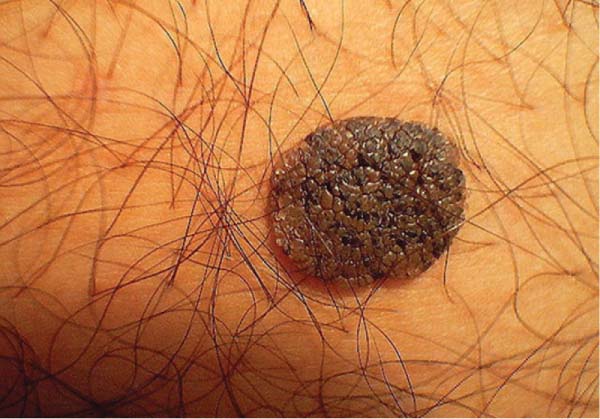
 Seborrheic keratosis (SK): Tan-brown-black color, sharply circumscribed, round/oval shape. Can be flat or minimally raised and are often scaly. A key characteristic is their “stuck-on” appearance. They most commonly appear on the face, trunk, and upper extremities and are more common with increasing age. SK has no malignant potential (Fig. 72.1).
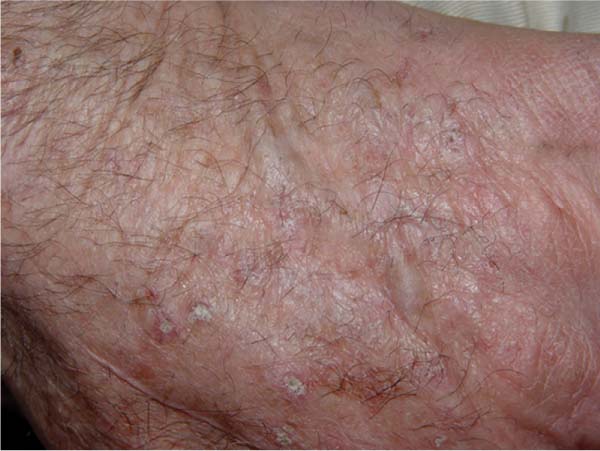
 Actinic keratosis (AK): Yellow-brown scaly patches found most often on sun-exposed skin. A key characteristic is their rough “sandpaper” like texture, often appreciated on light palpation rather than clinical examination. AKs occur on exposed surfaces, most commonly the face, ears, dorsum of the hands, and legs. More commonly seen in middle-aged and elderly with a greater predominance in males and fair-skinned individuals with a history of sun exposure. Most importantly, AKs have a documented malignant potential and 10 to 20% of AKs will progress to squamous cell carcinoma (Fig. 72.2).
 Lentigo: Tan, brown to black, small, well circumscribed, evenly pigmented macules that do not darken with sun exposure, thus differentiating them from ephelides (freckles). The two most common are lentigo simplex and solar lentigo.
 Lentigo simplex: The most common form; may arise anywhere on the body. They most commonly arise in childhood and are unassociated with sun exposure. They have no malignant potential.
 Solar lentigo: Present in sun-exposed skin. Most common in middle-aged individuals and those with a history of sun exposure. They have no malignant potential, but are considered markers for individuals who have had previous sunburns and are therefore at increased risk for developing skin cancer (Fig. 72.3).
 Lentigo maligna: Subtype of melanoma in situ that develops in sun-damaged skin in the middle-aged and elderly. Typically a large, brown-black patch with indefinite borders. It has an estimated 5% lifetime risk of progression to lentigo maligna melanoma (Fig. 72.4).
 Nevi: Can be congenital or acquired
 Congenital nevus: Sharply demarcated pigmented areas that can vary greatly in size, configuration, and color. By definition, these lesions are present at birth. Most common locations include the trunk, extremity, or face. They can appear with or without hair. Small congenital nevi (< 1.5 cm in diameter) have an extremely low incidence of malignant degeneration. Although medium-sized congenital nevi (1.5 to 20.0 cm) have a slightly greater risk of malignancy, lifelong medical observation is recommended over immediate prophylactic excision. Large congenital nevi (> 20 cm) have a reported incidence of malignant transformation from 5 to 40%, and removal is recommended by many. It is also important to rule out leptomeninges involvement when these are encountered in the head and neck.
![]()
Stay updated, free articles. Join our Telegram channel

Full access? Get Clinical Tree


