, Vincent Y. W. Lin2 and Joseph M. Chen2
(1)
Department of Otorhinolaryngology, Medical University of Vienna, Vienna, Austria
(2)
Department of Otolaryngology Head & Neck Surgery, Sunnybrook Health Sciences Center, Toronto, Ontario, Canada
Electronic supplementary material
Supplementary material is available in the online version of this chapter at 10.1007/978-3-7091-1490-2_2. Videos can also be accessed at http://www.springerimages.com/videos/978-3-7091-1489-6.
Locating the mastoid antrum is one of the earliest steps in the dissection of a temporal bone:
The soft tissue from the external auditory canal (EAC) and the root of the zygoma should be released from the bone by carefully pushing it forward with the use of an elevator. This helps in identifying the suprameatal spine (spine of Henle) and the area behind it, named McEwen’s triangle (delineated by the temporal line, the posterosuperior segment of bony external auditory canal, and the line drawn as a tangent to the EAC).
 This maneuver is important to help estimate the thickness of the bone of the EAC, which needs to be thinned out extensively prior to drilling the facial recess (see Fig. 3.2).
First, identify the three structures that create a triangle of attack into the mastoid (Fig. 2.1, Video 2). The tracking of one landmark to the other forms the principle of temporal bone surgery.
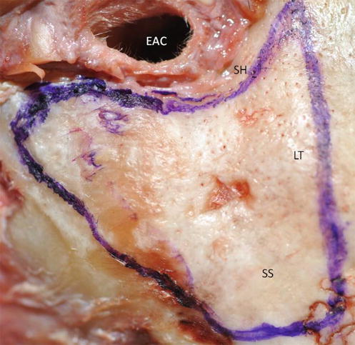
Fig. 2.1
Triangle of attack (EAC external auditory canal, SH spine of Henle, LT linea temporalis, SS sigmoid sinus)
Identifying these reliable landmarks is important in every case, but especially in cases with poor pneumatization:
The temporal line (inferior limit of temporalis muscle) as the approximate landmark of the middle fossa plate is drilled with a large cutting burr in an anterior to posterior direction. Be aware that the brain often hangs much lower than this line, especially in a sclerotic bone.
A second line is drilled parallel and just posterior to the external auditory canal.
The third line connects the first two lines and presents the probable posterior extent of pneumatization at the level of the sigmoid sinus. The sigmoid sinus can extend forward and be located superficially. Avoid injuring the sigmoid sinus and check its location on preoperative CT scans.
 The burr should be moved in a parallel fashion to the vital structures to be preserved: anterior-posteriorly versus the middle fossa plate, superior-inferiorly versus the EAC and superior-lateral to inferio-medial versus the sigmoid sinus (Fig. 2.2).
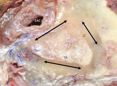
Fig. 2.2
The burr is moved parallel to vital structures (EAC external auditory canal, LT linea temporalis, SS sigmoid sinus)
Landmarks
Suprameatal spine (spine of Henle)
Root of zygoma
Triangle of attack:
Linea temporalis
EAC
Sigmoid sinus
Mastoid air cells
Middle fossa dura
Koerner’s septum
Stay updated, free articles. Join our Telegram channel

Full access? Get Clinical Tree


