Cornea and Sclera


8

Cornea and Sclera


Cornea


Normal Anatomy


I. Introduction


A. Multiple types of corneal disorders are discussed in this chapter. To appreciate the corneal disease state better, the normal anatomy must be understood. In vivo confocal microscopy (CFM) is a valuable tool for the study of corneal diseases. CFM findings complement and corroborate histopathological analyses.


B. The cornea (Fig. 8.1) is a modified mucous membrane (it can also be considered, in part, as modified skin).


The cornea often is affected in association with cutaneous disorders; for example, low corneal sensitivity, abnormal tear quality, decreased cellular cohesion, squamous metaplasia of the conjunctiva, and goblet cell loss have been described in the Hallopeau–Siemens subtype of dystrophic epidermolysis bullosa. Specific corneal abnormalities in this disorder may include recurrent corneal erosion and superficial punctate corneal erosions.


1. The cornea is covered anteriorly by a nonkeratinizing squamous epithelium of approximately five layers, representing modified skin epidermis.


Intermixed within the corneal epithelium are Langerhans’ cells (bone marrow-derived, CD Ia-expressing, dendritic-appearing cells) and occasional dendritic melanocytes. Langerhans’ cell histiocytosis has presented as a limbal nodule in an adult.


a. The deepest layer of epithelial cells, the basal layer, is the germinative layer and is attached to its neighboring basal cells and overlying wing cells by desmosomes.


b. The basal cell layer is also attached by hemidesmosomes to its own secretory product, a somewhat irregular, thin basement membrane.


Three major types of molecules are found in the basement membrane: type IV collagen, heparan sulfate proteoglycans, and noncollagenous proteins (e.g., laminin, nidogen, and osteonectin). The basement membrane represents an important physiologic barrier between the epithelium and the stroma.


c. The flattened, nucleated, superficial epithelial cells desquamate into the overlying trilaminar (mucoprotein, water, lipid) tear film. The mucoprotein layer serves to adhere the tear film to the epithelial microvilli.


d. Corneal epithelial basal cells are characterized by expression of the embryonic stem cell marker OCT4.


2. Corneal stem cells


a. Corneal stem cells, which express K3 keratin marker for corneal-type differentiation in contrast to conjunctival cells, reside in the transitional epithelium of the limbus. Other markers for stem cells are cytokeratin 15 (CK15) and CK14, which are expressed in the limbal basal epithelium, and P-cadherin (CDH3) and Wnt-4, which are expressed in basal and parabasal limbal epithelium in both adult and fetal corneas.


b. CK8 is strongly expressed in limbal epithelial basal cells. This marker is maintained during differentiation and migration of the limbal cells toward the central cornea.


c. Epithelial cells with stem cell features cover the early embryonic cornea but become confined to a “ridgelike” structure probably representing the early corneoscleral area later in development.


In healing large corneal abrasions that reach the limbus, the stem cells regenerate new corneal epithelium by a process called conjunctival transdifferentiation. First, the healing epithelium shows conjunctiva-like appearance, even to containing goblet cells, but then, slowly, it is transformed into a more cornea-like appearance without goblet cells.


d. Stem cells can become exhausted in multiple conditions that lead to their massive direct injury or to repetitive insults. Healthy corneas express K12 but not MUC1 cell markers, whereas conjunctivalized corneas from patients with limbal stem cell deficiency are characterized by the presence of MUC1 and the disappearance of K12.


e. The lacrimo-auriculo-dento-digital (LADD) syndrome is an autosomal-dominant disease with variable expression. Common ocular findings include hypoplasia or aplasia of tear glands, and lacrimal puncta or canaliculi, tear deficiency, recurrent or chronic conjunctivitis, keratoconjunctivitis sicca, and corneal ulceration secondary to sicca. Corneal stem cell deficiency and hypesthesia have also been described.


f. Similarly, ocular manifestations of keratitis–ichthyosis–deafness (KID) syndrome include lid abnormalities, corneal surface instability, limbal stem cell deficiency with secondary corneal complications, and dry eye.


C. Underlying the basal cell basement membrane is a thick, acellular, collagenous layer called Bowman’s membrane (by light microscopy) or Bowman’s layer [by transmission electron microscopy (TEM)].


Abnormalities of corneal epithelium can be demonstrated clinically by the use of fluorescein or rose Bengal. Fluorescein staining is enhanced when disruption of cell–cell junctions occurs, whereas rose Bengal staining is seen with deficiency of protection by the preocular tear film.


D. The bulk of the cornea, the stroma, consists of collagen lamellae secreted by fibroblasts called keratocytes that lie between the lamellae. The stromal lamellae are arranged much as a collapsed honeycomb with oblique lamellae, with the anteriormost lamellae (approximately one-third) being the most oblique (i.e., the least parallel) and the posterior (approximately two-thirds) being the least oblique (i.e., the most parallel) to one another.


1. Thy-1 expression is present in cultured corneal fibroblasts and myofibroblasts but not in fresh keratocytes. Thus, it may be used to differentiate these cell types.


2. The anterior third of the stroma is analogous to a highly modified dermis of the skin, and the posterior two-thirds of the stroma may be usefully considered analogous to a highly modified subcutaneous tissue of the skin.


a. Corneal thickness is highly heritable with clear ethnic differences.


b. α11 integrin probably plays an important role in early corneal development and in the scarring accompanying keratoconus.


E. An unusually thick basement membrane, Descemet’s membrane, secreted by the endothelium, lies between the stroma and the endothelial cells.


F. The posterior surface of the cornea is covered by a single layer of cuboidal cells, the corneal endothelium (mesothelium); no hemidesmosomes are present along these inverted cells. Endothelial cells decrease progressively with age even in healthy emmetropic eyes. This decrease can be expedited by accompanying disorders such as pseudoexfoliation syndrome. The final common result of endothelial insufficiency is corneal edema.


1. Varying abnormalities in aquaporin distribution have been found in pseudophakic and aphakic bullous keratopathy, and in Fuchs’ corneal dystrophy, suggesting the possibility of variations in the mechanism for fluid accumulation in each disorder.


2. Corneal endothelial cell count is reduced and the coefficient of cellular variability is increased in type II diabetes mellitus, but central corneal thickness is not increased.


3. Corneal endothelial cell numbers are decreased, and they express the characteristic mutant DRPLA protein in dentatorubropallidoluysian atrophy.


4. Corneal endothelial cells express some mesothelial cell markers, which may indicate a still unknown function that may be shared by corneal endothelium and mesothelial cells.


G. The cornea is one of the most unusual structures in the body in that it has no blood vessels and is transparent. Any pathologic lesions, therefore, are seen easily clinically as an opacification in the cornea.


H. The cornea is well innervated.


Approximately 20% of corneal nerves respond exclusively to noxious mechanical forces (mechanoreceptors); 70% are stimulated by extreme temperatures, exogenous irritant chemicals, and endogenous inflammatory mediators (polymodal nociceptors); and 10% are cold-sensitive and increase their discharge with moderate cooling of the cornea (cold receptors).


Increased tortuosity of corneal nerves as documented by CFM is associated with severity of somatic neuropathy in diabetes mellitus.


Congenital Defects


Absence of Cornea


Absence of the cornea is a very rare condition usually associated with absence of other parts of the eye derived from the primitive invaginating ectoderm (e.g., the lens).


Abnormalities of Size


I. Microcornea (<11 mm in greatest diameter; Fig. 8.2)


A. The eye is usually structurally normal.


Microcornea may be associated with other ocular anomalies such as are found in microphthalmos with cyst, trisomy 13, and the Nance–Horan syndrome (X-linked disorder typified by microcornea, dense cataracts, anteverted and simplex pinnae, brachymetacarpalia, and numerous dental anomalies; there is provisional linkage to two DNA markers—DXS143 at Xp22.3–p22.2 and DXS43 at Xp22.2).


B. The condition may be inherited as an autosomal-dominant trait.


C. Histologically, the cornea is usually normal except for its small size.


A lack of myofilaments and desmin in the cytoplasm of the anterior layer of iris pigment epithelium suggests that congenital microcornea may result from a defect of intermediate filaments.


II. Megalocornea (>13 mm in greatest diameter; see Fig. 8.2)


A. Most megalocorneas present as an isolated finding, are bilateral and nonprogressive, and do not, in themselves, produce symptoms (except for refractive error).


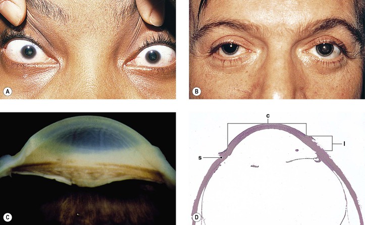
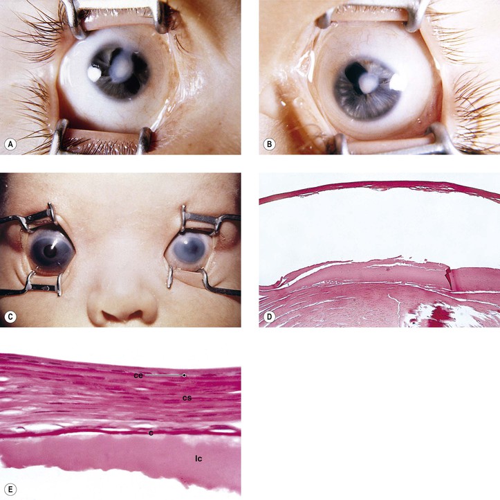
Cataract and subluxated lens commonly develop in adulthood. Glaucoma may result secondary to the subluxated lens. Rarely, megalocornea is associated with renal cell carcinoma.


B. Other ocular findings include arcus juvenilis, mosaic corneal dystrophy, cataracts, and pigmentary glaucoma.


Megalocornea, usually an isolated finding, may also be associated with ichthyosis, poikiloderma congenitale, Down’s syndrome, mental retardation, dwarfism, Marfan’s syndrome, craniostenosis, oxycephaly, progressive facial hemiatrophy, osteogenesis imperfecta, multiple skeletal abnormalities, nonketotic hyperglycemia, and tuberous sclerosis.


C. The condition usually has a recessive X-linked (in the region, Xq21–q26) inheritance pattern, but may be autosomal dominant or recessive.


D. Histologically, the cornea is usually normal except for its large size, especially in the limbal region.


Congenital Corneal Opacities


The main theories of causation are arrested development during embryogenesis, intrauterine inflammation, and trauma.


In a study of 72 eyes of 47 patients with congenital corneal opacities, Peters’ anomaly was the most common cause (40.3%), followed by sclerocornea (18.1%), dermoid (15.3%), congenital glaucoma (6.9%), microphthalmia (4.2%), and birth trauma and metabolic disease (2.8%). 9.7% were idiopathic.


Clinicopathologic Types—General


I. Facet


A. A facet (often the result of an embedded corneal foreign body) is a small, superficial spot seen by focal illumination as a distortion of the corneal light reflex or by slit lamp as a focal increased separation of the anteriormost two lines of corneal relucence.


B. Histologically, epithelium of increased thickness fills in the gap of previously abraded epithelium. Bowman’s membrane, and sometimes the very anteriormost corneal stroma, is focally absent. No scar tissue is present.


II. Nebula (Fig. 8.3)


A. A nebula is a slight, diffuse, cloudlike opacity with indistinct borders.


B. Histologically, scar tissue is found predominantly in the superficial stroma.


III. Macula


A. A macula is a well-circumscribed, moderately dense opacity.


B. Histologically, the scar is dense and involves the corneal stroma.


IV. Leukoma (Fig. 8.4; see also Fig. 8.10)


A. A leukoma is a white, opaque scar (e.g., see discussion of Peters’ anomaly, later).


B. Histologically, a large area of stromal scarring is present.


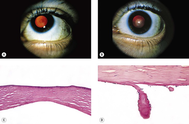
When iris is adherent to the posterior surface of the cornea beneath a region of corneal scarring, the resulting condition is called an adherent leukoma.


Clinicopathologic Types—Specific


I. Anterior embryotoxon


A. Anterior embryotoxon is synonymous with arcus juvenilis.


B. It may be present at birth or develop in early life, and clinically it appears identical to an arcus senilis (gerontoxon).


C. The condition may be associated with elevated serum lipids or cholesterol.


D. Histology—same as arcus senilis (see Fig. 8.20)


II. Corneal keloid


A. Corneal keloid presents as a hypertrophic scar involving the entire cornea.


If ectatic and lined by uveal tissue (iris), it is called a congenital corneal staphyloma.


B. Overabundant production of corneal scar tissue after trauma seems to be the cause of corneal keloid.


C. Although frequently noted at birth, probably secondary to intrauterine trauma, traumatic corneal keloids can occur at any age. They have been associated with Lowe’s syndrome, Rubinstein–Taybi syndrome, fibrodysplasia ossificans progressiva, and other developmental ocular disorders.


D. Histologically, abundant scar tissue in disarray replaces most or the entire cornea. Proliferating myofibroblasts (immunopositivity with α smooth-muscle actin and the intermediate filament vimentin), activated fibroblasts, and haphazardly arranged fascicles of collagen may be seen.


III. De Barsy syndrome is characterized by anomalous facial appearance, generalized cutis laxa, mental retardation, hypotonia, hyperreflexia, growth retardation, and corneal opacification and degeneration of Bowman’s membrane.


A. Light microscopic examination shows epithelial thickening, absence of Bowman’s membrane, and attenuation of stroma, particularly centrally. Hypercellularity and scarring of the anterior superficial stroma may also be seen.


B. Electron microscopy demonstrates replacement of the normal architecture of Bowman’s layer with a paucity of longitudinal and oblique collagen fibers. Although elastic fibrils are not usually present in the normal cornea, small bundles of elastic microfibrils are present in amorphous deposits that are immunopositive for elastin. Abnormal banding of Descemet’s membrane with normal-appearing endothelium may also be seen.


IV. Autoimmune polyendocrinopathy–candidiasis–ectodermal dystrophy (APECED)


A. APECED is an autoimmune disorder that mainly affects endocrine glands with manifestations such as adrenocortical failure, hypoparathyroidism, insulin-dependent diabetes mellitus, and pernicious anemia. Patients have chronic mucocutaneous candidiasis and ectodermal dystrophies.


B. Ocular manifestations include anterior keratopathy characterized by early epidermalization, destruction of Bowman’s membrane and the anterior stroma, which are replaced by vascularized scarring, proliferating fibroblasts, and chronic inflammation. The deeper stroma, Descemet’s membrane, and endothelium are not affected.


V. Central dysgenesis of cornea1


A. Peters’ anomaly (Fig. 8.5; see also section Fetal Alcohol Syndrome in Chapter 2)


1. Peters’ anomaly consists of bilateral central corneal opacities (leukomas) associated with abnormalities of the deepest corneal stromal layers, including local absence of endothelium, and Descemet’s and Bowman’s membranes.


a. Abnormalities of extracellular matrix may be present.


b. Proliferating myofibroblasts (immunopositivity with α smooth-muscle actin and the intermediate filament vimentin), activated fibroblasts, and haphazardly arranged fascicles of collagen may be seen.


2. It is associated with anomalies of the anterior segment structures (corectopia, iris hypoplasia, anterior polar cataract, and iridocorneal adhesions). Congenital corneal staphyloma has been seen.


3. The cause may be a defect of neural crest, ectoderm, and perhaps mesoderm, resulting in failure or delay in separation of the lens vesicle from surface epithelium. The causative event may occur in the migratory neural crest cells between weeks 4 and 7 of gestation.


4. Associated systemic abnormalities include congenital heart disease, external ear abnormalities, cleft lip and palate, central nervous system abnormalities, hearing loss, and spinal defects.


Peters’ anomaly may be part of a syndrome that includes microcephaly with cortical migration defects, and multiple intestinal atresias. It is believed to be a multiple vascular disruption syndrome.


5. Peters’ anomaly is usually inherited as an autosomal-recessive trait, but autosomal dominance or no inheritance pattern may also occur.


Similar findings have been reported in cerebro-ocular myopathy syndrome.


6. Histologically, endothelium and Descemet’s and Bowman’s membranes are absent from the cornea centrally, usually along with varying amounts of posterior stroma.


a. The corneal lamellae are more compact and more irregularly packed than normal corneal lamellae.


b. Immunohistochemistry shows an increase in fibronectin and collagen type VI.


c. Lens abnormalities are present (usually an anterior polar cataract); associated abnormalities of the iris and other structures may also be present. Internal ulcer of von Hippel is similar to Peters’ anomaly in that patients show the typical corneal abnormalities, but it differs in that no lens abnormalities are present.


B. Localized posterior keratoconus (Fig. 8.6)


1. Localized posterior keratoconus consists of a central or paracentral, craterlike corneal depression associated with stromal opacity. The depression involves the posterior corneal surface.


2. Unlike Peters’ anomaly, endothelium and Descemet’s membrane are present.


3. Neural crest–mesenchymal maldevelopment, infection, and trauma are proposed causes.


4. Histologically, the posterior curve of the cornea is abnormal, the overlying collagen of the corneal stroma is in disarray, and Bowman’s membrane may be absent centrally.


VI. Peripheral dysgenesis of the cornea and iris2


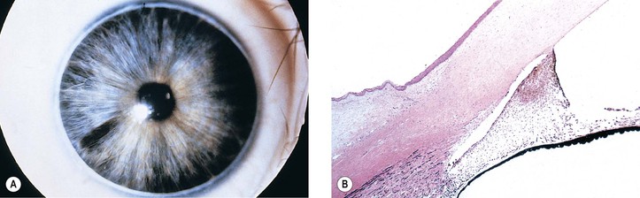
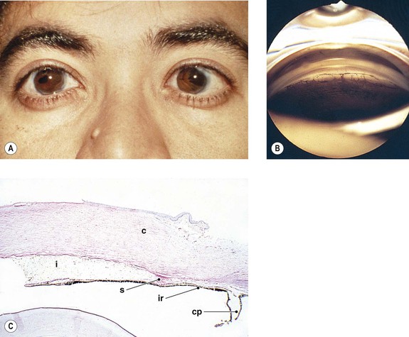
Peripheral dysgenesis of the cornea and iris includes a wide spectrum of developmental abnormalities, ranging from posterior embryotoxon (Axenfeld’s anomaly) to extensive anomalous development of the cornea, iris, and anterior chamber angle associated with systemic abnormalities (Rieger’s syndrome).3 An associated congenital glaucoma may occur, but the presence or absence of glaucoma does not necessarily depend on the degree of malformation. The abnormalities may be congenital, noninfectious, and noninherited (e.g., as part of trisomy 13 and partial trisomy 16q); congenital and inherited (e.g., Rieger’s syndrome); or congenital and infectious (e.g., rubella syndrome).


A. Posterior embryotoxon or embryotoxon corneae posterius (Axenfeld’s anomaly; Axenfeld–Rieger anomaly; Fig. 8.7)


1. Recognized clinically as a bow- or ring-shaped opacity in the peripheral cornea, posterior embryotoxon is an enlarged ring of Schwalbe located more centrally than normally, often seen in an otherwise normal eye or one that shows only a few mesodermal strands of iris tissue bridging the chamber angle to attach to the “displaced” Schwalbe ring.


2. Posterior embryotoxon may be accompanied by glaucoma.


3. Although most cases are not inherited, dominant and recessive autosomal pedigrees have been reported; the former often has prominent iris involvement.


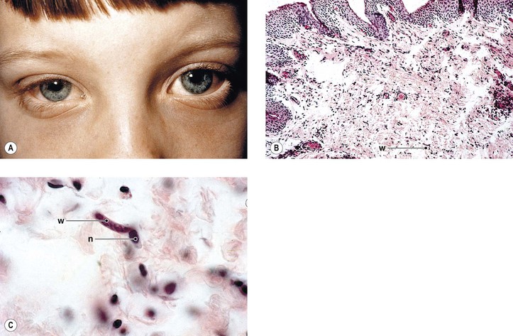
Axenfeld–Rieger anomaly has been linked to chromosome 6p25 (FKHL7 gene). Posterior embryotoxon (along with microcornea, mosaic iris stromal hypoplasia, regional peripapillary retinal depigmentation, congenital macular dystrophy, and anomalous optic discs) may be associated with arteriohepatic dysplasia (Alagille’s syndrome), an autosomal-dominant intrahepatic cholestatic syndrome. Posterior embryotoxon, iris abnormalities, and diffuse fundus hypopigmentation, together with neonatal jaundice, are highly characteristic of Alagille’s syndrome, which also has a strong association with optic drusen. Another association may be with oculocutaneous albinism.


B. Axenfeld–Rieger syndrome (Rieger’s syndrome; Fig. 8.8)


1. The syndrome includes Axenfeld’s anomaly together with more marked anomalous development of the limbus, the anterior chamber angle, and the iris (ectopia of the pupil, dyscoria, slit pupil, severe hypoplasia of the anterior layer of the iris, and iris strands bridging the anterior chamber angle).


Axenfeld–Rieger’s syndrome can be found as part of the SHORT syndrome (short stature, hyperextensibility of joints or hernia, ocular depression, Axenfeld–Rieger’s syndrome, and teething delay). Axenfeld–Rieger’s syndrome is different from iridogoniodysgenesis, which does not have a linkage to the 4q25 region (see later).


2. Glaucoma may be present (approximately 60% of cases).


3. Facial, dental, and osseous abnormalities are present. Associated neurocristopathy has been reported.


4. It is inherited as an autosomal-dominant trait, and it probably represents abnormal embryonic development of the cranial neural ectoderm.


Genetic causes of Axenfeld–Rieger syndrome include mutations, deletions, or duplications of the forkhead-related transcription factor FOXC1, as mutations of the homeodomain (HD) protein PITX2 (PITX2 gene; chromosome 4q25). Axenfeld–Rieger syndrome caused by a deletion of the paired-box transcription factor PAX6 has been reported. Axenfeld syndrome and Peters’ anomaly caused by a point mutation (Phe112Ser) in the FOXC1 gene has been reported, as has familial anterior segment dysgenesis syndrome, which includes iris and corneal abnormalities and cataracts, showing random aggregates of small-diameter filaments that stain positively for cytokeratin.


VII. Endothelial dystrophy, iris hypoplasia, congenital cataract, and stromal thinning (EDICT) syndrome is an autosomal-dominant syndrome that has been mapped to chromosome 15q22.1–q25.3.


VIII. Sclerocornea


A. This condition, usually bilateral, may involve the whole cornea or only its periphery, with superficial or deep vascularization. The cornea appears white and is difficult to differentiate from sclera.


B. Nystagmus, strabismus, aniridia, cornea plana, horizontally oval cornea, glaucoma, and microphthalmos may be present.


C. Congenital cerebral dysfunction, deafness, cryptorchidism, pulmonary disease, brachycephaly, and defects of the face, ears, and skin may also be seen.


D. The condition occurs in three ways: sporadic, isolated cases; familial cases in siblings but without transmission to other generations; and as a dominantly inherited disorder.


E. It has been noted in association with 22q11.2 deletion syndrome, which encompasses the phenotypes of DiGeorge (velocardiofacial) and Takao (conotruncal–anomaly–face) syndromes, and has included posterior embryotoxon, retinal vascular tortuosity, eyelid hooding, strabismus, and astigmatism.


F. Histologically, the most frequent findings are increased numbers of collagen fibrils of variable diameters, a decrease in the diameters of collagen fibrils from the anterior to the posterior layers, and a thin Descemet’s membrane.


Sclerocornea has been described in Mietens’ syndrome. It is also found in microphthalmia with linear skin defects syndrome, in which it has been present bilaterally. Sclerocornea is mainly a clinical descriptive term, and a distinct clinicopathologic entity of sclerocornea probably does not exist.


IX. Limbal (corneal; epibulbar) dermoids (Fig. 8.9)


A. Limbal dermoids are unusual congenital anomalies that contain mesoblastic tissues covered by epithelium. X-linked recessive inheritance has been reported.


B. They usually occur at the temporal or superior temporal limbal area, but they may involve the entire cornea.


1. Rarely, they may extend through the sclera into the uvea.


2. Dermoids are choristomas, congenital rests of benign tissue elements in an abnormal location. Other choristomas in this region include dermolipomas, lacrimal gland choristomas, osseous choristomas, and complex choristomas.


3. Corneal keloid may mimic dermoid recurrence following surgical excision of the latter lesion.


C. Histologically, they may be cystic or solid; are covered by corneal or conjunctival epithelium; and contain choristomatous tissue (tissue not normally found in the area) such as epidermal appendages, fat, smooth and striated muscle, cartilage, brain, teeth, and bone.


D. Goldenhar’s syndrome (Goldenhar–Gorlin syndrome, oculoauriculovertebral dysplasia; see Fig. 8.9)


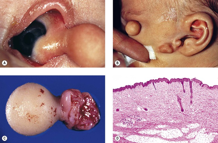
Goldenhar described the triad of epibulbar dermoids, auricular appendages, and pretragal fistulas in 1952. Eleven years later, Gorlin showed the added association with microtia and mandibular vertebral abnormalities (i.e., oculoauriculovertebral dysplasia).


1. Goldenhar–Gorlin syndrome is a bilateral condition characterized by epibulbar dermoids, accessory auricular appendages, aural fistulas, vertebral anomalies, and hypoplasia of the soft and bony tissues of the face. Sometimes it is associated with phocomelia and renal malformations.


Upper-eyelid colobomas commonly occur, but lower-eyelid pseudocolobomas are more often associated with the Treacher Collins–Franceschetti syndrome. Epibulbar choristoma, similar to that seen in Goldenhar’s syndrome, has been seen in association with nevus sebaceus of Jadassohn.


2. The condition is usually sporadic (frequency approximately 1 : 3000), not inherited, and occasionally may be related to first-trimester maternal intake of a teratogenic agent.


3. Histologically, the epibulbar dermoids appear the same as those found elsewhere.


Encephalocraniocutaneous lipomatosis (congenital neurocutaneous syndrome including epibulbar choristomas and connective tissue nevi of the eyelids) should be considered, along with the sebaceous nevus and the Goldenhar–Gorlin syndromes, in the differential diagnosis of epibulbar choristomas.


X. Dentatorubropallidoluysian atrophy


A. This neurodegenerative disorder is characterized by choreoathetoid movements, myoclonic seizures, cerebellar ataxia, and dementia.


B. Decreased corneal endothelial density may be the only ocular finding.


C. The definitive diagnosis may be made on DNA analysis.


Endothelial abnormalities have been reported in an African American family having a syndrome also characterized by abnormal craniofacial features and absence of the roof of the sella turcica. Other findings include abnormalities in the maintenance of retinal bipolar cells and of bipolar cells of the auditory system. One variation and one mutation of the homeobox transcription factor gene, VSX1 (RINX), characterize this family.


Inflammations—Nonulcerative


Epithelial Erosions and Keratitis


I. Epithelial erosion may be secondary to various causes—including traumatic, toxic, radiation-induced (e.g., ultraviolet), or inflammatory (e.g., rubeola) keratitis—or to inherited corneal dystrophies such as lattice and Reis–Bücklers, and numerous other conditions. Epithelial erosions may recur in an apparently spontaneous manner termed Recurrent Corneal Erosion Syndrome (RCES) (Table 8.1).


A. The condition is characterized by damage to the corneal epithelial cells, best seen after fluorescein staining of the cornea.


II. Epithelial keratitis may be caused by the same entities that cause epithelial erosions.


A. It is characterized by large areas of epithelial damage that can be seen grossly without the aid of fluorescein.


B. Thygeson’s superficial punctate keratitis is a recurrent corneal disease of unknown cause, characterized by focal epithelial lesions, bilaterality, intact corneal sensation, and no accompanying conjunctivitis.


1. Patients have symptoms of tearing, irritation, and photophobia.


2. The disorder is chronic and may last for 11 years.


C. Microsporidial keratitis is usually a disorder of immunocompromised individuals; however, it has been reported to involve apparently immunocompetent patients (see Chapters 3 and 4).


III. Histologically, epithelial erosion and keratitis show prominent basal cell edema of the epithelium, absent hemidesmosomes, and separation of the cells from their basement membrane.


Stromal (Interstitial) Keratitis


I. Viral causes


A. Herpes simplex virus (HSV; see later in this chapter)


B. Herpes zoster virus (see Chapter 4)


C. Epstein–Barr virus (see Chapter 3)


II. Bacterial causes


A. Syphilis (Fig. 8.10; see also Fig. 8.3B)


1. Widespread inflammatory infiltrate of the corneal stroma, especially of the deeper layers, is characteristic of luetic keratitis.


2. An associated anterior uveitis is present in the early stages.


3. The congenital form


a. Usually it is bilateral and develops in the second half of the first decade or in the second decade of life. It is rare before five years of age but uncommonly even may be present at birth.


b. Initially, the cloudy cornea is a result of inflammatory cell infiltration associated with an anterior uveitis that is followed by ingrowth of blood vessels just anterior to Descemet’s membrane.


Sarcoidosis, tuberculosis, leprosy, syphilis, and Cogan’s syndrome can all produce a deep interstitial keratitis with deep stromal blood vessels.


c. The acute inflammation may last two or three months, followed by a regression over many months.


d. The corneal changes are frequently associated with Hutchinson’s teeth and deafness (i.e., Hutchinson’s triad).


4. The acquired form is a late manifestation with an average time of appearance of 10 years after the primary luetic infection, and it is usually unilateral and often limited to a sector-shaped corneal area.


5. Histology


a. The cornea is edematous and infiltrated by lymphocytes and plasma cells. Blood vessels are present just anterior to Descemet’s membrane. With healing, the edema and inflammatory cells disappear, the stroma becomes scarred, but the deep stromal blood vessels persist.


b. In congenital chronic interstitial keratitis, the regenerating corneal endothelium produces excess basement membrane (Descemet’s) in a variety of forms, producing thickening, linear cornea guttata, ridges or networks of transparent material (glasleisten), and even networks and strands that project into the anterior chamber.


B. Lyme disease (see Chapter 4)


C. Tuberculosis (see Chapter 4)


III. Parasitic causes


A. Protozoal—leishmaniasis and trypanosomiasis can cause a chronic interstitial keratitis.


B. Nematodal—onchocerciasis (Fig. 8.11)


1. Onchocerciasis is one of the leading causes of blindness in the world, affecting approximately 18 million children and young adults in endemic areas in Africa and Central and South America.


Uveitis and peripheral anterior and posterior synechiae commonly cause secondary angle-closure glaucoma. Chorioretinitis secondary to posterior involvement also occurs. The glaucoma and chorioretinitis, along with the keratitis, are common causes of the blindness. The ubiquitous bacteria, Wolbachia, colonizes the major pathogenic filarial nematode parasites of humans, including Onchocerca volvulus, and may contribute significantly to the inflammatory reaction within the eye.


2. Onchocerciasis manifests itself as a severe disease of the skin and eyes (river blindness).


3. In the acute phase of the infestation, nummular or snowflake corneal opacities form a superficial punctate keratitis. A stromal punctate interstitial keratitis may also occur.


With careful slit-lamp examination, the microfilariae can sometimes be seen in the aqueous fluid in the anterior chamber.


4. Healing induces scar tissue to form in the corneal stroma along with a corneal pannus; the cornea can become completely opaque.


5. Optic neuritis and chorioretinitis may also occur and lead to blindness, especially in heavily infested young people.


6. The adult nematode worms, Onchocerca volvulus, produce microfilariae that migrate through skin and subcutaneous tissue (not blood or lymph) to reach ocular tissue.


The small black fly, Simulium species, ingests the microfilariae from an infected person and transmits them to the next human it bites. Immunologic cross-reactivity of a recombinant antigen of O. volvulus to a host ocular component of 44,000 M antigen suggests that intraocular presentation of the cross-reactive parasite antigen by microfilariae is essential for development of the ocular disease. Other filarial nematodes that may involve ocular structures include Loa loa and organisms that cause filariasis (e.g., Wuchereria bancrofti and Brugia malayi).


7. Histologically, the tiny worm is found along with an infiltrate of lymphocytes and plasma cells.


IV.

Stay updated, free articles. Join our Telegram channel

Jun 19, 2016 | Posted by in OPHTHALMOLOGY | Comments Off on Cornea and Sclera

Full access? Get Clinical Tree

Get Clinical Tree app for offline access