(1)
Department of Pathology, Duke University Medical Center, Durham, NC, USA
Abstract
The conjunctiva is a mucous membrane. It lines most of the anterior surface of the globe to the limbus, and it lines the posterior surface of the eyelids. The conjunctiva is responsible for allowing the eyelids to move over the surface of the cornea without causing damage to the cornea.
A wide range of pathology can affect the conjunctiva, and a solid foundation in general surgical pathology will allow you to recognize most lesions.
This chapter will introduce some basic pathology of the conjunctiva, especially some of the lesions that an ophthalmic pathologist might be specifically consulted upon to review.
Conjunctiva Basics
The conjunctiva is composed of a stratified nonkeratinizing squamous epithelium and an underlying substantia propria. The sclera is deep to the conjunctiva (Fig. 3.1).
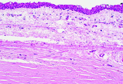
Fig. 3.1
Normal conjunctiva. The relationship of the bulbar conjunctiva (top half of the image) to the underlying sclera (bottom half of image) is shown
Goblet cells are present within the conjunctival epithelium. The goblet cells produce mucin for the corneal tear film layer. Finding goblet cells is a good clue that the tissue is the conjunctiva (Fig. 3.2).
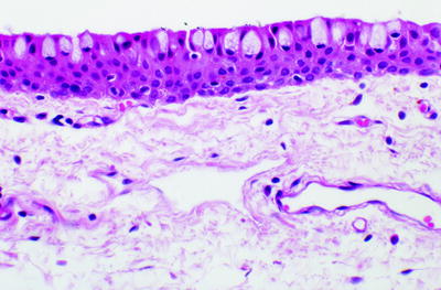
Fig. 3.2
Higher-magnification image of conjunctiva showing intraepithelial goblet cells and the underlying substantia propria
The conjunctiva is a continuous membrane but anatomically is divided into three parts:
Palpebral conjunctiva: lines the posterior surface of the eyelids.
Bulbar conjunctiva: lines the sclera of the anterior globe and extends to the pericorneal (limbus) surface.
Forniceal conjunctiva: The fornix is the junction between the palpebral conjunctiva and the bulbar conjunctiva. The fornices are visible in the exenteration image: Follow the palpebral conjunctiva along the back of the eyelids to where the conjunctiva turns back toward the cornea (see Fig. 1.2).
Although it might resemble skin, the conjunctiva is not skin. It is not considered to have an epidermis and a dermis; rather, it has an epithelium and a substantia propria.
In some instances, skin terminology and concepts in dermatopathology might not apply to the conjunctiva. For example, you would say “subepithelial melanocytic nevus” rather than “intradermal melanocytic nevus.”
In a full thickness eyelid wedge biopsy, the palpebral conjunctiva will be the deepest layer (Fig. 3.3).
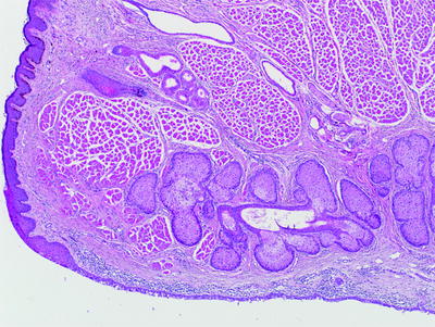
Fig. 3.3
Full thickness eyelid margin. The palpebral conjunctiva is the deepest layer seen at the bottom of the image, deep to the sebaceous glands of the tarsal plate
The caruncle is a modified fold of the conjunctiva in the medial canthus region which also contains adnexal structures including sebaceous glands and hair follicles (Figs. 3.4 and 3.5).
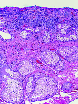
Fig. 3.4
Caruncle tissue with adnexal structures including sebaceous glands. A subepithelial melanocytic nevus is seen
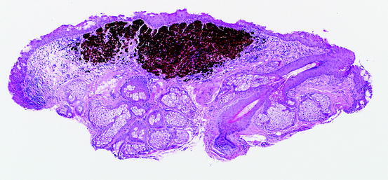
Fig. 3.5
Caruncle nevus. A pilosebaceous unit is seen toward the right side of the image
Accessory lacrimal gland tissue is occasionally present in a conjunctiva biopsy (Fig. 3.6).
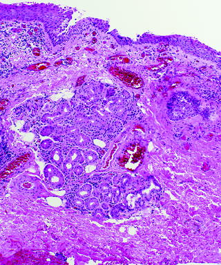
Fig. 3.6
Conjunctiva with accessory lacrimal gland tissue
Conjunctiva Pathology: Pigmented Lesions
Pigmented lesions of the conjunctiva are either melanocytic or nonmelanocytic in origin. Similar to pigmented lesions of the skin – especially the melanocytic lesions – these can be some of the most challenging lesions to diagnose and classify.
Listed here are some of the differential diagnostic considerations for conjunctival pigmented lesions:
Complexion-associated pigmentation (Fig. 3.7)
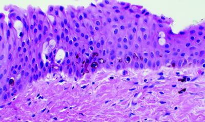
Fig. 3.7
Conjunctiva showing pigmentation within the basal epithelium. This could either be interpreted as complexion-associated melanosis, an ephelis (freckle), or acquired melanosis; you must correlate the biopsy with the clinical history
Ephelis (freckle)
Nevus. Two nevi familiar to ophthalmic pathologists are:Get Clinical Tree app for offline access
1.
The compound or cystic compound nevus, which has a characteristic cystic component (Figs. 3.8, 3.9, 3.10)
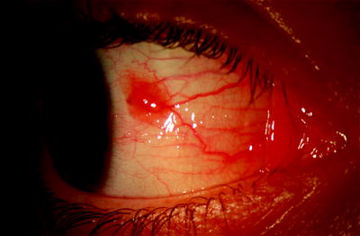
Fig. 3.8
Clinical image of a compound nevus of the conjunctiva
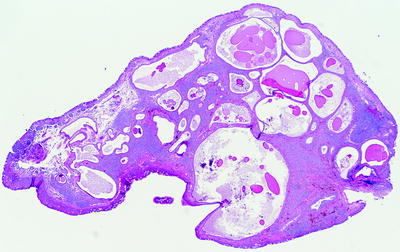
Fig. 3.9
Compound nevus of the conjunctiva. Note the multiple dilated cystic spaces
< div class='tao-gold-member'>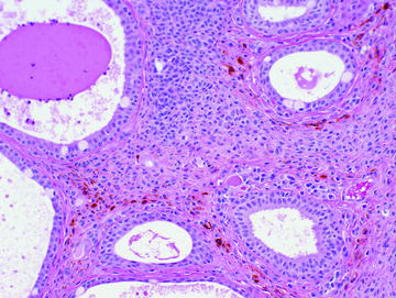 Only gold members can continue reading. Log In or Register to continue
Stay updated, free articles. Join our Telegram channel

Full access? Get Clinical Tree


