Chapter 13
CONJUNCTIVA

The conjunctiva, as the most superficial layer of the eye and inner eyelids, is frequently involved in ocular injuries. Although it has little intrinsic structural strength, it does provide significant protection against low-momentum foreign bodies (see Chapter 24) and chemical agents; careful examination of its entire surface should therefore always be a part of the examination.
EXAMINATION
Penlight
The penlight is used first to inspect the bulbar and palpebral conjunctiva; before opening the lids, ensure that no open globe injury is present (see Chapter 8). Follow the steps outlined below:
• With the patient looking upward, gently evert the lower lid and evaluate the lower bulbar/palpebral surfaces and the inferior fornix.
• With the patient looking downward, raise the upper lids anda inspect the superior bulbar surface.
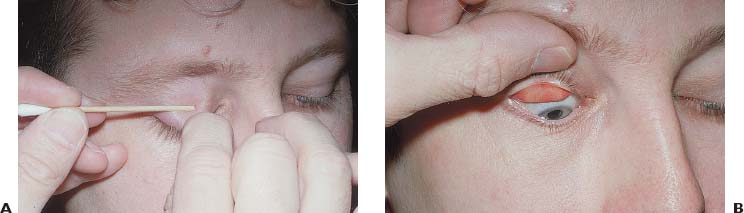
FIGURE 13–1 (A and B) Eversion of the upper lid (see the text for details).
To examine the superior tarsal conjunctiva, the upper lid needs to be everted:
• grasp the lashes gently;
• place a small instrument (e.g., cotton-tipped applicator) on the skin of the upper lid at the location of the lid crease;
• with gentle posterior pressure from the applicator handle and anterior traction on the lashes, the lid can be everted and the applicator handle removed; this results in more patient comfort and in a flatter palpebral surface (see Fig. 13–1).
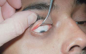
FIGURE 13–2 Double eversion of the upper lid. To visualize the fornix, the everted upper lid must be further lifted away from the globe. Here a Desmarres lid speculum is inserted behind the tarsal plate to allow inspection of the superior fornix and bulbar conjunctiva.
To inspect the superior fornix move the upper lid anteriorly, away from the globe, with an instrument (double eversion, see Fig. 13–2). A small dental mirror may be helpful.1
PEARL… Double eversion of the upper lid is required to inspect the superior fornix.
Slit-lamp
Slit-lamp evaluation of the conjunctiva proceeds similarly to the penlight inspection. using medium-power magnification, the entire conjunctival surface should be examined for foreign bodies, lacerations, or areas of epithelial loss. The upper lid should be everted and the superior palpebral surface also inspected.
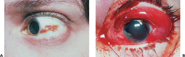
FIGURE 13–3 (A) Subconjunctival hemorrhage may be spontaneous or the result of trauma. In this patient, the hemor-rhage was spontaneous. (B) Extensive subconjunctival hemorrhage due to trauma. The examiner needs to consider the pos-sibility of globe rupture or laceration.
PEARL… The conjunctival surface can be stained with fluorescein dye to detect epithelial denudation. Rose bengal stain-ing can help in detecting small foreign bodies lodged in the fornix or adherent to the palpebral conjunctiva.
SPECIFIC INJURIES
Subconjunctival Hemorrhage
It appears as a bright red patch of conjunctival tissue with distinct or feathered borders (Fig. 13–3A). If it is severe, the conjunctiva may become elevated and prolapse through the palpebral fissure; the entire bulbar conjunctiva may be involved (Fig. 13–3B). Generally resolving spontaneously in 7 to 10 days, its color evolves from bright red to yellow green. occasionally, when the hemorrhage involves the perilimbal conjunctiva, blood breakdown products can be seen in the anterior peripheral corneal stroma as a greenish discoloration.
Hemorrhage under or into the conjunctiva can occur:
• as a result of even minor ocular trauma;
• spontaneously; or
• in association with a variety of conditions including Valsalva maneuvers (see Chapter 33), primary conjunctival amyloidosis,3 inverted positioning,4 dancing;5 and, by far the most common, systemic hypertension.b
The management of a traumatic subconjunctival hemorrhage is hopeful expectancy, although it must be ensured that the hemorrhage does not indicate or conceal a deeper or more extensive injury.


