Conjunctiva
Normal Anatomy
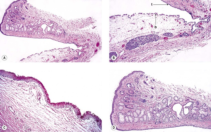
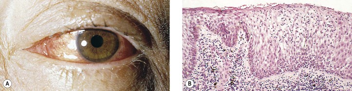
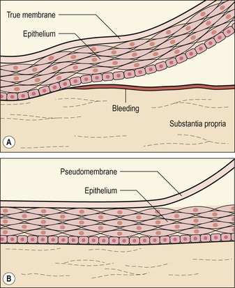
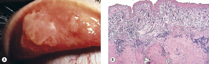
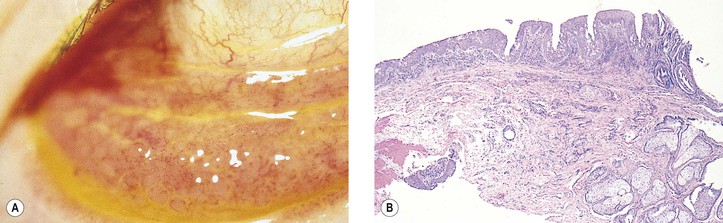
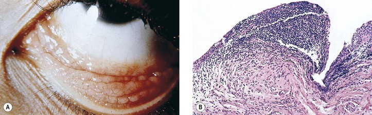
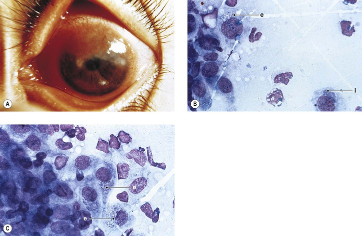
I. The conjunctiva (Fig. 7.1) is a mucous membrane, similar to mucous membranes elsewhere in the body, whose surface is composed of nonkeratinizing squamous epithelium, intermixed with goblet (mucus) cells, Langerhans’ cells (dendritic-appearing cells expressing class II antigen), and occasional dendritic melanocytes.
A. Idiopathic stem cell deficiency is rare, most commonly found in women, and may be familial in some cases. Patients exhibit severe photophobia and, on clinical examination, have corneal vascularization accompanied by loss of the limbal palisades of Vogt, hazy peripheral corneal epithelium, and the presence of conjunctival goblet cells by impression cytology.A conjunctivalized pannus may develop on the cornea of those with total limbal stem cell deficiency. Characterization of this tissue demonstrates that it is not corneal, as evidenced by failure to stain for cornea-specific K12 mRNA and protein, but rather, it is conjunctival, as evidenced by the presence of goblet cells, the weak expression of K3, and the strong expression of K19.
B. The homeostasis of the conjunctiva is dependent, in part, on the maintenance of a normal tear film, which is composed of lipid, aqueous, and mucoid layers (the mucoid layer is most closely apposed to the corneal epithelium and the lipid layer is at the tear film : air interface). Multiple disorders are associated with abnormal tear composition, quantity and/or quality, and secondary ocular surface changes.
1. Tear film abnormalities have been documented in association with cigarette smoking, pseudoexfoliation syndrome, and pseudoexfoliation glaucoma, and they are reflected in abnormal conjunctival impression cytology and altered goblet cell morphology.
2. The pattern of human leukocyte antigen (HLA)-DR expression in mild and moderate dry eyes appears to reflect disease progression, and it suggests that inflammation may be a primary cause of ocular surface damage.
3. Squamous metaplasia of the ocular surface epithelium and ocular tear function abnormalities have been associated with interferon and ribavirin treatment for hepatitis C. Similarly, conjunctiva in β-thalassemia exhibits goblet cell loss and conjunctival squamous metaplasia.
4. Inflammation plays a significant role in the pathogenesis of dry eye.
5. Complete androgen-insensitivity syndrome may promote meibomian gland dysfunction and increase the signs and symptoms of dry eye. In patients with dry eyes, the degree of conjunctival metaplasia, characterized by increased stratification, epithelial cellular size, and a general loss of goblet cells, correlates with the clinical severity of their disorder.
6. Mucin gene expression levels, particularly MUC1, are decreased in dry eye and are biomarkers that can be evaluated using impression cytology specimens.
7. Marx’s line represents a narrow line of epithelial cells posterior to the tarsal gland orifices along the lid marginal zone, averaging 0.10 mm in width, and is stained with lissamine green dye. It is believed to be the natural site of frictional contact between the eyelid margin and the surfaces of the bulbar conjunctiva and cornea, rather than the edge of the tear meniscus or location of the edge of the lacrimal river.

II. The conjunctival epithelium rests on a connective tissue, the substantia propria.
III. The conjunctiva is divided into three zones: tarsal, fornical–orbital, and bulbar.
A. The substantia propria of the tarsal conjunctiva adheres tightly to the underlying tarsal connective tissue, whereas the substantia propria of the bulbar conjunctiva (and even more so the fornical–orbital conjunctival substantia propria) adheres loosely to the underlying tissue (the fornical–orbital conjunctiva being thrown into folds).
B. The periodic acid–Schiff (PAS) stain-positive goblet cells are most numerous in the fornices, the semilunar fold, and the caruncle. The latter is composed of modified conjunctiva containing hairs, sebaceous glands, acini of lacrimal glandlike cells, globules of fat, occasionally smooth-muscle fibers, and rarely cartilage.
C. The tarsal conjunctiva meets the keratinized squamous epithelium of the skin on the intermarginal surface of the lid near its posterior border.
Congenital Anomalies
Cryptophthalmos (Ablepharon)
Epitarsus
I. Epitarsus consists of a fold of conjunctiva attached to the palpebral surface of the lid or lids of one or both eyes. The fold has a free edge, and both surfaces (front and back) are covered by conjunctival epithelium.
II. Histologically, the folded conjunctival tissue looks like normal conjunctiva except for the occasional presence of islands of cartilage.
Hereditary Hemorrhagic Telangiectasia (Rendu–Osler–Weber Disease)
I. It is a generalized vascular dysplasia characterized by multiple telangiectases in the skin, mucous membranes, and viscera, with recurrent bleeding and an autosomal-dominant inheritance pattern.
II. Dilated conjunctival blood vessels, frequently in a star or sunflower shape, may appear at birth but are not usually fully developed until late adolescence or early adult life.
III. Histologically, abnormal, dilated blood vessels are seen in the conjunctival substantia propria.
Congenital Conjunctival Lymphedema (Milroy’s Disease, Nonne–Milroy–Meige Disease)
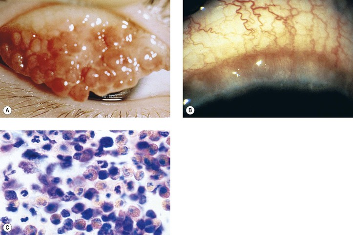
I. This condition of hypoplastic lymphatics is characterized by massive edema, mainly of the lower extremities and rarely of the conjunctiva, and has an X-linked recessive inheritance pattern.
A. Mutations in the kinase domain of the vascular endothelial growth factor receptor-3 (VEGFR3) gene cause Milroy disease.
B. Late-onset hereditary lymphedema may be associated with distichiasis (lymphedema–distichiasis syndrome) and has an autosomal-dominant inheritance pattern, mapped to 16q23 and to mutations in the FOXC2 gene.
II. The disease is thought to be due to a congenital dysplasia of the lymphatics, resulting in chronic lymphedema.
III. Histologically, dilated lymphatic channels and edematous tissue are seen.
Laryngo-Onycho-Cutaneous (LOC or Shabbir) Syndrome
I. LOC is an autosomal-recessive epithelial disorder characterized by cutaneous erosions, nail dystrophy, and exuberant vascular granulation in certain epithelia, especially the conjunctiva and larynx.
II. It is confined to the Punjabi Muslim population and is caused by an unusual N-terminal deletion of the laminin α3a isoform, thereby demonstrating that the laminin α3a N-terminal domain is a key regulator of the granulation tissue response. The protein product is secreted by basal keratinocytes of stratified epithelia, and it has been postulated that LOC results from a dysfunction of keratinocyte–mesenchymal communication.
Vascular Disorders
Sickle-Cell Anemia
II. The characteristic findings (marked in SS disease and mild in SC disease), however, are multiple, short, comma-shaped or curlicued conjunctival capillary segments, mostly near the limbus, often seemingly isolated from the vascular network (Paton’s sign).
III. Histologically, the capillary lumen is irregular and filled with sickled erythrocytes.
Conjunctival Hemorrhage (Subconjunctival Hemorrhage)
I. Intraconjunctival hemorrhage (see Fig. 5.30) into the substantia propria, or hemorrhage between conjunctiva and episclera, most often occurs as an isolated finding without any obvious cause.
III. Histologically, blood is seen in the substantia propria of the conjunctiva.
Lymphangiectasia
I. Abnormal diffuse enlargement of lymphatics appears clinically as chemosis. Localized, dilated lymphatics appear clinically as a cyst or a series of cysts, the latter commonly in the area of the interpalpebral fissure.
II. When involvement is diffuse, the cause is not usually known.
III. Histologically, the lymphatic vessels are abnormally dilated.
Diabetes Mellitus
See section Conjunctiva and Cornea in Chapter 15.
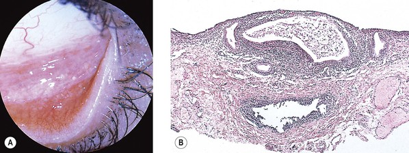
Hemangioma and Lymphangioma
See also Chapter 14.
I. Acquired sessile hemangioma of the conjunctiva (Fig. 7.2)
A. Mean age at diagnosis is 58 years (range, 31–83 years); usually a coincidental finding.
B. Flat collection of intertwining, mildly dilated blood vessels usually on the bulbar conjunctiva.
C. Feeding artery and draining vein seen with leakage of fluorescein dye from deeper, but not more superficial, vessels.
D. Lesion is nonprogressive without systemic disease associations.
E. Histopathologic examination shows two or three layers of dilated, congested blood vessels that otherwise appeared to be normal.

Inflammation
Basic Histologic Changes
I. Acute conjunctivitis (Fig. 7.3)
A. Edema (chemosis), hyperemia, and cellular exudates are characteristic of acute conjunctivitis.
B. Inflammatory membranes (Fig. 7.4)
1. A true membrane consists of an exudate of fibrin–cellular debris firmly attached to the underlying epithelium by fibrin that characteristically, on attempted removal, the epithelium is stripped off and leaves a raw, bleeding surface.
2. A pseudomembrane consists of a loose fibrin–cellular debris exudate not adherent to the underlying epithelium, from which it is easily stripped, usually without bleeding.
3. Ligneous conjunctivitis (Fig. 7.5) is an unusual bilateral, chronic, recurrent, membranous or pseudomembranous conjunctivitis of childhood, most commonly in girls, of unknown cause. The condition persists for months to years and may become massive.
a. The conjunctivitis is characterized by woodlike induration of the palpebral conjunctiva, chronicity, and rapid recurrence after medical or surgical treatment. Severe corneal complications may occur.
b. Similar lesions may also occur in the larynx, vocal cords, trachea, nose, vagina, cervix, and gingiva. Rarely, the middle ear may exhibit a similar histopathologic process.
c. Severe type I plasminogen deficiency has been linked to ligneous conjunctivitis.
d. Histologically, the conjunctival epithelium is thickened and may be dyskeratotic. The subepithelial tissue consists of an enormously thick membrane composed primarily of fibrin, albumin, immunoglobulin G (IgG), and an amorphous eosinophilic material containing a sprinkling of T and B lymphocytes and plasma cells.
C. Ulceration, or loss of epithelium with or without loss of subepithelial tissue associated with an inflammatory cellular infiltrate, may occur with acute conjunctivitis.
D. A phlyctenule usually starts as a localized, acute inflammatory reaction, followed by central necrosis and infiltration by lymphocytes and plasma cells.


II. Chronic conjunctivitis (Fig. 7.6)
A. The epithelium and its goblet cells increase in number (i.e., become hyperplastic).
B. The conjunctiva may undergo papillary hypertrophy (Fig. 7.7), which is caused by the conjunctiva being thrown into folds. Papillary hypertrophy is primarily a vascular response.
1. The folds or projections are covered by hyperplastic epithelium and contain a core of vessels surrounded by edematous subepithelial tissue infiltrated with chronic inflammatory cells (lymphocytes and plasma cells predominate).
2. The lymphocyte (even lymphoid follicles) and plasma cell infiltrations are secondary.
C. The conjunctiva may undergo follicle formation. Follicular hypertrophy (Fig. 7.8) consists of lymphoid hyperplasia and secondary visualization.
E. Chronic inflammation during healing may cause an overexuberant amount of granulation tissue to be formed (i.e., granuloma pyogenicum; see Fig. 6.11).
F. The conjunctiva may be the site of granulomatous inflammation (e.g., sarcoid; see Chapter 4).
H. Clinical and/or histopathologic demonstration of tarsal conjunctival disease may be evidenced by (1) conjunctival hyperemia and granuloma formation, areas of necrosis, or active fibrovascular changes in the tarsus or conjunctiva; or (2) an inactive fibrovascular scar associated with subglottic stenosis and nasolacrimal duct obstruction in patients with Wegener’s granulomatosis (WG).


III. Ligneous conjunctivitis (see earlier, this chapter).
A. Ocular cicatricial pemphigoid (benign mucous membrane pemphigoid, pemphigus conjunctivae, chronic cicatrizing conjunctivitis, essential shrinkage of conjunctiva)
1. This is a rare, T-cell immune-mediated, bilateral (one eye may be involved first), blistering, chronic conjunctival disease. It may involve the conjunctiva alone or, more commonly, other mucous membranes and skin in elderly people.
2. The disease results in shrinkage of the conjunctiva (secondary to scarring), trichiasis, xerosis, and, finally, reduced vision from secondary corneal scarring.
3. Approximately 22% of patients who have systemic, nonocular, mucous membrane pemphigoid develop ocular disease.
a. Subepithelial conjunctival bullae rupture and are replaced by fibrovascular tissue containing lymphocytes (especially T cells), dendritic (Langerhans’) cells, and plasma cells.
1) The epithelium has an immunoreactive deposition (immunoglobulin or complement) along its basement membrane zone. The presence of circulating antibodies to the epithelial basement membrane zone can also be helpful in making the diagnosis. Such immunohistochemical confirmation is important because the clinical characteristics of ocular mucous membrane pemphigoid and pseudopemphigoid are similar, which may lead to a clinical misdiagnosis.Increased expression of connective tissue growth factor has been demonstrated in the conjunctiva of patients with ocular cicatricial pemphigoid, and it is probably one of the factors involved in the pathogenesis of the typical conjunctival fibrosis in the disorder. Macrophage colony-stimulating factor has increased expression in conjunctiva in ocular cicatricial pemphigoid, and there is a positive correlation between its expression and the accumulation of macrophages in conjunctival biopsies in patients with pemphigoid.
2) The vascular and inflammatory components lessen with chronicity, resulting in contracture of the fibrous tissue with subsequent shrinkage, scarring, symblepharon, ankyloblepharon, and so forth. The use of the immunoperoxidase technique in biopsy material may increase the diagnostic yield in clinically suspected cases.
3) Expression of macrophage migration inhibitory factor is increased in cicatricial pemphigoid and may help regulate the inflammatory events in this disorder.
4) Elevated numbers of conjunctival mast cells are present in ocular cicatricial pemphigoid, as well as in atopic keratoconjunctivitis and Stevens–Johnson syndrome.
5) The histopathologic alterations in the ocular surface from abnormal tear film vary considerably depending on the nature of the precipitating ocular condition.
a) Dryness secondary to facial nerve palsy is an aqueous-deficient process resulting in a relatively pure squamous metaplasia response.
b) Ocular cicatricial pemphigoid is primarily a mucous-deficient syndrome and results in hypertrophy and hyperplasia of the ocular surface epithelium.
c) Patients with primary Sjögren syndrome, which involves deficiency of both the aqueous and mucin tear components, start with a squamous metaplasia process but display hypertrophy and hyperplasia at later stages of the disease.
B. Secondary scarring occurs in many conditions. Examples include chemical burns, erythema multiforme (Stevens–Johnson syndrome), old membranous conjunctivitis (diphtheria, β-hemolytic Streptococcus, adenovirus, and primary herpes simplex), trachoma, trauma (surgical or nonsurgical), paraneoplastic pemphigus, and pemphigus, vulgaris and deliberate chronic use of high-dose topical hydrogen peroxide Cicatricial conjunctivitis may be a manifestation of porphyria cutanea tarda.
C. Conjunctival involvement in toxic epidermal necrolysis has been reported in association with autoimmune polyglandular syndrome type I, which is defined as the presence of two of the following diseases: Addison’s disease, hypoparathyroidism, and chronic mucocutaneous candidiasis.
D. Ocular complications of autoimmune polyendocrinopathy syndrome type I are characterized by reduced tear production (63%) that can result in corneal scarring and vision loss. Other ocular complications include lens opacities (18%), hypotrichosis (12%), anisometropic amblyopia (5.9%), and myopia (5.9%).
Specific Inflammations
Infectious
I. Virus—see subsection Chronic Nongranulomatous Inflammation in Chapter 1.
A. As an alternative to viral culture, the most sensitive and specific methods of confirming adenovirus conjunctival infection are PCR (100%), IgM detection (92.9%), and direct antigen detection by fluorescent stain (85.8%).
II. Bacteria—see sections Phases of Inflammation in Chapter 1 and Suppurative Endophthalmitis and Panophthalmitis in Chapter 3. Also see Chlamydiae below.
III. Chlamydiae cause trachoma, lymphogranuloma venereum, and ornithosis (psittacosis).
A. They are gram-negative, basophilic, coccoid, or spheroid bacteria.
B. The chlamydiae are identified taxonomically into order Chlamydiales, family Chlamydiaceae, genus Chlamydia, and species trachomatis and psittaci.
1. Trachoma, caused by the bacterial agent C. trachomatis, is one of the world’s leading causes of blindness and primarily affects the conjunctival and corneal epithelium. Healing is marked by scarring or cicatrization.
2. In vivo confocal microscopy can be used clinically to quantify inflammatory and scarring changes in the conjunctiva in trachoma in which dendritic cells are closely associated with the scarring process.
3. Histology of MacCallan’s four stages:
a. Stage I: Early formation of conjunctival follicles, subepithelial conjunctival infiltrates, diffuse punctate keratitis, and early pannus
1) The conjunctival epithelium undergoes a marked hyperplasia, and its cytoplasm contains clearly defined, glycogen-containing intracellular microcolonies of minute elementary bodies and large basophilic initial bodies (epithelial cytoplasmic inclusion bodies of Halberstaedter and Prowazek). The subepithelial tissue is edematous and infiltrated by round inflammatory cells.
2) Fibrovascular tissue from the substantia propria proliferates and starts to grow into the cornea under the epithelium, destroying Bowman’s membrane; the tissue is then called an inflammatory pannus.
b. Stage II: Florid inflammation, mainly of the upper tarsal conjunctiva with the early formation of follicles appearing like sago grains, and then like papillae. The follicles cannot be differentiated histologically from lymphoid follicles secondary to other causes (e.g., allergic).
1) The corneal pannus increases, and large macrophages with phagocytosed debris (Leber cells) appear in the conjunctival substantia propria.
c. Stage III: Scarring (cicatrization): in the peripheral cornea, follicles disappear, and the area is filled with thickened, transparent epithelium (Herbert’s pits); as the palpebral conjunctiva heals, a white linear horizontal line or scar forms near the upper border of the tarsus (von Arlt’s line). Cicatricial entropion and trichiasis may result.
d. Stage IV: Arrest of the disease

D. Inclusion conjunctivitis (inclusion blennorrhea)
1. Inclusion conjunctivitis is caused by the bacterial agent C. trachomatis (oculogenitale).
2. It is an acute contagious disease of newborns quite similar clinically and histologically to trachoma, except the latter has a predilection for the upper rather than the lower palpebral conjunctiva and fornix.
3. Histologically, a follicular reaction is present with epithelial cytoplasmic inclusion bodies indistinguishable from those of trachoma.
E. Lymphogranuloma venereum (inguinale)
1. Lymphogranuloma venereum, caused by C. trachomatis, is characterized by a follicular conjunctivitis or a nonulcerating conjunctival granuloma, usually near the limbus and associated with a nonsuppurative regional lymphadenopathy. The clinical picture is that of Parinaud’s oculoglandular syndrome (see later).
2. Histologically, a granulomatous conjunctivitis and lymphadenitis occur, the latter containing stellate abscesses. Elementary bodies and inclusion bodies cannot be identified in histologic sections.
IV. Fungal—see the subsection Fungal, section Nontraumatic Infections in Chapter 4.
V. Parasitic—see the subsection Parasitic, section Nontraumatic Infections in Chapter 4 and see Chapter 8.
VII. Parinaud’s oculoglandular syndrome (granulomatous conjunctivitis and ipsilateral enlargement of the preauricular lymph nodes) consists of a granulomatous inflammation and may be caused most commonly by cat-scratch disease but also by Epstein–Barr virus infection, tuberculosis, sarcoidosis, syphilis, tularemia, Leptothrix infection, soft chancre (chancroid—Haemophilus ducreyi), glanders, lymphogranuloma venereum, Crohn’s disease, and fungi.
Noninfectious
I. Physical—see subsections Burns and Radiation Injuries (Electromagnetic) in Chapter 5.
II. Chemical—see subsection Chemical Injuries in Chapter 5.
III. Allergic
A. Allergic conjunctivitis is usually associated with a type 1 hypersensitivity reaction and can be subdivided into acute disorders (seasonal allergic conjunctivitis and perennial allergic conjunctivitis) and chronic diseases (vernal conjunctivitis, atopic keratoconjunctivitis, and giant papillary conjunctivitis).Mast cells play a central role in the pathogenesis of ocular allergy. Their numbers are increased in all forms of allergic conjunctivitis, and they may participate in the process through their activation, resulting in the release of preformed and newly formed mediators. Chronic conjunctivitis may be accompanied by remodeling of the ocular surface tissues.
B. Vernal keratoconjunctivitis (vernal catarrh, spring catarrh; Fig. 7.10)
1. Vernal keratoconjunctivitis is a bilateral, recurrent, self-limited conjunctival disease occurring mainly in warm weather and affecting young people (mainly boys).
a. It is of unknown cause, but it is presumed to be an immediate hypersensitivity reaction to exogenous antigens.
b. The disease is associated with increased serum levels of total IgE, eosinophil-derived products, and nerve growth factor.
c. The cells infiltrating the conjunctiva in vernal conjunctivitis include lymphocytes, eosinophils, mast cells, and natural killer (NK) cells.
2. Vernal conjunctivitis may be associated with, or accompanied by, keratoconus (or, more rarely, pellucid marginal corneal degeneration, keratoglobus, or superior corneal thinning).
3. Involvement may be limited to the tarsal conjunctiva (palpebral form), the bulbar conjunctiva (limbal form), or the cornea (vernal superficial punctate keratitis form), or combinations of all three. It is mediated, at least in part, by IgE antibodies produced in the conjunctiva.
a. The tarsal conjunctiva may undergo hyperplasia of its epithelium and proliferation of fibrovascular connective tissue along with an infiltration of round inflammatory cells, especially eosinophils and basophils. Papillae that form as a result can become quite large, clinically resembling cobblestones.
b. The epithelium and subepithelial fibrovascular connective tissue of the limbal conjunctival region may undergo hyperplasia and round-cell inflammatory infiltration, with production of limbal nodules.
c. In the larger yellow or gray vascularized nodules, concretions, containing eosinophils, appear clinically as white spots (Horner–Trantas spots).
d. Degeneration and death of corneal epithelium result in punctate epithelial erosions that are especially prone to occur in the upper part of the cornea.
C. Inflammatory cells (eosinophils and neutrophils) in brush cytology specimens from the tarsus correlate with corneal damage in atopic keratoconjunctivitis. In atopic blepharoconjunctivitis, the tear content of group IIA phospholipase A2 is decreased without any dependence on the quantity of different conjunctival cells.
Other characteristic ocular surface pathologic changes in atopic keratoconjunctivitis include inflammation, decreased corneal sensitivity, tear film instability, and changes in conjunctival epithelial mucins 1, 2, and 4 mRNA expression.
E. Contact blepharoconjunctivitis
F. Phlyctenular keratoconjunctivitis
A. Graft-versus-host disease (GvHD) conjunctivitis
1. A significant percentage (perhaps 10%) of patients who have had an allogeneic (an HLA-identical donor, e.g., a sibling) bone marrow transplantation develop a distinct type of conjunctivitis, representing GvHD of the conjunctiva.
2. It presents with pseudomembrane formation secondary to loss of the conjunctival epithelium.
3. In approximately 20% of these cases, the corneal epithelium also sloughs.
B. Wegener’s granulomatosis (WG) should be considered when conjunctival inflammation is recurrent and not typical of other conjunctival inflammatory conditions. Based on assessment of the presence of major basic protein and eosinophil cationic protein, it has been suggested that activated eosinophils in the sclera or conjunctiva of patients with ocular limited WG may predict the progression to complete WG.
C. Inflammatory pseudotumor, characterized by the presence of aggregates of chronic inflammatory cells (lymphocytes, plasma cells, neutrophils, and fibroblasts) without noncaseating epithelioid granuloma formation, has been reported to occur simultaneously in the conjunctiva and lung.
D. Rarely, conjunctival ulceration may be a manifestation of Behçet’s disease, and it is characterized on histopathologic examination by disrupted epithelium, infiltration by both acute and chronic inflammatory cells, and high endothelial venules. Immunohistologic studies of the inflammatory infiltrate reveal primarily T-cell populations admixed with several B cells and CD68+ histiocytes.
V. Neoplastic processes (e.g., sebaceous gland carcinoma) can cause a chronic nongranulomatous blepharoconjunctivitis with cancerous invasion of the epithelium and subepithelial tissues.
A. Sebaceous carcinoma may involve the conjunctival epithelium in 47% of cases, of which the superior tarsal and forniceal conjunctiva are involved in 100%; inferior tarsal conjunctiva, 68%; inferior forniceal conjunctiva, 64%; superior bulbar conjunctiva, 68%; and inferior bulbar conjunctiva, 57%. The caruncle is involved in 54% and the cornea in 39%. Metastasis occurs in 11%.
B. Impression cytology may be useful in the detection of conjunctival intraepithelial invasion by sebaceous gland carcinoma; however, full-thickness biopsies are necessary to confirm the diagnosis.
Conjunctival Manifestations of Systemic Disease
Deposition of Metabolic Products
I. Cystinosis (Lignac’s disease)—see Fig. 8.50.
II. Ochronosis—see Chapter 8.
III. Hypercalcemia—see Chapter 8.
IV. Addison’s disease: Melanin is deposited in the basal layer of the epithelium.
V. Mucopolysaccharidoses—see Chapter 8.
VI. Lipidosis—see Chapter 11.
VII. Dysproteinemias
A. Bilirubin salts are deposited diffusely in the conjunctiva and episclera but not usually in the sclera unless the jaundice is chronic and excessive; even in the latter case, the bulk of the bilirubin is in the conjunctiva (scleral icterus, therefore, is a misnomer).
B. Rarely, the icterus can extend into the cornea.
X. Malignant atrophic papulosis (Degos’ syndrome)—see Chapter 6.
XII. Marfan syndrome with ectopia lentis: Consistent, qualitative abnormalities in conjunctival fibrillin-1 staining pattern can be seen in the conjunctiva.
XIII. Chronic renal failure requiring hemodialysis: Squamous metaplasia of the conjunctival epithelium and corneoconjunctival calcification may be seen.

Full access? Get Clinical Tree

