Many surgeons now combine phacoemulsification with vitrectomy for a variety of reasons. This approach adds significant complexity but is indicated in certain situations. There is a widespread but incorrect notion that vitrectomy inevitably leads to cataract. It is widely appreciated that vitrectomy leads to progression of preexisting nuclear sclerosis, likely due to ascorbic acid depletion and resultant permanent increase in the partial pressure of oxygen in the vitreous cavity by 7 to 12 mm Hg (Holekamp, Chang, Steffanson). BSS Plus (Alcon) has been available for three decades (Edelhauser) and has eliminated the development of posterior subcapsular cataract during the procedure, yet many surgeons use BSS or, even worse, lactated Ringer’s solution. Other surgeons choose inappropriately to add a variety of compounds including bicarbonate, dextrose, antibiotics, and epinephrine, all of which can lead to posterior subcapsular cataract, especially if infrequent mixing errors do not occur. Bicarbonate is unnecessary and inappropriate to use with BSS Plus because it is correctly buffered without additives. Dextrose was added three decades ago when diabetic patients were often markedly hyperglycemic during surgery, but the widespread availability of serum glucose monitoring during surgery has eliminated this issue. Contact of the posterior lens with a gas bubble over a period of several days leads to posterior subcapsular cataract; this occurs only if the patient does not maintain the correct position and when there is discontinuity in the anterior vitreous cortex. Patient education is very important. Some of what is described as poor compliance by the patient should be attributed to poor patient education. The vast majority of younger patients with a clear lens undergoing vitrectomy will retain a clear lens for decades if BSS Plus without additives is used and gas bubble contact with the lens is avoided. In short, cataract is not inevitable after vitrectomy.
Optimal visualization is essential for vitrectomy, especially if epiretinal or internal limiting membrane (ILM) dissection, drainage of subretinal fluid through retina breaks, or retinopexy is required. Posterior subcapsular cataracts interfere with visualization more than nuclear sclerotic cataracts. If the surgery is elective, as is typically the case with macular surgery, cataract surgery can be performed 1 month before vitrectomy if the cataract is likely to interfere with visualization during vitrectomy. A potential problem with pre–pars plana vitrectomy (pre-PPV) cataract surgery is the accurate determination of axial length. The Zeiss IOL Master uses the retinal pigment epithelium (RPE) for measurement in contrast to A-scan ultrasound that uses the ILM. Measurement from the RPE is not a problem with epimacular membranes (EMMs) or vitreomacular traction syndrome; however, the A-scan ultrasound axial length may be reduced when these conditions are present or increased when macular holes are present. Fixation is an additional issue with macular disease; it may be difficult to determine if the axial length is measured in the fovea or an extramacular region with either technology.
Cataract surgery performed as a separate procedure after PPV permits more accurate axial length measurement. The procedure can be performed before or shortly after PPV if cataract interferes with examination of the retina and a retinal detachment (RD) is present. For this scenario, the options are phaco-vit or pars plana lensectomy with complete removal of the posterior capsule. Although endocapsular lensectomy with retention of the anterior capsule followed by intraocular lens (IOL) implantation in the ciliary sulcus is possible, this typically results in severe capsular opacification. Therefore, the anterior capsule could not be utilized as a barrier for silicone oil posterior retention. The lensectomy, complete removal of the capsule with forceps and inferior iridectomy, approach has been advocated for these cases, but phaco-vit approach is now favored because it enables the preservation of a silicone oil barrier and optimal correction of aphakia.
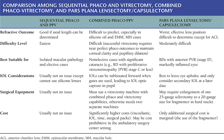
SILICONE OIL ISSUES
Silicone oil has a different index of refraction than vitreous. Because the posterior surface of most IOLs is convex, instead of plano, the usual IOL power calculations cannot be used. Although many surgeons think silicone oil must be removed after a certain number of months, this is simply not true. The incidence of silicone oil–related glaucoma is approximately 11% (1), silicone oil is not toxic to the retina, and corneal complications are even less common. A not uncommon scenario is PPV plus silicone oil to accomplish reattachment, removal in several months followed immediately by redetachment, and reoperation with replacement of the silicone oil. There is simply no need to remove oil contained behind a posterior chamber lens; the refractive effect is approximately four diopters (D), and contrary to popular belief, oil does not cause decreased vision. If there is a full fill behind a posterior chamber intraocular lens (PCL), change in focus with changing head position as well as emulsification is minimized. IOL calculations must anticipate the ultimate retention or removal of oil. There is a greater impetus to removal of oil in younger patients.
GENERAL PHACOEMULSIFICATION CONCEPTS
Many key phaco concepts are similar to vitrectomy principles. Richard Mackool and others emphasize the importance of maintaining optimal visualization, frequent adjustment of microscope focal plane, and stability (intraocular pressure [IOP] maintenance, nonleaking wounds, rigid aspiration system), which are equally essential for vitrectomy.
Phaco and vitrectomy are both safer and more effective when a closed system without leaks is utilized. The principal author has long recommended using 45 mm Hg, unless perfusion pressure is compromised, just as Mackool advocates elevated infusion pressure for phaco. Two-handed methods are best for both procedures both for optimal access and for surgical manipulation. A spatula, chopper, or nucleus manipulator is used to hold back the capsule and manipulate and position lens material similar to using the endoilluminator to hold back retina or peel vitreous with bimanual spreading technique.
High vacuum, a flared tip, and low flow (for sculpting the nucleus) promote efficiency without surge. This concept is the same as the principal author’s port-based flow-limiting concept for PPV. The author developed linear (proportional) control of vacuum for vitrectomy, and this was subsequently applied to all phaco machines for aspiration control of both flow and vacuum. Unfortunately, many PPV surgeons make little use of linear control, fully depressing the pedal and changing vacuum settings on the console. Mackool emphasizes avoiding position 2 (aspiration) or 3 (aspiration and ultrasound) unless actually intending to remove material at that time. The surgeon may elect to engage the “continuous irrigation” mode in the Constellation and Infinity machines, which maintains constant irrigation despite foot pedal position and avoids unintended position 0 of the foot position. The continuous irrigation mode can be toggled on and off when entering and exiting the eye to save irrigation fluid.
There are also essential differences between phaco and PPV; the phaco tip should be positioned near the center of the chamber and lens fragments mobilized to the phaco tip by the use of an appropriate aspiration flow rate to protect the capsule and iris. In marked contrast, the vitreous cutter should be brought to the vitreous; vitreous should not be drawn to the port by high vacuum.
GENERAL PHACOEMULSIFICATION PRINCIPLES
A small, tapered (reduced incision width), nonleaking sideport incision should be constructed. This can be done with the Alcon I-Knife. The entry should be created at a location that will be 2 to 2.5 clock hours from the phaco incision prior to making the latter incision. The separation between the sideport and the keratome incision should be refined to allow for surgeons “comfortable hand position.” The globe can be stabilized with an index finger placed against the nasal conjunctiva during the creation of these incisions.
For microincisional surgery, the temporal clear corneal incision should be made with a 2.2-mm Alcon metallic keratome with a superior bevel (diamond knives can result in inaccurate incision size if not inserted and removed at the exact same meridian). The initial entry should be made with the blade at approximately 10-degree angle to the cornea. After entering the cornea for 0.25 to 0.5 mm, the angle of the blade is lowered so that it is parallel to the cornea. When the blade has penetrated the cornea so that the 2-mm mark on its anterior surface reaches the external incision, the tip of the blade is directed slightly downward (parallel to the iris) and the anterior chamber is entered.
The anterior capsule should be stained with trypan blue, and this can be done efficiently with one injection from a syringe that sequentially delivers air, trypan blue, and then BSS to remove as much of the trypan blue as possible from the chamber prior to viscoelastic injection. Sequential injections of Viscoat (anterior) and Provisc or other hyaluronic acid–based ophthalmic viscoelastic device (OVD) posterior to the Viscoat is done to replace the air with clear viscous material.
The continuous curvilinear capsulorhexis (ccc) should be initiated centrally with a bent needle, forceps, or cystotome. Capsulorhexis forceps (Mackool Microincisional Capsulorhexis Forceps) are then used to create the ccc. It is important to regrasp the elevated flap at a position that is relatively close to the margin of the advancing tear during the creation of the capsulorhexis.
Hydrodissection using a flat 25-gauge cannula to elevate the anterior capsule prior to injection of BSS results in the separation of lens material from the capsule (capsular-cortical cleavage). Hydrodissection is performed in at least two locations, usually 180 degrees apart. After each injection, the nucleus should shift forward and should be repositioned by gentle depression with the cannula prior to repetition of the hydrodissection maneuver. Viscodissection is then accomplished by injecting Viscoat beneath the anterior capsule for 1,800 opposite the phaco incision. In eyes with evidence of zonular laxity, viscodissection should be performed for 360 degrees.
The nucleus can now be rotated in order to verify that the hydrodissection or viscodissection has been successful; however, this maneuver is not mandatory if the surgeon is confident that separation has been successful.
A Mackool chopper or other chopper or spatula is introduced through the sideport incision, and the phaco tip is inserted through the primary incision without the need to fixate the latter with forceps. Viscoelastic material is aspirated from the surface of the lens in order to prevent obstruction of the phaco tip by OVD. The nucleus is then sculpted and divided by either chopping or other nuclear division methods. Mackool prefers to sculpt a central bowl of approximately two phaco tips (2 mm) in width prior to performing phaco chop, and he stabilizes the nucleus during sculpting by placing the Mackool chopper over the edge of the equatorial nucleus opposite the phaco incision.
Note that a nonleaking phaco incision is extremely important to reduce intraocular turbulence and excessive fluid flow through the eye. Rotation of instruments utilizing the incisions as if they were an oarlock prevents distortion of the cornea and compression of the phaco sleeve against the vibrating tip (this reduces the risk of incision burn). The authors prefer the Alcon 450 mini-flared tip with enhanced Ultrasleeve and torsional phacoemulsification.
Chopping of the nucleus is an excellent, efficient technique with the exception of nuclei that are elastic or extremely dense (red-black nuclei). In the former case, high-vacuum levels can be used to remove the peripheral lens material until only a small central nuclear plate remains. The latter can then be elevated with a spatula and removed.
Red-black nuclei are best divided into two heminuclei by sculpting a long, very deep central trough prior to cracking with a spatula and phaco tip.
Whenever possible, use both instruments to rotate the nucleus and/or quadrants in order to reduce the amount of stress placed on the lens zonules. Try to avoid the placement of the phaco tip in the peripheral regions of the posterior chamber, and after one or two nuclear quadrants have been removed, a spatula should be placed posterior to the phaco tip during subsequent nucleus removal in order to prevent anterior movement of the lens capsule (infusion misdirection syndrome) and possible capsule aspiration.
A 900 I/A tip with a small (0.25 mm) port permits access to cortex for 360 degrees. In this manner, cortex can be removed with the port of the I/A tip facing anteriorly.
Note that the PPV can be performed prior to insertion of the IOL. This is highly desirable as a more uniform view of the peripheral vitreous cavity is obtained when not looking through the edges of the optic after completion of the vitrectomy. The only risk of delaying the introduction of the IOL is the possibility of inadvertent capsular tear during anterior vitrectomy, which is prevented by the presence of the IOL creating tension on the capsule and diminishing its mobility. The capsular sac is then filled with Provisc prior to injecting the single-piece acrylic IOL via a Monarch injector and D cartridge. Do not remove the viscoelastic until the haptics have released from the optic. If necessary, their release can be facilitated by using a chopper to place traction on a haptic while the optic is stabilized with the I/A tip. After the haptics have opened, the viscoelastic can be aspirated with the 900 I/A tip, followed by stromal hydration of both the primary and the sideport incisions.
PHACO-VITRECTOMY TECHNIQUES
Combined phacoemulsification with intraocular lens (IOL) implantation and vitrectomy is a safe technique when used in the appropriate patients (2). The threshold for the use of iris expander is lower than for standard phaco. For example, a case with a pupil size of 4 mm or less may be done without problems as standard phaco since anterior chamber infusion during phaco maintains pupillary size but may develop marked miosis when the anterior chamber infusion is terminated at the end of the phaco. Early use of iris expanders prevents severe miosis and a very difficult vitrectomy. If iris expanders are used, do not remove until the completion of the vitrectomy.
Iris prolapse through the cataract incision leads to miosis during vitrectomy. Keratome wounds should be made 2 × 2 mm, which is longer than some phaco surgeons use, in order to prevent iris prolapse.
Staining of the anterior lens capsule with trypan blue is usually required. The red reflex can often be poor in cases that need combined phaco-vitrectomy approach, usually from coexisting vitreous hemorrhage or RD. The need for capsular staining may be underestimated if the only criterion used is the appearance of the lens. Since adequate continuous curvilinear capsulorhexis (CCC) is critical in combined phaco-vitrectomy approach, it is best to use trypan blue staining in the majority of cases.
Viscoelastic can be left in after phaco to maintain chamber depth and pupil size. In the setting of combined vitrectomy, IOP elevation in the early postoperative period is rare. If silicone oil is to be used at the end of the vitrectomy, the surgeon should remove the viscoelastic from the anterior chamber immediately before the oil fill, and it should be replaced with air. This prevents oil emulsification from the viscoelastic interaction.
The cataract incision should be sutured with 10-0 nylon in all phaco-vitrectomy cases. This prevents failure of the self-sealing clear corneal incision mechanism if hypotony occurs during the vitrectomy or postoperatively.
If the posterior lens capsule is not clear at the end of the phaco and this interferes with visualization during the vitrectomy procedure, a small posterior capsulectomy can be performed with the vitreous cutter to improve visualization of the retina after the IOL is well positioned. The vitreous cutter is ideal for the construction of a posterior continuous curvilinear capsulorhexis. This rarely leads to problems other than potential fogging of the IOL if air is used as vitreous infusion or allowing silicone oil access to the AC. YAG laser capsulotomy is ineffective in the presence of silicone oil.
Higher levels of postoperative inflammation are typical in phaco-vitrectomy cases. Very frequent dosing of topical steroids in the early postoperative period (e.g., every hour) with a rapid taper to qid after 1 or 2 weeks postoperatively often prevents inflammatory complications.
Even if the visual potential of an eye is poor, there are still benefits of placing an IOL in an eye that will require long-term silicone oil retention is planned. The IOL can form a barrier to the forward movement of the silicone and prevent emulsification glaucoma and keratopathy. In addition, aphakic eyes with long-term silicone oil fill containing an inferior RD may develop neovascular glaucoma and require Ahmed valve placement. In these eyes, if the eye is aphakic, the silicone oil can exit the vitreous cavity through the valve into the subconjunctival space, causing an inadequate intraocular oil fill that, in turn, causes progression of the previously contained RD.
PHACOEMULSIFICATION PRINCIPLES FOR SILICONE OIL–FILLED EYES
Because silicone oil “floats” over aqueous, oil will frequently push forward on the posterior lens capsule causing something similar to “positive pressure” during phaco. This is likely the main cause of complications during the procedure. A tight seal of the cataract incision around the phaco probe significantly decreases turbulence and intraoperative incision leaks and stabilizes capsular position and chamber depth. The height of the infusion bottle with gravity-fed infusion should be elevated to the maximum to maintain lens capsule inflation during the case. If a small bubble of silicone oil enters the AC through intact zonules, it is best to ignore it. All cortical fragments must be removed without damaging the capsule. In standard phaco, one may leave small cortical fragments without problems. If a cortical fragment is left in a silicone oil case, it may not be reabsorbed as expected and can cause inflammation and oil emulsification. It is best to use bimanual I/A or angulated, that is, 90-degree, I/A tip to remove subincisional cortex. If a small posterior capsular tear occurs during the last phase of the I/A, immediate introduction of an OVD or IOL can seal the hole and prevent further oil bubbles from coming into the AC. If a large capsular tear is encountered, insertion of a multipiece acrylic IOL into the ciliary sulcus and capture of the lens optic through the capsulorhexis can be done to isolate the AC from the oil; if this cannot be done, it may be best to avoid IOL placement and proceed with total capsular removal to prevent leaks of oil into the AC that can lead to silicone oil glaucoma and keratopathy.
ISSUES REGARDING INTRAOCULAR LENS CALCULATIONS FOR SILICONE OIL–FILLED EYES
Silicone oil impedes ultrasound A-scan measurements by slowing of ultrasound waves and decreasing the intensity of the posterior spike. If media clarity allows measurements to be made with the IOL Master (which utilizes laser optical measurements instead of ultrasound), this is the best option. Otherwise, calculations based on the contralateral eye measurements, prior refractions, etc. must be used. Therefore, do biometry with IOL Master soon after initial surgery with silicone prior to the onset of cataract and other possible retinal complications such as pucker or recurrent RD, which may affect measurement.
Because the refractive index of silicone oil is higher than water, the refractive power of the IOL/silicone interface is lower than the refractive power of the IOL/vitreous interface (the refractive power is proportional to the ratio between refractive indexes on both sides of the refractive interface, based on Snell’s law). In effect, silicone oil diminishes the power of the IOL, creating a hyperopic outcome. The higher the lens power of the IOL placed, the higher the total loss of its power in a silicone oil–filled eye, since the difference is based on a percentage loss of power. For this reason, highly myopic eyes tend to be closer to emmetropia than hyperopic eyes when IOLs are placed in silicone oil–filled eyes, because they require lower power IOLs. Theoretically, the IOL design that provides the most reliable refractive results in silicone oil–filled eyes is a planoconvex lens (the plano side faces the silicone oil interface). This neutralizes the refractive interface between IOL and silicone and places all the refractive power on the front surface of the IOL. This design may be difficult to obtain and is usually made of polymethyl methacrylate (PMMA) and is not available with modern, foldable acrylic material. Another source of error in refractive outcomes in silicone oil eyes relates to the completeness of the oil fill. If there is any degree of underfill, a layer of aqueous can wedge between the IOL and the silicone oil (Fig. 8.1). This creates multiple refractive surfaces and prismatic effects that can be impossible to predict or correct with an IOL. If the goal is long-term fill with silicone oil, the IOL can be calculated for an emmetropic or mildly myopic result. If the goal is eventual removal of the silicone oil, placement of an IOL that is appropriate for the eye after oil removal should be done. The majority of the cases that require silicone oil and phacoemulsification with IOL placement are incapable of achieving excellent acuity due to the underlying retinal pathology; there is normally no need to achieve a precise refractive result. In the event of major refractive surprises or if the visual acuity happens to be very good, the IOL can always be replaced within the first month or two after implantation.
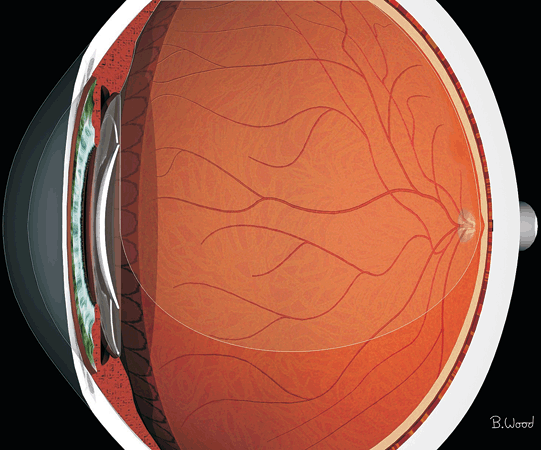
Figure 8.1  A layer of aqueous wedged between the IOL and the silicone oil can create multiple refractive surfaces and prismatic effects that can be impossible to predict or correct with an IOL.
Stay updated, free articles. Join our Telegram channel

Full access? Get Clinical Tree


