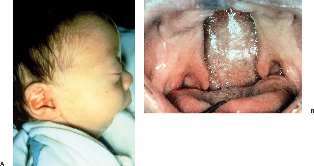
84 A 38-week-old infant was delivered vaginally with initial Apgar scores of 8 and 10, and he developed intermittent respiratory problems in the delivery room. Desaturations occurred when the child was breathing quietly while in a supine position. On admission of the neonate to the newborn nursery, a physical examination demonstrated a large U-shaped cleft of the palate, mandibular hypoplasia, and glossoptosis. With the child in a side-lying position, saturations were maintained at 98% with no retractions. Feedings were difficult, with frequent choking episodes. Cleft palate feeders were used along with positioning to improve the efficiency of feeding and minimize desaturations. Over 1 week, a sleep study was obtained that showed no airway obstruction when the child was either prone or on his side. A genetics consultation was obtained that confirmed the diagnosis of Pierre Robin sequence, with no associated syndrome. Feeding abilities improved. Appropriate weight gain was documented over the week. The child was discharged home with close follow-up, and no additional problems developed. The palate was repaired when the boy was 9 months of age. Ventilation tubes were inserted for a history of chronic otitis media. Mandibular growth continued. He was followed up by the craniofacial anomalies team over the years for coordinated orthodontic and craniofacial care. No additional treatment was required for the retrognathic mandible. Normal resonance was achieved following his palatoplasty, which reconstructed the velum and restored normal velopharyngeal sphincter function (Fig. 84.1). 1. The embryologic sequence involved in the normal closure of the palate includes (1) an intrinsic force that enables the palatal shelves to change from a vertical to a horizontal orientation, (2) migration of the tongue in an anteroinferior direction away from the shelves, and (3) palatal fusion. The configuration of the skull base and the size and migration of the tongue and mandible may be responsible for the development of an isolated cleft of the palate. 2. Cleft palate is associated with Pierre Robin sequence. The sequence consists of (1) mandibular hypoplasia, (2) glossoptosis, and (3) midline cleft of the palate. The sequence usually occurs as an isolated triad of abnormalities but occasionally constitutes part of more complex entities such as trisomy 18 or Stickler syndrome. The cause of cleft palate in Pierre Robin sequence is thought to be from retropositioning or underdevelopment of the mandible, leading to upward displacement of the tongue and causing arrest of the developing palatal shelves, eventually leading to cleft palate. Airway obstruction in patients with Pierre Robin sequence may be life threatening. Fig. 84.1 The patient showed the typical features of Pierre Robin sequence. Note the severely retrodisplaced mandible (A) and the palatal clefting (B). 3. A cleft palate should alert the clinician to the possibility of other anomalies or associated syndromes. A genetic consultation is advisable. 4. The incidence of clefts of the lip, palate, or both represent one of the most frequently occurring congenital deformities. The frequency is higher in whites (1.34 per 1000 births) than in blacks (0.41 per 1000 births). The chances for a future offspring to have an isolate cleft palate are (1) one sibling has a cleft with no parent with a cleft, 2%; (2) one sibling has a cleft with one parent with a cleft, 17%; and (3) no sibling with a cleft with one parent with a cleft, 7%. 5.
Cleft Palate
History
Differential Diagnosis—Key Points
![]()
Stay updated, free articles. Join our Telegram channel

Full access? Get Clinical Tree


