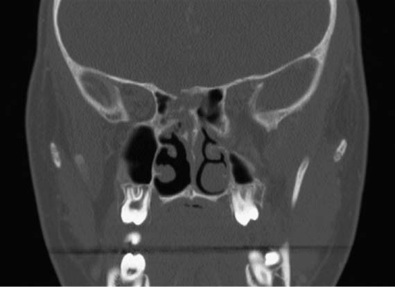
47 A 27-year-old man was involved in a low-speed motor vehicle collision as a front passenger and was brought urgently to the emergency department for evaluation. After his respiratory status was deemed stable and his cervical spine was cleared, he underwent a maxillofacial computed tomography (CT) scan that demonstrated a comminuted nasal fracture with septal deviation to the patient’s right side. Evidence was seen of a bony dehiscence of the skull base at the right posterior ethmoid sphenoid junction (Fig. 47.1). The otolaryngology service was consulted for both the nasal fracture and mild bloody rhinorrhea that had failed to resolve spontaneously despite direct pressure and topical nasal decongestants. A directed history elicited no medical history, no previous maxillofacial trauma (until the motor vehicle accident), and no prior sinonasal surgeries. He was not on medications at the time of the collision, and he denied drug or environmental allergies. He denied visual or auditory deficits, symptoms of nasal obstruction, or anosmia. Fig. 47.1 Coronal computed tomography image demonstrates a traumatic encephalocele extending into the right ethmoid and sphenoid sinuses. On physical examination, the patient was afebrile with stable vital signs. He was awake, alert, and fully oriented. He demonstrated several superficial skin lacerations and abrasions as well as a C-shaped deformity of the nasal septum. He also had a small amount of blood-tinged clear drainage emanating from both nares, which was more obvious when the patient leaned forward. Drainage that had seeped onto his pillowcase showed a “halo” sign. Rigid nasal endoscopy at the bedside did not demonstrate an obvious intranasal source. The remainder of his head and neck examination was unremarkable. Some of the fluid was captured in a test tube and sent for laboratory studies. He was observed in the hospital for an additional 2 days with bed rest precautions. The rhinor-rhea resolved spontaneously within 48 hours of the initial consultation. The patient was subsequently discharged in stable condition, with closed reduction of the nasal fracture scheduled as an outpatient procedure. 1. In this case, the patient clearly had a nasal fracture, and mild epistaxis would be expected. However, as it did not stop with the usual measures, seemed more clear than frankly bloody, and was exacerbated by leaning forward, the possibility of a post traumatic cerebrospinal fluid (CSF) leak needs to be considered. 2. CSF is formed in the choroid plexus within the lateral, third, and fourth ventricles of the brain at a rate of 20 mL per hour. The total volume of CSF in an adult is 90 to 150 mL. Normal CSF pressure is 140 mm H2O (10–15 mm Hg) in adults and 40 mm H2O in children, although pressure varies with the cardiac and respiratory cycles, activity level, and changes in head position. Maintaining CSF pressure requires a delicate balance between CSF secretion and resorption. 3. Between 1 and 3% of all closed-head injuries result in CSF leaks. Roughly 80% of all CSF leaks manifest as rhinorrhea, whereas the remaining patients demonstrate otorrhea. The most common cause of CSF rhinorrhea is accidental head trauma (44%), followed by iatrogenic injury from endoscopic sinus surgery, skull-base surgery, septoplasty, neurosurgery (29%), and tumors (22%). Congenital and “spontaneous” CSF leaks account for the remaining 15% of cases. Spontaneous CSF leaks, which have no obvious inciting factor or cause, are most commonly encountered in obese, middle-aged women, who usually complain of headaches, pulsatile tinnitus, and disequilibrium. This may be a manifestation of benign intracranial hypertension or another cause of increased intracranial pressure (ICP). This elevation exerts pressure on the anterior skull base, with thinning and remodeling of bone, particularly in areas such as the lateral sphenoid recess and cribriform plate. Once a defect occurs, the dura begins to herniate (meningocele), which can then incorporate brain parenchyma as it enlarges (encephalocele) (Fig. 47.2). 4. The most common location for CSF rhinor-rhea is the cribriform plate (35%), followed by the sphenoid sinus (26%), anterior ethmoid (18%), frontal sinus (10%), posterior ethmoid (9%), and inferior clivus (2%). The site of the leak is important in determining the best surgical approach for repair when surgery is indicated. Fig. 47.2
Cerebrospinal Fluid Rhinorrhea
History
Differential Diagnosis—Key Points
![]()
Stay updated, free articles. Join our Telegram channel

Full access? Get Clinical Tree


