15 BRAIN TRAUMA
Low Vision in the Primary Care Practice
Paul B. Freeman
ICD-9: 369.00
 THE DISEASE
Establishing a practice emphasis of providing care for patients with visual impairments must begin with a philosophy of care as the basis for treatment. In providing vision rehabilitation, a practitioner is helping an individual who has been told (or feels that) nothing more can be done to improve sight abilities and function, with either conventional glasses, contact lenses, or medical and/or surgical intervention. Independent of age, the “uncorrectable” decrease in vision can create disabilities, which can lead to frustration and depression. Hopefully, the process of rehabilitative care will lead to a visually impaired patient who feels more independent, feels more self-confident, and possesses a feeling of self-worth, all of which can minimize the risk of depression.
Providing visual rehabilitative care may necessitate introducing a new paradigm in an established practice, which will affect scheduling, staff responsibilities, and paper work. For many eye care practices, the patient flow pattern might need to be modified for these patients, as well as the philosophical view of patient care by both staff and doctors. As an example, many patients who need vision rehabilitation will require multiple visits, and the time involved will vary based on the patient’s abilities and motivation (similar in some respects to a specialty contact lens fitting). For our purposes in this presentation, the discussion covers integration of vision rehabilitative services within an existing practice from a pool of established patients, as well as drawing patients from the outside community.
The Patient
Visually impaired patients can come from a number of different sources. Initially, one might look at the demographics in a community to determine the need and depth of commitment to a practice that emphasizes vision rehabilitative care. Patient referrals can be initiated within a primary care practice by discussing the parameters of vision rehabilitation with the doctors and staff, that is, a chief visual complaint that cannot be resolved with eye glasses or contact lenses. External to the practice, one might look to health care professionals, including optometrists, ophthalmologists, primary care physicians, diabetologists, podiatrists, geriatricians, physiatrists, and occupational and physical therapists. One could also look to organizations that support the care of visually impaired people, including aging agencies and services for the visually impaired, those for the elderly (which include assisted living and nursing home facilities), as well as schools and educational support systems. This is not an exhaustive list, but it does point out the need to reach out beyond the eye care community.
While a standard eye care lane will have most of the optical and diagnostic equipment necessary to evaluate the visually impaired patient, some additional diagnostic and rehabilitative equipment is desirable. The basic components are listed here and can be added to as the practice population increases; more complete information on the options for testing and rehabilitative equipment can be found in the additional readings and Web sites at the end of this chapter:
- A moveable distance chart such as The Designs for Vision distance number chart or Early Treatment Diabetic Retinopathy Study chart
- Multiple near charts including single letter charts, word charts, single sentence charts, and paragraph charts
- Availability of low powered telescopes for distance evaluation, that is, 2.2×, 3×, 4×
- Trial lens microscopes in powers from +12 to +32
- Hand magnifiers in powers from +10 to +32
- Portable electronic systems for initial assessment (devices such as the Pebble, Nemo, Quicklook, Traveller)
Equipment utilized in rehabilitative care can be as limited or as extensive as the practitioner desires to support the need to offer patients all the various options that might be available to accomplish specific tasks. The categories of devices (concentrating primarily on the rehabilitation of central vision loss) can be categorized as follows:
For near activities
- Microscopes
- Telemicroscopes
- Hand magnifiers
- Stand magnifiers
- Electro-optical magnifiers
For distance activities
- Telescopes
- Electro-optical magnifiers
For glare and contrast
- Various filters, including Corning lenses, “FitOvers,” etc.
The Evaluation Process
The evaluation begins with a comprehensive history, followed by a comprehensive eye examination and vision enhancement determination, a summary of the findings and the direction of rehabilitative care, and rehabilitative activities using magnification options where appropriate.
Significant History
The history taking for a visually impaired patient is essentially no different than taking a history of any patient, and it includes the initial complaint, general health questions, and previous eye care. Due to the nature of the patient’s circumstances, the history is generally more detailed; it can include more depth of questioning into the basis for the complaint as well as overall visual functioning related to previous visual abilities (for acquired vision loss) or new goals (for congenital vision loss). As an example, questions with an overarching theme might include the following:
1. How do you spend your day now, and how is that different with what you did previously?
2. What would you like to do now, or how would you like to spend your days now?
3. What are your plans for the future?
Note that the first two questions are general and open ended and give the interviewer some direction and room to further query any of the answers to gain a better understanding of what abilities (activities) were lost, as well as what activities are now important to regain. For most patients, especially the elderly, clinical experience has shown that the primary reason for distress of someone who is visually impaired is the inability to read. In my experience, this is followed by the inability to write, recognize people, identify medications (which can have serious, even deadly consequences), and drive (this must be correlated with the visual requirements for driving in that state). The answer to the third question is somewhat telling, especially for seniors, in that this may indicate feelings of hopelessness and depression, and although we are not mental health care professionals, the responses to the last question may warrant further investigation by a qualified professional. An example of a history form for a visually impaired patient can be found in the Art and Practice of Low Vision and Functional Assessment of Low vision.
The Treatment
The components of the basic examination are no different than any primary care comprehensive ophthalmic evaluation, even for the established patient. The difference is how the clinician proceeds after the endpoint of a conventional lens prescription, that is, 20/200 best corrected visual acuity at both distance and near. From there, and prior to dilation (which I believe is integral to the evaluation, independent of any previous eye and vision evaluations, unless contraindicated), the doctor must calculate the appropriate magnification to begin the rehabilitative process of regaining the previously identified functional activities. Most often, just demonstrating improvement in vision will lift the spirits of the patient, and that alone can build confidence to move forward. However, the final prescription does not have to be determined at that visit. Using the example of 20/200, the first step would be to demonstrate an improvement in distance vision by using a low-powered, easy-to-view-through telescope, such as a 2.2× Galilean system. Simply improving the vision to 20/100 or slightly better will begin to change the patient’s misperception that “nothing more can be done to improve the vision.” Then calculating an initial near lens for reading should follow. This demonstration is more critical than that for distance vision, keeping in mind that reading is most often the primary reason for the visit. Using the criteria of a 20/50 demand (measured at 16 in or +2.50 D of accommodation) as threshold vision for standard reading, one must first determine the amount of magnification necessary to read that size print, that is, 200 size letter seen/50 size letter desired or 200/50 = 4×. Deciding how to achieve that magnification will then be based initially either on relative size magnification, where the smaller print is enlarged to meet the desired threshold at or around the patient’s habitual working distance, or by moving the target closer to make the image size of the small target achieve the visual angle necessary to appear to be four times larger. For the latter, use the modified formula: Magnification = D/2.5 (M = D/2.5), where D is the dioptric power necessary to focus that magnified image, and 2.5 is the referenced dioptric power of where the target is habitually held for the threshold vision found (using conventional lenses). In this situation, the initial lens choice would be a +10.00 D add, and the working distance would be approximately 10 cm. (A modification to this lens, however, should be considered if there is distortion with the Amsler grid. Clinically, it has been found that doubling the calculated power will minimize distortion, which is far more disturbing than the close working distance required for the print to be in focus.) The next step is to place the calculated lens in the trial frame, over the distance (refractive) lens prescription (if one is measured), and have the patient bring reading material to the focal length of the lens. Reading should progress systematically from identifying in sequence, single letters, words, an isolated sentence, and a sentence in a paragraph. At any point if this becomes difficult, this process should be stopped as the patient should leave this activity on a positive note.
Summary and “Home Work”
Summarizing the evaluation, once again, is similar to summarizing an evaluation where a discussion of a pathology and prognosis is reviewed. Questions about stability of the condition and possible visual endpoints should be covered. Where the rehabilitative summarization differs is that there is now visible proof that the patient has the potential to regain an activity that heretofore was considered lost. The various equivalent magnification forms for reading, as an example (microscope, hand magnifier, stand magnifier, telemicroscope, electronic magnifier), should then be reviewed with realistic expectations of how the desired task will be accomplished. Because a microscopic lens (the lens power calculated previously for near) is typically a good starting point, a home exercise to acclimate the patient to an unfamiliar short working distance can be given, even before working with lenses. An instructional set adapted and modified from the Art and Practice of Low Vision follows as an example:
- Find a dimly lit area
- Sit comfortably
- Take deep slow breaths
- Tense then relax, starting with the face
- Visualize a comfortable surrounding
- Bring your hands up to your face with your eyes closed. Visualize reading the numbers 1 to 30 and when you are done put your hands down
- Feel that the activity is effortless
- Do this activity 12 times during the day
Rehabilitative Visit(s)
The number of visits required to ultimately establish a successful vision rehabilitative outcome is dependent on the patient’s needs, the magnification options, and the motivation and expectation(s) of the patient. Familiarizing the patient with many of the devices will foster an appreciation of magnification, working distance, and the useful field of view of the device(s), as well as how specific device(s) will fit into the patient’s lifestyle. Whether the magnification should be provided as a fulltime device (i.e., bifocal microscope), or a device to be used on an intermittent basis (i.e., full diameter microscope, only for specific activities, and not to be worn while walking), is best determined after there is some familiarity with the various options and their strengths and limitations, based on rehabilitative activities. Then, equivalent powered systems, including handheld stand, and electro-optical magnification can be explored as indicated.
Compensation
The philosophy of compensation for these services will be dependent on the fiscal philosophy of the practice. One must look at the amount of time necessary to achieve the patient’s ultimate goal(s). Consideration must be given to whether the patient originates from within the existing office (established patient) or if the patient is referred into the practice (new patient). Coding will be consistent with that distinction.
- Initial evaluation: comprehensive ophthalmic evaluation (often covered by many insurances), refraction (coverage varies based on insurance), and functional vision evaluation based on optical or nonoptical therapeutic treatment options (often not covered by most) insurances
- Rehabilitative visits: coding depends on how these visits are structured and who does the therapy
- Device fees: calculated based on office philosophy
Where to begin to find low vision devices
http://www.enhancedvision.com/index.cfm
http://www.designforvision.com/
http://www.walterslowvision.com/
http://www.chadwickoptical.com
Visual Field Expansion
Paul B. Freeman
ICD-9: 368.40
 THE DISEASE
Rehabilitating someone with a visual field loss is analogous to teaching someone to safely walk backward. In both situations, the prime consideration is safety, with the identification and discrimination of targets in the environment not always critical to the primary goal of safely going from point A to point B. Walking backward, as an example, requires one initially to have an awareness of one’s own body space and the physical ability to periodically peer around and look in the direction to which one is intent on going. It is interesting to point out that the area behind one (i.e., the nonseeing area) is not perceived as black or dark, but rather is simply a space that is not seen all the time. Typically, this does not interfere with walking backward. In fact, this is an excellent way to explain the basic concept of a visual field loss. This is important to communicate to those who work with patients with a visual field loss, especially to family members, as unfortunately the most common way to visually demonstrate the loss is by portraying the missing field as black.
Along with viewing intermittently, the individual must have the ability to scan the environment, to remember where things are in relation to the other things in the environment as well as the self, and then to move in such a fashion as to arrive at the goal-oriented spot without contacting any of the objects in the path. In addition, one must have the ability to predict potential activity and movement in the environment based on the visual memory of where objects are initially and where their ultimate positions will be, based on direction and velocity of movement, and then to calculate whether any paths will intersect.
Etiology
A visual field loss typically occurs as a result of an acquired brain injury such as a stroke, or trauma from a motor vehicle or pedestrian accident, fall, fight, etc. Depending upon where the insult occurs anatomically, the visual field loss can be monocular, binocular, lateral, superior, inferior, quadrant(s)-specific, or a symmetrical or an asymmetrical constriction. In addition, in some situations, this visual field loss may be only one of the constellation of problems that an individual might have. To complicate the rehabilitative process, the individual can also be monocular, can have uncorrectable (with standard eyeglasses or contact lenses) central visual field losses, can have variable refracted issues to deal with such as traumatic myopia, or can be diplopic, to name but a few additional ophthalmic problems. Moreover, this individual might have psychological and emotional problems superimposed, including anger, agitation, depression, frustration, inattention, or a general lack of understanding of the problem at hand due to cognitive dysfunctions. Expressive communication may be a challenge due to aphasia. And finally, this individual might be apraxic, or have difficulty with purposeful movement. In summary, it is not unusual that affected patients have more challenges than “only” a visual field loss.
Ancillary Tests
An evaluation of an individual with a visual field loss should consist of a comprehensive ophthalmic eye and vision evaluation. Specific to the visual field loss, one might initially perform a single or simultaneous confrontation visual field, having the patient be more attentive to the location of the examiner’s hands rather than discrimination of the number of fingers being held up. This is a good starting point for justifying additional, more formal visual field testing. Formal visual field testing should then be performed at both distance and near. Distance monocular visual field testing, especially to determine the lateral extent of the visual fields, should be done using a Goldmann visual field protocol using three isopters (e.g., I4e, III4e, V4e) to definitively determine the extent of the visual field and its density. However, to determine the binocular visual field, I have found that using the Esterman protocol on a Humphries perimeter is a good way to estimate the functional impact of a visual field loss. At near, using the standard protocol for Amsler grid testing is a good method for determining the impact of the visual field loss on near-range activities such as reading or eating.
The Treatment
Rehabilitation can consist of compensatory strategies in combination with optical and nonoptical components. Optically, one can use prisms or mirrors to help an individual to be aware of space and objects within the visual field loss. For constricted visual fields, the optics of minus lenses or reversed telescopes can be used. It is most critical when using any type of optical device that the patient (as well as caregivers and family members) be continually reminded that the use of any of these devices will not “strengthen” or “restore” the natural visual field but rather is a strategy to improve awareness of that visual field loss area. Because, in my experience, use of mirrors is very challenging for most patients, the discussion here will concentrate on prisms for hemianopic visual field losses.
There are three approaches to prescribing prisms. One is using a (press on) prism placed on a spectacle lens on the side of the visual field loss, with the base in the direction of the visual field loss, and with the apex edge of the prism at or near where sight begins (this can ultimately be ground into the lens). The exact placement of the apex edge of the prism, as well as the power of the prism, is determined by the patient’s comfort, the ability of the patient to laterally scan into the prism, and the consideration of how much angular displacement is desired when looking into the prism, remembering that the linear displacement will vary depending on how far away the objects being viewed are. Keep in mind that chromatic aberration and distortion are proportional to the prismatic power; that is, the stronger the prism, the more the aberration and distortion when viewing into the prism. Additionally, one must then consider whether that prism should be monocular (on the temporal aspect of the lens on the visual field loss side with the base out) or binocular (on the temporal aspect of the lens and the nasal aspect of the fellow lens, both on the half of the lens of the visual field loss side and on the base toward the visual field defect). A second technique to prescribing prisms is to cover the entire lens with the prism, still with the base of the prism in the direction of the visual field loss. This technique is used to shift the entire visual world in the direction where vision is optimum and is typically done binocularly. A third use of prisms is simultaneous viewing through sector prisms placed superiorly and inferiorly on the lenses and covering both sighted and nonsighted aspects of the patient’s visual world.
My preference is the first option, to use prisms which cover only half the lens, monocularly, and placed on the spectacle lens within the visual field loss (base toward the defect). I only want the patient to glance into the prism briefly to detect anything that might be in that nonseeing field; if the patient views through the prism for any length of time, typically double vision will be experienced. To be successful, I have found that the patient should first practice eye movements and behavior necessary to see into the prism. The following sequence is used prior to placing the prism on the glasses:
- Sit in a comfortable position, and every 8 to 10 seconds look over into the nonsighted visual field and describe what is being seen.
- Stand in a comfortable position, and every 8 to 10 seconds look over into the nonsighted visual field and describe what is being seen.
- Walk in a controlled environment, and look over into the nonsighted visual field and describe what is being seen.
Occasionally, patients find this activity challenging and require additional visual support. Borrowing from vision therapy activities for eye movement skills can often be beneficial. One technique that has proven to be useful is flashlight tag, where both the patient and therapist (who can be any member of the family as well) play tag with the light from the flashlight projected on a wall, with the therapist moving the light projected on the wall first and the patient visually following and “tagging” the therapist’s light. The distance from the wall can be variable, but the initial activity should be started from the sighted visual field, moving the light gradually into the nonseeing visual field, and in a systematic (somewhat predictable) way so as not to confuse the patient. As the patient becomes more adept at this activity, the therapist can randomize the light placement, sequence, and speed.
Once this can be done comfortably and easily, prismatic lenses can be applied to the glasses. Fresnel press on lenses are relatively inexpensive, are easily applied and removed, and can be adjusted on the glasses to accommodate changes in scanning ability. Prior to cutting a Fresnel lens, I have found it helpful to use a clip on system with a rigid prism, which can be placed over the lenses to determine if the prism will be acceptable to the patient. (There are situations where the initial scanning training without the prism is so effective that the patient becomes aware of the nonsighted side, so that prismatic lenses are either not needed, of limited value, or actually annoying to the patient.) However, once the prism is determined to be of benefit, a Fresnel lens should be appropriately applied to the spectacle lenses (keeping in mind the position and the power are variables and should be decided upon between the patient and the doctor). The following is the training sequence used with the prisms, and a similar to the activity done without them. The concept is to look into the prism for awareness and not to view through the prism while looking straight ahead.
- Sit and view into the prism every 8 to 10 seconds and describe what is being seen. (While viewing into the prism, the patient might be encouraged to quickly reach for an object seen through the prism. This demonstrates the concept of image displacement.)
- Stand and view into the prism every 8 to 10 seconds and describe what is being seen.
- Walk, in a controlled environment, and view into the prism every 8 to 10 seconds and describe what is being seen. (Again, occasionally encourage the patient to touch an object, such as a door handle, while walking by and viewing through the prism, to reinforce the concept of image displacement.)
- Walk, in an uncontrolled environment, and view into the prism every 8 to 10 seconds and describe what is being seen.
I have also found that for this activity, it is useful for the partner to be on the full visual field side, so that the patient is talking to someone who is visibly there and must also be made to feel the need to rely on scanning to relate that information to the partner. The partner should, however, be cognizant of situations that can put the patient in harm’s way and act appropriately. The positioning of the partner can vary.
The above strategies are best used with lateral prism for left or right hemianopsia, but there are situations where the prism can compensate for lower (and less frequently upper) visual field loss, and the same protocol would apply.
Nonoptical therapy for hemianopic visual field losses consists primarily of developing an awareness of the environment, oftentimes using “anchors” or perimeter guards to delineate the full extent of the range of the visual field necessary for the activity. As an example, with a right hemianopia, one is always reading into a visual field loss, not knowing where the end a line of print might be. By marking the right side of the page (e.g., with a colored strip of paper or a ruler), the reader can then read until the mark is reached, allowing more attention to the task and less time worrying about where the endpoint is. Functionally, this often seems to improve comprehension and reading enjoyment. Conversely, with a left visual field loss, marking the left side of the page allows the reader to know how far to scan back to the next line. Other techniques for reading can consist of placing the entire reading material within the sighted area or rotating the reading material in such a fashion that it is along the midline (rotating the page 90 degrees), so that the material being read from top to bottom is on the sighted side.
Eating is another task which requires an awareness of personal space. By visibly marking off the extent of the eating area on the nonsighted side, the patient is instructed to view the “anchor” periodically to make sure that there is no food, drink, etc. that is “hidden” in the visual field loss area. Oftentimes, patients will remark that prior to this activity, food or drink was not always finished. Obviously with this strategy, everything needs to be placed within the perimeter of the patient’s personal space. This strategy of using an “anchor” to mark the extent of the area within the nonseeing visual field is useful for any activity where the patient is stationary, especially within a few feet, like playing cards.
Caregivers and families of individuals with extensive or hemianopic visual field loss should also be aware of how they can best interact without unsettling the patient. Approaching the patient from the sighted side is much more positive than emerging from the visual field defect. An analogy might be having someone approach a “normally” sighted person from behind without any warning; one can appreciate how annoying and unsettling that would be. Likewise, a patient with a hemianopia should be positioned, so that most visual activity is within the sighted visual field. This could apply to the positioning of a favorite chair for watching TV, a patient’s bed with respect to the door, or a student in the classroom.
Constricted visual field losses are less common, but no less devastating. Patients with these losses often report feeling like they are walking down a very narrow corridor and may benefit from minus lenses or reversed telescopes to increase awareness of the environment. The technique for using these optics requires an appreciation of the positions of objects (relative to each other) in the environment viewed though the lens or reversed telescopic system which minifies to allow for more information to fit into a limited visual space (the size and area is dependent on specific system). Remembering the relationships and then removing the optics (when holding either a minus lens or a reversed telescope, or viewing through the carrier in a head mounted reversed telescope) and scanning the environment for confirmation are the basis behind increased awareness of what is missing due to the constricted visual field. This allows for safe passage through the area just viewed. Many of the same nonoptical concepts used for the hemianopic patient can then also be modified for an individual with a constricted visual field.
Working with a patient with a vision field loss is more about teaching awareness and reaction to information within the nonseeing area, and safety, than about cultural activities such as reading or watching TV. That said I would be remiss in not at least briefly discussing visual field loss and driving. There are two considerations: the legal aspects of the patient with the visual field loss driving and the ability of that person to safely drive. Visual field requirements vary from state to state and some states have no visual field requirements. Certainly, if the patient does not meet the legal visual requirements, the discussion about driving is relatively straightforward. However, when there are limited or no visual field requirements, a combination of lenses, prisms, and external mirrors to increase visual field awareness, as well as response time of the patient must all be taken into account for the safety not only of the driver but also of anyone who might come into proximity of the driver.
Patients who suffer a visual field loss will typically not regain sight in the affected area to any significant degree. However, with both optical and nonoptical rehabilitative strategies, these patients can become more safe and independent in their environment, a goal which is well worth pursuing.
Where, other than local labs (for Fresnel lenses) to look for visual enhancement devices
http://www.chadwickoptical.com
http://www.gottliebvisiongroup.com/visual_field_awareness_system.htm
http://www.designforvision.com/
Visual Dysfunction in Traumatic Brain Injury
Kenneth J. Ciuffreda and Neera Kapoor
ICD-9: 850
 THE DISEASE
Pathophysiology
Traumatic brain injury (TBI) refers to an acquired brain injury that results when an external force causes either an open‐ or a closed-head insult, with the former frequently being fatal (e.g., gunshot to the head). It is of sudden onset and is not progressive, congenital, degenerative, developmental, or genetic. Although not progressive per se, some secondary effects occur within hours to days of the insult: the release of neurotoxic compounds eventually produces ischemia and hence produces further brain cell damage. TBI is categorized as mild, moderate, or severe based on the degree of initial lost/altered state of consciousness of the patient, posttraumatic amnesia, loss of consciousness, neuropsychological testing, neuroimaging, and the Glasgow Coma Scale.
Due to the pervasiveness of the injury, its consequences are broad and can adversely affect one’s sensory, motor, perceptual, cognitive, psychological, and/or behavioral states. More specifically, TBI may have visual sequalae, as 6 of the 12 cranial nerves impact vision either directly (cranial nerves II, III, IV, and VI) or indirectly (cranial nerves V and VII).
Etiology
Due to the coup-contrecoup nature of the acceleration and deceleration type of TBI, there is both direct and indirect insult to the brain, and hence its pervasive impact on the individual. There is the primary injury at the region of impact, and the secondary injury opposite to the initial impact, as well as rotational forces on the brain causing shearing effects resulting in contusions and blood leakage. Hence, the global nature of the injury produces massive diffuse axonal injury (DAI) and brain cerebral edema, which have an adverse effect on the brain’s physiological control and complex information processing, including that of vision and related systems (e.g., the vestibular system). While still experimental, a recent brain imaging technique, namely diffuse tensor imaging involving analysis of water flow patterns in the brain, holds promise for more accurate visualization and quantitative assessment of DAI in TBI.
The Patient
Clinical Symptoms
The general symptoms of TBI (mainly the mild form) are of three broad categories: (1) physical, such as nausea, dizziness, headache, fatigue, and sleep disturbances; (2) cognitive, such as attention, executive functions, language, memory, concentration, and processing speed; and (3) emotional/behavioral, such as anger, disinhibition, and irritability.
The visual symptoms of TBI (especially the mild form) are listed in Table 15-1. The visual signs of TBI (especially the mild form) are listed in Table 15-2.
TABLE 15-1 Oculomotor and Visual Symptoms in TBI
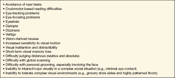
From Suchoff IB, Ciuffreda KJ, Kapoor N., eds. Visual and Vestibular Consequences of Acquired Brain Injury. Santa Ana, CA: OEP Foundation Press, 2001.
Stay updated, free articles. Join our Telegram channel

Full access? Get Clinical Tree


