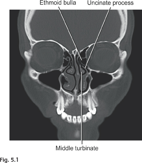
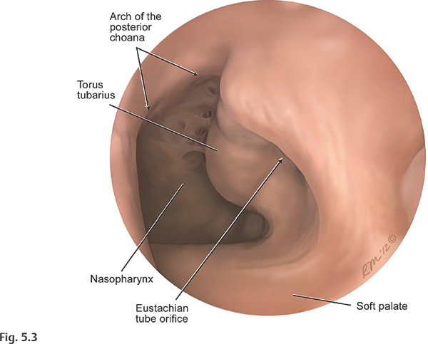
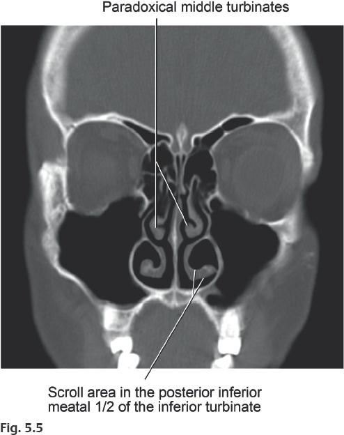
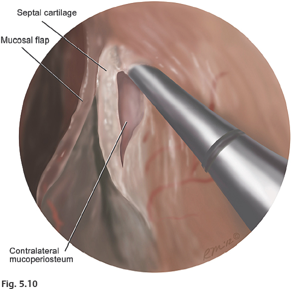
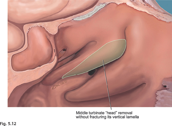
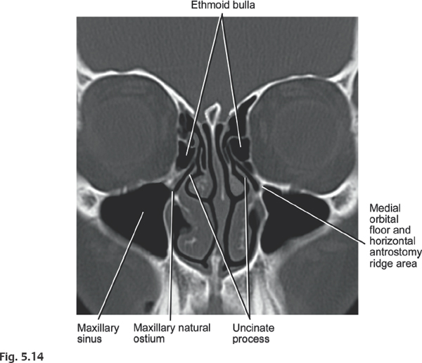
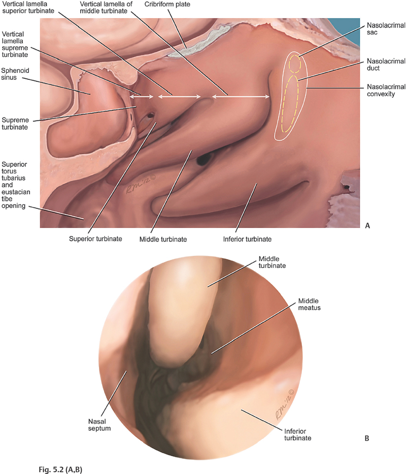
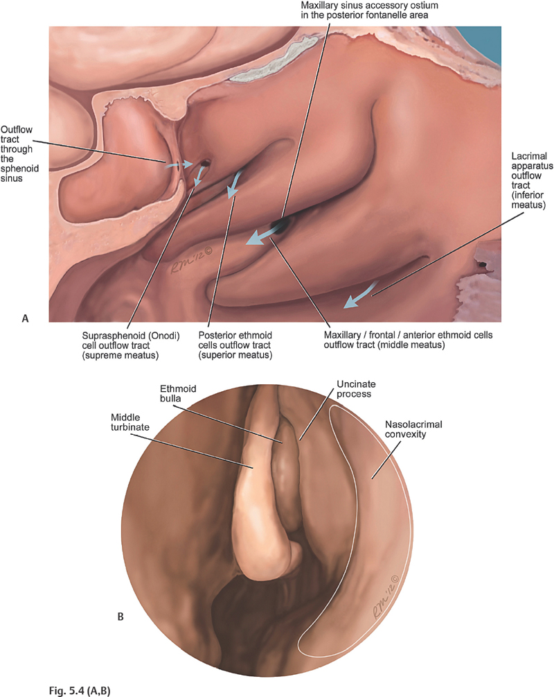
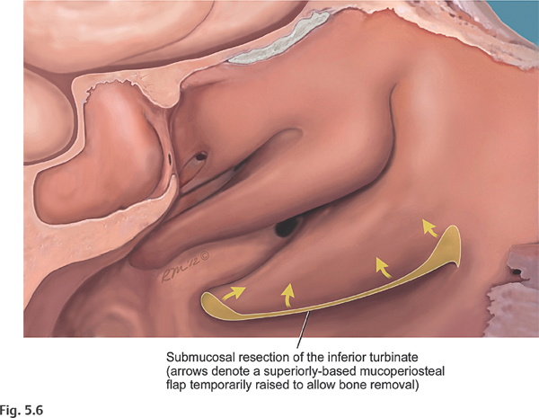
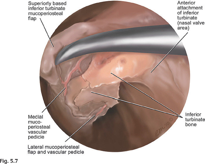
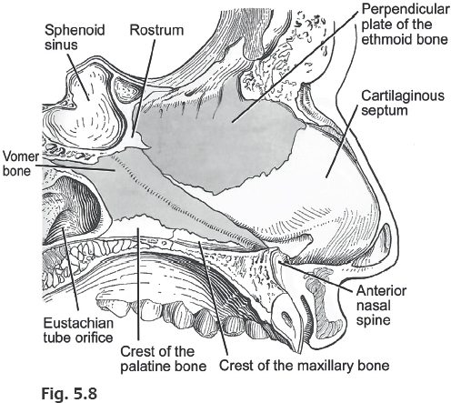
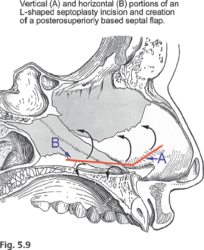
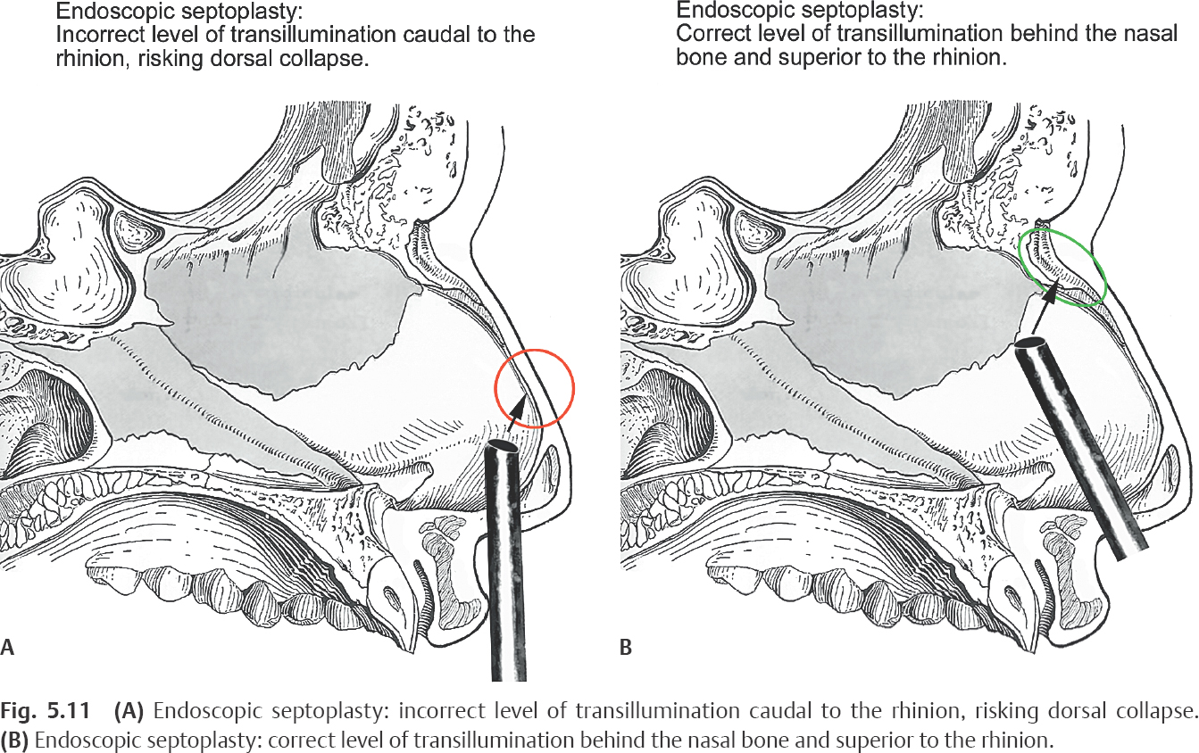
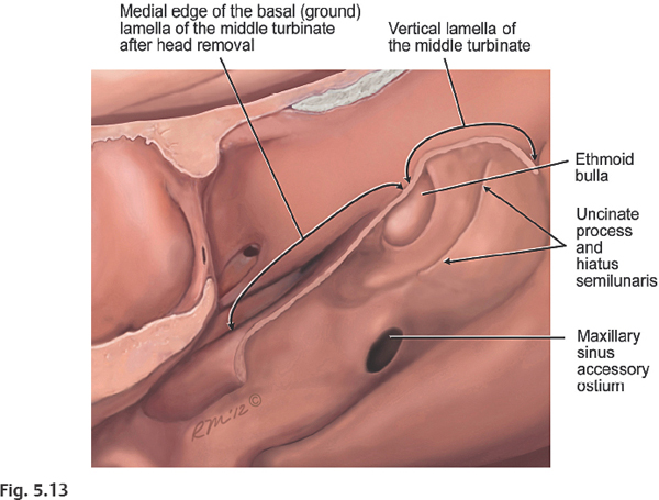
5 Key Landmarks (Fig. 5.1) The surgeon starts examining the nasal fossa by passing a 30-degree telescope posteriorly (looking laterally) along the junction of the inferior and middle turbinates and adjacent to the nasal septum (Fig. 5.2A,B). The structures at the posterior nasal choana (i.e., the posterior nasopharyngeal wall, eustachian tube opening and torus tubarius, posterior choanal arch, posterior septum, and posterior ends of the middle and inferior turbinates) are routinely identified before proceeding with endoscopic surgery of the paranasal sinuses (Fig. 5.3). Early identification of these structures establishes the antero-posterior dimensions of the nasal airway, provides a drainage route for blood into the nasopharynx, and facilitates the introduction of endoscopic surgical instrumentation and telescopes. The anterior ostiomeatal complex (ethmoid bulla, uncinate, and surrounding recesses and drainage outflow track for the maxillary, frontal, and suprabullar ethmoid air cells), can be seen by gentle medial displacement of the middle turbinate, toward the nasal septum (Fig. 5.4A,B). A separate contralateral suction may be used for the continuous evacuation of accumulated blood and debris from the nasopharynx. When bilateral polyp disease is present, a bilateral nasal polypectomy is performed first, to reestablish the anteroposterior dimensions of the nose, as well as to facilitate the placement of a contralateral nasopharyngeal suction. Suction-irrigation is performed as necessary. Monopolar or bipolar suction cautery is helpful, if discrete bleeding vessels are encountered during surgery. However, excessive cauterization should be avoided to minimize crusting and prolonged healing in these areas. Key Landmarks (Fig. 5.5) An endoscopic inferior turbinoplasty and/or submucous resection bone may be indicated when there is poor endoscopic visualization of the nasal and posterior choanal structures, or symptomatic nasal obstruction due to turbinate hypertrophy.1,2 Frequently, inferior turbinate bone enlargement may contribute to the turbinate hypertrophy and resultant nasal obstruction resistant to medical treatment.3 Using a 30-degree telescope, a microdebrider is used to perform an inferolateral incision along the inferior edge of the inferior turbinate. Alternatively, this incision can be performed with a sickle knife or a cutting forceps. Mucosal flaps are then raised on the medial and lateral surfaces of the inferior turbi-nate, and the turbinate bone is partially removed in a piecemeal fashion (Figs. 5.6 and 5.7). To minimize the chance of secondary maxillary sinusitis, care should be taken to avoid fracturing the inferior turbinate lamellar attachment to the lateral nasal wall, adjacent to the maxillary natural ostium.4 For additional airway space, the lateral mucosal flap in the inferior meatus and “scroll” area (if present) is trimmed as needed to remove redundant mucosa (Fig. 5.5). At the completion of the procedure, the medial and lateral mucosal flaps of the inferior turbinate are reposed along the entire anteroposterior extent of the inferior turbinate. This minimizes the chance of prolonged crusting due to exposed bone (osteitis) or de-epithelialized surfaces. Key Landmarks (Fig. 5.8) A significant septal spur or deviation may preclude adequate endoscopic visualization or adversely affect nasal airway patency. In these cases, an endoscopic septoplasty may be indicated.5–8 Using a 30-degree telescope looking slightly superomedi-ally, an ipsilateral L-shaped or T-shaped incision is performed in the septal mucosa. The vertical portion of this incision (‘A’ in Fig. 5.9) is performed immediately anterior to the deviated area to facilitate cartilage or bone removal. The horizontal portion of the incision ‘B’ in Fig. 5.9) is made perpendicular to the vertical incision at the junction of the floor and nasal septum or just slightly superior to this point, depending on the extent of the deviation. The incision should be made only through the mucosa on the ipsilateral side (Fig. 5.9). Through this incision, a posterosuperiorly based mucoperiochondrial flap is elevated on the ipsilateral side. The incision is then carefully advanced (with a periosteal elevator) through the septal cartilage where the contralateral mucoperichodrium is identified, elevated, and preserved to avoid the chance of a permanent septal perforation (Fig. 5.10). The septal spur or deviated portion of the nasal septum is then removed. Occasionally, it is necessary to remove a strip of the perpendicular plate bone just posterior to the coronal plane of the rhinion to free up a caudal deflection and break the cartilage “spring” caudal to this area. Nevertheless, dorsal and caudal struts of septal cartilage are always preserved to avoid the chance of septal collapse and saddle-nose deformity. Periodic transillumination should reveal a bright light posterosuperior to the rhinion, where it is safe to remove cartilage or bone without the risk of dorsal collapse (Fig. 5.11A). Transillumination caudal to the rhinion implies that the surgeon is removing cartilage too close to the dorsal strut with impending loss of dorsal support (Fig. 5.11B). Key Landmarks When the middle turbinate is enlarged, a middle turbinoplasty may be indicated.9,10 Middle turbi-nate enlargement may be due to mucosal hyper-trophy or a concha bullosa. Middle turbinate reduction may be indicated to improve access to the middle meatal structures, sphenoethmoidal recess and sphenoid ostium. Although controversial, it may be also indicated to relieve headache caused by contact between the enlarged middle turbinate and the septum. A conservative reduction of the middle turbinate head can be performed whereby visualization of the middle or superior meatal structures is improved without adversely affecting olfaction, ostial drainage from the anterior ethmoids or frontal sinuses, or the patient’s airway.11,12 The procedure is performed using a tru-cut biting forceps starting anteriorly and moving posteriorly toward the tail of the middle turbinate. The posterior attachment of the resected portion of the middle turbinate is usually freed with a microdebrider and cauterized. Care is taken to sharply resect the middle turbinate head while avoiding fracturing or de-epithelializing the vertical lamella of the turbinate adjacent to the olfactory cleft (Fig. 5.12). In case of a concha bullosa, reduction of lateral portion of the middle turbinate can be also performed using powered instrumentation. However, the oscillations of the microdebrider may inadvertently fracture the vertical lamella of the middle turbinate, rendering it unstable. In the absence of a stable basal lamella (discussed in later sections), this may result in lateralization of the middle turbinate with maxillary, ethmoid air cells, or frontal sinus obstruction. If this occurs, the ethmoid cavity can be temporarily lightly packed with resorbable or nonresorbable packing, to keep the middle turbinate from lateralizing. Alternatively, an absorbable suture may be placed between the middle turbinate and nasal septum. The mucosal membranes on the medial aspect of the middle turbinate as well as around its “axilla” are preserved to avoid scarring around the olfactory cleft or frontal recess/orbital wall, respectively (Fig. 5.13). Key Landmarks (Fig. 5.14) Using an angled probe, the uncinate process, hiatus semilunaris, and infundibulum are identified. The uncinate process is gently back-fractured with the angled probe and carefully removed with a back-biting forceps or powered instrumentation to expose the lateral (orbital) wall of the infundibulum and the maxillary sinus natural ostium (Fig. 5.15A,B,C). Care is taken to conserve the mucosal membranes of the lateral infundibular wall. The tail or posteroinferior remnant of the uncinate may occlude the natural ostium. Thus, this remnant must be identified and removed to clearly see the natural ostium of the maxillary sinus. The superior border of the natural ostium demarcates the junction of the medial orbital floor (MOF) with the lamina papyracea (i.e., the junction of the floor and the me-dial wall of the orbit) (Figs. 5.16 and 5.17).
Basic Endoscopic Sinonasal Dissection
 Intranasal Examination
 Inferior and middle turbinates
 Nasal septum
 Posterior choanal arch
 Eustachian tube opening
 Nasolacrimal convexity
 Inferior Turbinoplasty and Submucous Resection of the Inferior Turbinate
 Inferior turbinate “scroll” area
 Middle turbinate anterior and posterior (tail) attachment
 Lamellar attachment to the lateral nasal wall
 Septoplasty
 Perpendicular plate of the ethmoid bone
 Anterior nasal spine
 Cartilaginous septum
 Rhinion
 Middle Turbinoplasty
 Middle turbinate anterior attachment and “axilla”
 Posterior attachment or tail
 Vertical lamella
 Basal or ground lamella
 Maxillary Sinusotomy
 Uncinate process inferior attachment
 Medial orbital floor (MOF)
 Horizontal, transitional, and vertical antrostomy ridge
 Posterior fontanelle area and posterior third of the inferior turbinate
Ento Key
Fastest Otolaryngology & Ophthalmology Insight Engine