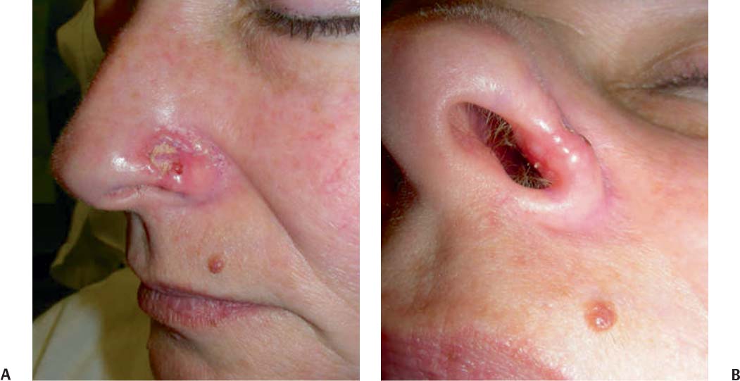
39 A 42-year-old white woman had a 1-year history of an enlarging lesion involving her left nasal ala. During the past 3 months, the lesion bled intermittently. Medical history is significant for a 10 pack-year cigarette smoking history. Family history is notable for melanoma in her father. Physical examination revealed a well-developed, well-nourished middle-aged white woman who appeared older than her stated age. Examination of her face revealed significant photoaging. Involving her entire left nasal ala and extending into the nasal vestibule was an ill-defined, approximately 3.0×2.5 cm brightly erythematous, telangiectatic, crusted, focally ulcerated plaque with pearly borders (Fig. 39.1). The remainder of the physical examination was within normal limits. Basal cell carcinoma (BCC) subtypes and the differential diagnosis: 1. Nodular BCC. The most common subtype, commonly found on sun-exposed areas (head, neck), often appears as a telangiectatic pearly papule with rolled borders and frequent ulceration. Differential diagnosis includes dermal nevus, seborrheic keratosis, amelanotic melanoma, and sebaceous carcinoma. 2. Pigmented BCC. This subtype appears as a hyperpigmented, sometimes pearly papule. Differential diagnosis includes compound nevus, blue nevus, appendageal tumor, seborrheic keratosis, and nodular melanoma. 3. Superficial BCC. This subtype appears most commonly as an erythematous patch or plaque on the trunk. Differential diagnosis includes a single plaque of dermatitis or psoriasis, Bowen disease, Paget disease, tinea corporis, and squamous cell carcinoma. 4. Morpheaform (sclerosing) BCC. This aggressive variant appears as a white or pink plaque. Differential diagnosis includes scar tissue and morphea. 5. Fibroepithelioma of Pinkus. This subtype often appears as a pink papule on the trunk. Differential diagnosis includes acrochordon and dermal nevus. A biopsy is the diagnostic procedure of choice. Biopsy can be performed either by shave biopsy, incisional (punch) biopsy, or excisional biopsy with primary closure. The shave biopsy is usually preferred, with the exception of morpheaform BCC or recurrent BCC in a scar, in which a punch biopsy is more effective. If an incisional biopsy is performed, it is important to include the thickest portion of the nodule and the periphery to show maturation and the deepest area of invasion. The result of the biopsy in this patient revealed nodular BCC invading deep into the subcutaneous tissues. Given the invasive nature of this tumor and the high risk of this location, the following additional evaluation should be considered: Fig. 39.1 Patient photos. (A) Entire left nasal. (B) Nasal vestibule. 1. A complete otolaryngology examination should be performed before removal of the primary tumor so that an estimate of the tumor size and possible spread can be evaluated. 2. If any signs of bone, orbital, or sinus invasion are evident, a preoperative high-resolution CT scan of the orbit and paranasal sinuses should be performed up through the skull base to rule out orbital, sinus, or intracranial invasion. If intracranial invasion is a concern, MRI may be helpful to evaluate dural invasion. If the tumor is extensive enough to warrant a craniofacial resection or orbital exenteration, a metastatic workup that includes a chest radiograph and bone and liver serology should be obtained at the minimum. More focal evaluations can be performed if any signs or symptoms of distant metastasis are evident. Metastatic BCC is exceedingly rare, occurring in less than 0.01% of cases. BCC of the left nasal ala extending into the nasal vestibule
Basal Cell Carcinoma
History
Differential Diagnosis—Key Points
Test Interpretation
Diagnosis
Medical Management
Stay updated, free articles. Join our Telegram channel

Full access? Get Clinical Tree


