Audiologic Tests for Otologic Surgery


Audiologic Tests for Otologic Surgery


Dianne H. Meyer

Edward L. Applebaum


This chapter describes audiologic tests used in the diagnosis and measurement of hearing disorders. The first section reviews basic audiologic procedures, including pure-tone and speech audiometry, acoustic immittance, auditory brainstem response (ABR), and evoked otoacoustic emissions (EOAEs). The following sections discuss the application of these basic procedures, as well as other specialized tests, in the evaluation of conductive, sensorineural, and central auditory disorders. Two patient groups, young children and patients with nonorganic hearing loss, require modified procedures, and these are discussed in the last sections. An effective audiologic evaluation depends on careful test selection and knowledgeable interpretation of the results.


BASIC AUDIOLOGIC PROCEDURES


Pure-tone and Speech Audiometry

The evaluation of all complaints of hearing loss begins with the determination of air- and bone-conduction pure-tone thresholds, the speech recognition threshold (SRT), and word recognition scores. These basic measurements reveal the severity of hearing loss within the tested frequencies and indicate whether the loss is conductive, sensorineural, or mixed. By convention, routine audiometric measurements are made only for the frequencies in the range of 250 to 8,000 Hz, even though normal hearing may encompass a far greater spectrum (20 to 20,000 Hz). Some patients’ complaints of hearing problems and tinnitus may not be diagnosed if the losses occur in frequencies not routinely tested. It is possible to obtain thresholds at frequencies above 8,000 Hz, but specialized equipment and calibration procedures are required.

Speech audiometry refers to measurements of the SRT and word recognition ability. Taken together, these two measures serve as a check on the pure-tone results, help in the diagnosis of retrocochlear problems, and indicate how the hearing loss impacts on the patient’s day-to-day communication. The SRT is the patient’s threshold for spondee words, and it should agree closely with pure-tone thresholds at 500, 1,000, and 2,000 Hz. Word recognition is a suprathreshold test that indicates how well the patient is able to understand speech when it is made loud enough to hear easily. Word recognition is expressed as the percentage of phonetically balanced words correctly recognized at a specified level above the SRT (usually 40 to 50 dB above the SRT). In general, better pure-tone thresholds result in higher word recognition scores.

An essential part of audiometric testing is masking. As a general rule, masking must be used whenever it is possible for the test signal to cross the skull and be heard in the nontest ear. For air-conduction thresholds, this possibility exists when the signal to the test ear exceeds bone conduction sensitivity in the nontest ear by an amount equal to or greater than interaural attenuation. With standard audiometric earphones, attenuation across the skull is 40 dB; with insert phones, the attenuation is 70 to 90 dB, depending on the depth of insertion (1,2). For bone conduction testing, interaural attenuation is essentially nonexistent. Therefore the nontest ear must be masked whenever an air bone gap is present. These masking principles apply for both pure-tone and speech audiometry.

Some hearing disorders are characterized by distinctive audiometric configurations, such as the high-frequency notch associated with noise exposure. Other audiometric patterns, such as the degree of symmetry between ears, may determine if additional evaluation of the hearing loss is needed.

Pure-tone and speech audiometry should be done for all patients before and after otologic surgery, unless the patient is unable to cooperate. To allow for adequate healing after surgery, the first postoperative test usually is deferred until about 6 weeks after the operation. If a postoperative complication is suspected, testing is done sooner. Follow-up audiometry may be done as early as 1 to 2 weeks after myringotomy with placement of ventilating tubes and still be indicative of the hearing improvement resulting from the surgery.


Acoustic Immittance Measurements

Acoustic immittance measurements evaluate how well energy flows through the outer and middle ear systems. The basic measurements include tympanometry and acoustic reflexes, both of which are often considered routine parts of an audiologic evaluation. Immittance measurements are sensitive to middle ear problems, and they help to differentiate cochlear from retrocochlear disorders. The measurements are made by delivering a pure-tone signal through a probe that fits snugly in the ear canal. The sound pressure level (SPL) of this “probe tone” is monitored while the air pressure is varied in the external ear canal. Any changes from the normal SPL patterns are related to the functional integrity of the ear.

Several classification systems have been proposed for tympanograms, some descriptive in nature and others that analyze shapes based on multiple probe tone frequencies and resistance and reactance components. 
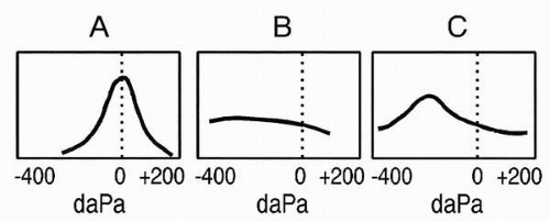
Figure 3.1 shows a commonly used classification that categorizes tympanograms on the basis of shape and tympanometric pressure peak (3). These basic types are based on adult ears and are associated with a low-frequency probe tone, usually 226 Hz. Type A is a normal tympanogram with peak immittance at or near 0 decaPascals (daPa). Variations of type A include type As, in which the peak is shallower than normal, and type AD, in which the peak is higher than normal. Type As may be found in cases of ossicular fixation, whereas type AD may be found with ossicular discontinuity or tympanic membrane abnormality. The type B, or “flat,” tympanogram depicts very little or no change in immittance with variation in air pressure and is found in cases of middle ear effusion. The type C tympanogram has a negative peak, indicating negative air pressure in the middle ear space.

Tympanometry is ordered when there is a question about the mobility of the tympanic membrane, the air pressure within the middle ear space, or the status of the ossicular chain. It provides useful information about middle ear effusions that may not be obvious clinically, such as an effusion behind a thick or opacified tympanic membrane that impairs otoscopic evaluation of the middle ear. Tympanometry is done to determine the presence of persistent, abnormally high or low middle ear pressures in patients with complaints of aural fullness. It is valuable in differentiating between abnormally fixed or interrupted ossicular chains behind an intact tympanic membrane. In patients with rhythmic tinnitus or clicking sensations in their ears, tympanometry is used to determine if the tympanic membrane is being contracted abnormally by clonic middle ear muscle contractions.


FIG. 3.1 A commonly used classification system for tympanograms. Type A is a normal tympanogram with peak immittance at or near 0 daPa. The type B tympanogram indicates little or no change in immittance with variation in air pressure. The type C tympanogram indicates the presence of negative air pressure in the middle ear space. daPa, deca-Pascals.

The acoustic reflex, or stapedius muscle reflex, is a bilateral response that occurs in response to loud sounds. In normal ears the reflex occurs at 70- to 100-dB hearing level (HL) for pure-tone signals (4). Clinical measurements include reflex threshold, the lowest sound level that elicits the reflex, and reflex decay. Both contralateral and ipsilateral reflexes can be measured, thereby testing the entire reflex arc, including eighth nerve, low brainstem, and facial nerve pathways. Acoustic reflex testing is ordered when lesions of these structures are suspected, such as in acoustic neuromas and brainstem infarctions, and to help determine the site of involvement of facial nerve disorders.


Auditory Brainstem Response

As one of several groups of auditory evoked potentials, the auditory brainstem response does not directly measure hearing but does measure a process that is highly related to hearing sensitivity. The ABR measures electrical activity of the auditory nerve and auditory pathway to the mid-brainstem level. The ABR offers the advantages of being easy to measure in children and adults. It is sensitive to both auditory and neurologic disorders but does not require a behavioral response from the patient.

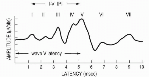
The ABR consists of a series of five to seven waves with latencies between 1 and 10 msec following stimulus presentation (Fig. 3.2). Waves I and II are generated by the peripheral auditory nerve, and waves III, IV, and V are generally related to the cochlear nucleus, superior olivary complex, and nuclei of the lateral lemniscus, respectively (5). Clicks are the best stimuli to elicit the ABR because of their abrupt onset and broad frequency spectrum. The click-evoked ABR provides information about the basal end of the cochlea and is associated with hearing sensitivity in the 2,000 to 4,000 Hz region. More frequency-specific information may be obtained with tone bursts, filtered clicks, and masking techniques.

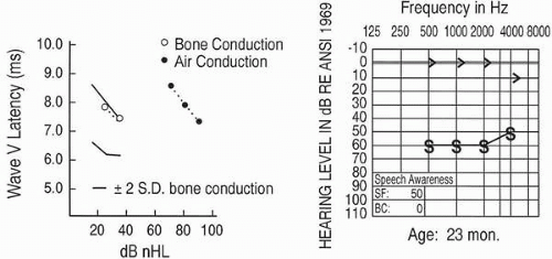
Clinical interpretation of the ABR is based on measurements of wave latencies, interpeak intervals (IPI), and wave V threshold (see Fig. 3.2). The 
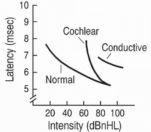
wave V latency-intensity function refers to the increase in wave V latency as stimulus intensity decreases. Changes in the shape of the latency-intensity function and in the amount that it deviates from the expected normal function are used to predict type and degree of hearing losses. Patterns associated with conductive
hearing loss and with high-frequency cochlear hearing loss are shown in Figure 3.3.


FIG. 3.2 An ABR waveform from a normal-hearing adult subject. The arrow illustrates measurement of the I to V interpeak interval (IPI).

The clinical applications of the ABR are varied. The procedure plays an important role in determining auditory thresholds of young infants and other difficult-to-test patients. It is useful in detecting the presence of lesions in the auditory pathway, such as in acoustic neuroma or multiple sclerosis. It also is valuable for intraoperative monitoring of the integrity of the ear and auditory pathway during intracranial surgery near these structures. Automated versions of the ABR are used in newborn hearing screening.


FIG. 3.3 Wave V latency-intensity functions. General patterns are shown for normal hearing, high-frequency cochlear hearing loss, and conductive hearing loss.


Evoked Otoacoustic Emissions

Evoked otoacoustic emissions are sounds recorded in the ear canal that are associated with normal cochlear function. They do not measure hearing sensitivity directly, but their presence is highly related to normal outer hair cell activity. Although they may occur spontaneously, the most common clinical application is to measure the emissions in response to an evoking stimulus. Transient evoked otoacoustic emissions (TEOAEs), which occur after the presentation of a brief stimulus such as a click, typically are measured if hearing thresholds do not exceed 30-dB HL. Distortion product otoacoustic emissions (DPOAEs) are produced by the ear in response to two simultaneous pure-tones. They are present if hearing thresholds do not exceed 50 dB HL (6). The middle ear system must be normal in order for EOAEs to be measured.

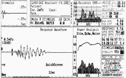
Clinical interpretation of EOAEs is based on amplitude of the emission at a specific frequency or across a frequency range. To be accepted as a response, the EOAE must have good replicability and must exceed the noise floor measured at the time of testing. Figure 3.4 shows a normal TEOAE response from an adult.

The measurement of EOAEs has many clinical applications, including differentiation between sensory and neural hearing loss, monitoring of cochlear function of patients treated with ototoxic drugs, tinnitus evaluation, and monitoring of noise-induced hearing loss. EOAEs are essential to the audiologic evaluation of young infants and other difficult-to-test patients. They are used extensively in newborn hearing screening.


FIG. 3.4 A transient evoked otoacoustic emission result for a normal-hearing adult patient. The response waveforms (A and B) are superimposed, reflecting high reproducibility (96%). The signal-to-noise ratio (SNR) shows a strong emission response from 800 to 3,200 Hz.


AUDIOLOGIC EVALUATION OF CONDUCTIVE HEARING LOSS

An audiologic evaluation for conductive hearing losses should include pure-tone air- and bone-conduction thresholds, speech audiometry, and immittance measurements. Careful masking must be used in all cases. The ABR may be used to estimate conductive hearing losses when patients are unable to give reliable behavioral responses. Patterns of air-conduction thresholds alone are not sufficient to diagnose conductive hearing losses, as was once thought.

Clinical indicators published by the American Academy of Otolaryngology-Head & Neck Surgery recommended pure-tone and speech audiometry be completed preoperatively and postoperatively for mastoidectomy, myringotomy and tympanostomy tubes, stapedectomy, and tympanoplasty procedures (7). In addition, the guidelines recommended tympanometry prior to myringotomy and tympanostomy tubes, and tympanometry plus acoustic reflex testing prior to stapedectomy surgery.


Pure-tone and Speech Audiometry

The expected finding on the pure-tone audiogram is the air-bone gap, indicated by better thresholds by bone conduction than by air conduction. Although air- and bone-conduction thresholds alone cannot identify a specific ear disease, they may reflect changes that have occurred in the middle ear transmission properties. A greater loss at the low frequencies suggests an increase in the stiffness of the middle ear system, whereas a greater loss at the high frequencies is associated with an increase in the mass of the system. An example of these relationships can be seen in the course of otitis media (8). In the early stages of the disease, as middle ear air pressure is reduced, the tympanic membrane and ossicular chain may become increasingly stiff, resulting in a rising air conduction audiometric configuration with normal bone conduction. Later, high-frequency thresholds may be affected as fluid accumulates and causes mass loading of the ossicular chain. The additional loss in sensitivity at the high frequencies results in a flat audiometric configuration.

Middle ear disorders also may influence bone-conduction thresholds. Changes in bone conduction can occur when the disease process alters the resonant characteristics of the ossicular system or middle ear space. For example, the “Carhart notch” associated with otosclerosis is a slight loss in bone-conduction threshold maximal in the frequency region around 2,000 Hz (9). The phenomenon occurs because otosclerotic stapes fixation eliminates the influence of normal ossicular resonance on bone-conduction thresholds at 1,500 to 2,000 Hz. Following surgery, this mild depression in bone-conduction thresholds is not seen, explaining why
there is sometimes an apparent improvement in bone-conduction thresholds (“overclosure” of the air-bone gap).

Speech audiometry with conductive hearing loss should result in SRTs that are in agreement with air-conduction thresholds in the most important speech frequencies (500 to 2,000 Hz). If bone-conduction responses are normal, word recognition scores should be 90% or better when test words are delivered at a sufficiently high sensation level, usually about 40 dB above the SRT. It also is possible to administer speech audiometry via bone conduction if the audiometer is calibrated for speech delivered through the bone receiver. This technique is especially useful with patients who show poor reliability in air- and bone-conduction pure-tone testing.

Expected audiometric results for conductive disorders are described in Table 3.1. It is apparent that different types of middle ear disease may have similar effects on the sound conductive system and therefore may result in similar audiometric configurations.


TABLE 3.1. Summary of audiometric and immittance findings for selected conductive hearing losses


Disease


Audiometric findings


Tympanogram


Acoustic reflex


Aural atresia


Maximum conductive loss


Cannot evaluate


Cannot evaluate


Cholesteatoma


Hearing loss varies, depending on the location and size of the cholesteatoma; occasionally hearing is normal (10)


Type AS, AD, or B, depending on ossicular chain involvement


Usually elevated or absent with probe and tone to the involved ear


Discontinuity of the ossicular chain


Flat configuration, 50-60 dB (11)


Type AD (low-frequency probe)


Usually absent with probe and tone to the involved ear


Glomus tumors


Conductive, mixed, or sensorineural loss; degree of loss ranges from normal to severe (12)


Variable, depending on ossicular chain involvement; may show vascular perturbations


With probe to involved ear, reflexes may be absent or impossible to measure due to pulsations


Malleus fixation


Usually a rising configuration due to increased stiffness; 40-60 dB; sensorineural component may be present (13)


Type B or AS


Absent with sound and probe to the involved ear


Otitis media with effusion


Audiometric contour depends on the stage of the disease and the relative stiffness and mass effects; usually ranges between 10 and 40 dB (14)


In early stages, type C, and then progresses to type B


Absent with sound and probe to the involved ear


Otosclerosis


Initially a rising configuration; as stapes fixation increases, the configuration flattens; maximum air-bone gap about 50 dB; bone conduction may show Carhart notch at 2 kHz (15)


Type A or AS


Usually absent with sound and probe to the involved ear; in early stages, reflexes may be present and characterized by negative deflections and an on-off effect


Perforation of the tympanic membrane (TM)


Hearing loss depends on size and location of the perforation; small perforations in the posterior half of the TM affect low-frequency hearing; perforations in the anterior area affect high-frequency hearing (16)


Type B


Absent with probe to the involved ear


Atelectasis


Variable, depending on extent of tympanic membrane collapse


Type C or B


Often absent


Tympanosclerosis


Mild to moderate loss, depending on amount and location of plaque deposits (17)


Type C or AS; type A if tympanic membrane is not involved


May be absent with probe to the involved ear


Audiometric Masking

Appropriate masking is important in the evaluation of all hearing losses, but it is critical with conductive hearing loss. Unrecognized errors can lead to useless and perhaps even harmful surgery on a nonhearing ear. Bilateral conductive loss presents a special audiometric testing problem referred to as the masking dilemma. In this situation the intensity level of the masking noise needed to mask the nontest ear is so high (due to the degree of air-conduction loss in that ear) that the masking noise itself may cross over via bone conduction and elevate the threshold in the test ear. The use of insert phones, rather than supra-aural earphones, often eliminates the need for masking and avoids many masking dilemmas.


Acoustic Immittance Measurements

Tympanometric and acoustic reflex findings in combination with a complete audiogram can confirm the presence of
conductive hearing loss and differentiate among types of conductive pathology. Table 3.1 includes the tympanogram type and acoustic reflex pattern typically found for selected conductive hearing disorders.

When interpreting tympanogram shapes, it must be remembered that in the presence of more than one middle ear lesion, the more lateral pathology will dominate. For example, the combination of otosclerosis and a monomeric tympanic membrane will likely result in a tympanogram with high compliance, reflecting the thin and flaccid tympanic membrane.

The acoustic reflex is extremely sensitive to middle ear pathology, even when conductive hearing loss is not apparent on the pure-tone audiogram. An air-bone gap of only 5 dB in the probe ear is enough to obscure measurement of the reflex, and an air-bone gap of greater than 30 dB in the stimulated ear will prevent measurement of the reflex in a normal hearing probe ear (18). A distinctive pattern occurs with early otosclerosis in that reflexes may show a negative, biphasic pattern known as the on-off effect (19).


Auditory Brainstem Response

The ABR may be needed to identify and quantify conductive hearing loss in patients unable to be evaluated by behavioral methods, such as newborns, infants, young children, and developmentally delayed or cognitively impaired patients. The effect of conductive pathology on the click-evoked ABR is to prolong the latencies of the component waves. As shown in Figure 3.3, the slope of the latency-intensity function with conductive hearing loss is parallel to the normal slope. The amount in decibels that the wave V latency-intensity function is shifted approximates the degree of conductive hearing loss. All the ABR waves are shifted approximately equally, so the interpeak intervals remain within normal limits.


FIG. 3.5 ABR and behavioral test results for a 23-month-old infant with bilateral microtia, complete atresia of the right external auditory canal, and partial atresia of the left external auditory canal. ABR results are shown for the left ear. Responses to bone conduction ABR were in the expected normal range and were consistent with behavioral bone conduction results. Responses to bone conduction ABR showed an expected shift in the latency-intensity function.

The bone-conduction ABR provides a more direct estimate of cochlear function. Bone-conduction ABR is especially valuable in infants with aural malformations in whom air-conduction and immittance measurements may be difficult or impossible to do. In newborns who fail hearing screening, ABR may help to identify conductive involvement.

Figure 3.5 shows air conduction and bone conduction for a 23-month-old infant with bilateral microtia, complete atresia of the right external auditory canal, and partial atresia of the left external auditory canal. Inner ear structures appeared normal on computerized tomography. There was a shift in the ABR latency-intensity function for air conduction, but the bone-conduction responses fell within the expected normal range. The ABR results supported the behavioral responses that indicated a 60-dB conductive hearing loss. This case also demonstrates how contralateral masking may be needed with ABR and that masking dilemmas may exist. Because of the patient’s absent or malformed external auditory canals, insert receivers could not be used to increase the interaural attenuation. The ABR results therefore reflected function of the better cochlea if a difference in hearing existed between the two ears. Bone-conduction ABR has a number of calibration and interpretive considerations, but it is invaluable as a procedure to identify or confirm conductive involvement in difficult-to-test patients.


AUDIOLOGIC EVALUATION OF SENSORINEURAL HEARING LOSS

Pure-tone and speech audiometry and immittance measurements are obtained for all patients with suspected pathology of the cochlea and/or eighth nerve. The ABR is included when a lesion of the eighth nerve is suspected and when behavioral thresholds are in question or cannot be done. Another
auditory evoked potential, electrocochleography (EcochG), is useful in the diagnosis of Ménière’s disease. Table 3.2 summarizes the audiometric results for selected sensorineural hearing losses.


Pure-tone and Speech Audiometry

If otoscopic findings and all immittance results are normal, it may not be necessary to obtain bone-conduction thresholds. Because outer and middle ear function are normal, air-conduction thresholds alone will reveal the severity and symmetry of the hearing loss. For SNHL such as endolymphatic hydrops, periodic pure-tone threshold testing is helpful in evaluating the benefits of treatment or progression of the disease.


TABLE 3.2. Summary of site of disorder and audiometric findings for selected sensorineural hearing losses

Only gold members can continue reading. Log In or Register to continue

Stay updated, free articles. Join our Telegram channel

Sep 23, 2016 | Posted by in OTOLARYNGOLOGY | Comments Off on Audiologic Tests for Otologic Surgery

Full access? Get Clinical Tree

Get Clinical Tree app for offline access